Igor D. Alexandrov
1,2* , Victor A.
Stepanenko
3and Margarita V.
Alexandrova
1,21Dzhelepov Laboratory of Nuclear Problems, Joint Institute for Nuclear Research, Dubna 141980, Moscow Region, Russia
2Vavilov Institute of General Genetics, Russian Academy of Sciences, 119991, Moscow, Russia 3Laboratory of Information Technologies, Joint
Institute for Nuclear Research, Dubna 141980, Moscow Region, Russia
Abstract
Data on the non-random distribution of radiation-induced inversion breakpoints were used as genet-ic markers for 2D and 3D modeling of the large-scale architecture of the chromosome 2 in
Drosophila sperm genome at the time of
irradia-tion. During modeling, Gmax program with applica-tion of splines, the addiapplica-tional objects and opera-tions as well as the methods of visualization of macromolecular biostructures were employed. A specific megarosette-loop model of spatial arrangement of chromatin fiber in the haploid sperm nucleus can account well for the experimen-tal data.
Key words: 2D/3D models, large-scale chromosome geometry, sperm genome, Drosophila.
Introduction
The development of modern chromosome fluores-cence in situ hybridization (FISH) methods (Agard et al., 1989; Trask et al., 1993) has permitted to perform 2D and 3D visualization of distinct chromosomes (or “chromosomal territories” (CTs), according to Theodor Boveri, 1909) as well as euchromatic (coding) and het-erochromatic (condensed centromeric) chromosomal regions in interphase nuclei of animal and human somatic cells (Croft et al., 1999; Edelmann et al., 2001; Habermann et al., 2001; Lukasova et al., 2002; Tanabe et al., 2002; Manders et al., 2003; Kreth et al., 2004).
These observations have also given an impetus to creation of vastly different current models for the large-scale geometric arrangement of interphase chro-mosomes in eukaryotic somatic cells (Sachs et al., 1995; Cremer and Cremer, 2001; Solovei et al., 2002) the basic propositions of which appear to strengthen a classical model (Rabl, 1885) of non-random radial (polar) configuration of somatic interphase chromo-somes in respect to both its major features, namely, that the chromosomes occupy distinct nuclear domains throughout the cell cycle and centromeric and telomeric regions are located at the opposite side of the nucleus (inner and peripheral parts of the cells nucleus, respectively, according to present definition).
This polar orientation of CTs has been clearly seen in most chromosomes and most cell types investigat-ed although non-random polar and random non-polar conformations may be persisted for different chromo-somes in the same cell nucleus (Skalnikova et al., 2000).
Unlike the data for diploid somatic cells, it is unknown so far what kinds of chromosome configura-tion patterns correspond to those in haploid sper-matids and mature male germ cells, especially, taking into account the unique chromatin remodeling (the his-tone-to-protamine transition) in late spermiogenesis (Sassone-Corsi, 2002; Kimmins and Sassone-Corsi, 2005).
*Correspondence Author:
Dzhelepoj Laboratory of Nuclear Problems, Joint Institute for Nuclear Research, Dubna 141980, Moscow Region, Russia e-mail: [email protected]
Received: June 18, 2007; Accepted: July 18, 2007.
39 spatial arrangement of the animal male germ cell
genome: II. 2d-3d Simulation and visualization of spatial
configuration of major chromosome 2 in Drosophila sperm
In order to clarify the issue as to whether so-called Rabl orientation of chromosomes still persists in mature sperm genome after spermatid differentiation, the pattern of radiation-induced intrachromosomal exchanges (inversions) in Drosophila melanogaster autosome 2 was studied after γ-rays or neutron irradi-ation of D-32 wild-type adult males.
The following two facts were taken into account to select such approach for clarification of the issue of interest. Firstly, no there is direct approaches to-day to examine the sperm chromosomes by the cytological or physical techniques without disruption of the existing organization. Secondly, analysis of radiation-induced chromosome exchanges which are assumed to devel-op from DNA double-strand breaks (DSBs) and ran-dom pairwise interactions (Savage, 1989) have been shown to be a useful tool for reconstruction of the large-scale chromosome geometry at the time of irra-diation of interphase somatic cells (Sachs et al., 1993; Ostashevsky, 2000) since the assuming complete ran-domness of DSB pairwise interaction resulting in the exchanges scored may be drastically modified with position of interacted DSBs in genome (Skalnikova et al., 2000). For example, two genetic loci with DSBs on the same chromosome which are well separated in most nuclei obviously cannot be close to each other in order to interact and vice versa, two genetic loci which are spatially close to each other in most nuclei must frequently interact yielding the high frequency of inver-sion expected.
Using this approach for Drosophila male germ cells, we showed previously that the chromosome dis-tribution of second inversion breakpoints relative to the “fixed” first one (genetic locus vg at the middle of 2R arm of autosome 2) is highly non-random showing most often contacts and interactions of this genetic locus with centromeric region and both telomeric ends of autosome 2 in mature sperms irradiated by γ-rays or fission neutrons (Alexandrov et al., 2007). Such non-randomness suggests that the global arrangement of chromosome 2 in sperm nucleus is not in line with the linear folded Rabl configuration but rather matches to the specific giant rosette-loop structure 2D and 3D models of which are proposed in this paper.
Preliminary basic assumption before modelling
Prior to modeling of the spatial configuration of Drosophila autosome 2 in sperm nuclei at the time of irradiation, it is necessary to define some basic assumptions relating to the presumptive chromosome structure in haploid sperm nucleus, the most salient
features of inversion induction pattern detected in irra-diated sperms, and the presupposed physical param-eters of DSBs interactions giving rise to inversions observed.
As it has already been intimated (Alexandrov et al., 2007), the cytological analysis of radiation-induced inversions has been carried out on the interphase poly-tene chromosomes of salivary gland somatic cells. These chromosomes well known to be formed during endonuclear polyploidization (about 1024 n in adult larvae) as a result of many times (28) repeated replica-tions of the elementary haploid parental interphase chromosomes ≈ 30 nm in diameter. The discrete ele-mentary chromatin fibers may be easy identified with the electron microscopy (Ananiev and Barsky, 1985). Moreover, each elementary fiber shows the same banding pattern (the picture of bands or chromomers) as the same polytene chromosome as a whole. Therefore, one can assume that the classical banding and section maps of Drosophila polytene chromo-somes can be extended to the haploid chromochromo-somes in sperm genome of this organism and used in the course of modeling.
According to our experimental data described in the first report, the vg gene (middle of 2R arm of auto-some 2) interacts most often, to give rise radiation-induced inversions, with centromeric heterochromatin (the first “hot” area), with several sections in 2R arm telomeric end (the second “hot” area) as well as in 2L arm telomeric end (the third “hot” area) and with neigh-boring genetic loci in the 48-49 and 50-51 sections of 2R arm. Therefore, on the large-scale geometry of Drosophila interphase chromosome 2 , the genomic separations of the vg gene from “hot” areas lie in the ranges ≈ 10; 8-13;29-32, and 0.3-3 Mb for areas above listed, respectively (Alexandrov et al., 2007). In other cases, the vg gene occasionally interacts with some genetic loci randomly dispersed along the autosome 2. To reconstruct the global chromosome architec-ture and, mainly, to clarify whether so-called polar Rabl configuration of chromosomes still persists in sperm genome, it is not of necessity to deal with acci-dental genetic loci involved in the vg inversions but it will be warranted to pool the relevant genetic loci in each “hot” area and to consider the latter as a “hot” node during modeling. The nodes are primarily a bio-logical entities and their postulated chromosome posi-tion can be selected to fit the situaposi-tion of interest.
The statistically non-random interactions of the vg gene with certain of the autosome 2 areas show that Spatial arrangement of male-cell genome
Alexandrov et al.
the gene under study and all “hot” nodes as its poten-tial “partners” are well close to each other in small “sensitive” nuclear volume the minimal radius of which should not exceed 5-50 nm if to take into account that the haploid genome is compacted within the sperm head to a volume of about 5% of that of a somatic cell nucleus (Sassone-Corsi, 2002) and a distance (r) of pairwise interaction of two DSBs in irradiated somatic cells lies in the range 0.1-1 ηm (Brenner, 1988). Thereat, since the centromeric heterochromatin is located on the nuclear envelope (Zhimulev, 1993), it can be considered as a kind of “anchor” for the entire
autosome 2 around of which the vg gene and other “hot” nodes are grouped.
Basic approaches to modelling
In order to provide a clear visual representation of the chromosome spatial model, the following capabilities were required: subsequent editing, rotation, scaling, displacement of the whole fiber and any chosen object in it, switching on/off of visualization of a selected sub-set of objects of the model, and the adjustment of additional illumination of the whole picture.
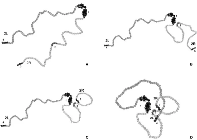
Figure 1. The consecutive steps of 2D modeling of the elementary chromosome 2 fiber macroarchitecture in Drosophila sperm genome. (A) A schematic views of the relaxed chromosome thread in polar Rabl configuration with the “hot” nodes (1,2,3,4) located at the appropriate chromosome regions (see Table 1). (B) The first step of modeling showing the close proximity of centromeric heterochromatin (1) with the vg gene region (2) and the potential “giant” loop giving rise to inversion after radiation-induced recombination between the “hot” nodes. (C) The next step of modeling indicating the proximity of the middle (2) and telomeric end (3) of 2R arm of chromosome 2. (D) The end result of modeling representing the proximity of the vg gene region (2) with the telomeric end (4) of 2L arm of chromosome 2 and the appropriate “giant” loop resulting in the inversion after irradiation. As an idealization, all loops are relaxed.
A B
For these purposes, the Gmax v.1.2 software and some supporting programs were used. This software has the mode of constructing the skeleton of a figure on the basis of splines which are convenient for the solution of the purpose in hand. In particularly, the method of B splines provides a capability of coloring, deforming, rotating, displacing, hiding, or fixing a selected marked subset of object without violation of the whole thread that essentially simplifies the process for modeling of the chromosome fiber (Zaikin et al., 2003).
2D model
Upon construction of the Two-dimensional model, the distribution and frequency patterns of the second inversion breakpoints interacting with the vg gene have been taken into account. For simplicity, the four chromosome “hot” nodes with which the vg gene more often interacts were stood out and the average distances of interaction of each “hot” node with the vg gene were calculated with regard to the number of interaction observed (Table 1) using the equation
where N is the total number of interactions for the four “hot” nodes; ni is the number of interactions with a
particular node, and 25 nm is an average distances of DSBs interactions calculated on the base of the data for somatic cells above mentioned.
After that, the planar projection with the centers of concentric circles in the region of heterochromatin is constructed manually and the reference points of the spline for the chromosome thread are arranged for the right and left arms at different sides, taking into account the frequency of interaction. A rather conven-ient mechanism of the visualization and editing of the spline reference points was used for monitoring; it was possible to change the thickness of the constructed thread. It should be specially noted that, at present, a major part of the chromosome thread are modeled in the relaxed state, i.e., by the smooth spline loop due to the absence of relevant data for a given step of mod-eling. After the visual chromosome thread model was created (Figure 1A), the loops were consecutively constructed: first, the area of the vg gene was “drawn” close to the heterochromatin (Figure 1B); then, the telomeric ends of the 2R and 2L arms of chromosome 2 were arranged relative to the vg gene taking into account the distances of interactions (Table 1). Because the second “hot” node and the vg gene are intimately linked together on the model scale selected these are considered as a single “hot” node during modeling.
As one can be seen from the visual 2D model con-structed (Figure 1D), on the large-scale geometry, an elementary fiber of the chromosome 2 in Drosophila sperm genome shows the specific rosette-loop config-uration. Thereat, the bases of loops are clustered with-in the presumed “sensitive” microvolume associated with the centromeric heterochromatin. This simple 2D model reflects a pattern of principle of the global chro-mosome arrangement for animal sperm genome which Spatial arrangement of male-cell genome
Alexandrov et al.
74
Number of “hot” nodes *
Contents of “hot” nodes Number of interactions with the
vg gene, n
Distance (r) of interaction, nm
1 Centromeric heterochromatin 21 40
2 48‐49 and 50‐51 sections, surrounding vg gene
18 44
3 56‐60 sections (2R telomeric end) 10 59
4 21‐25 sections (2L telomeric end) 7 71
Total (N) 56
Table 1. The average distance of interaction of the vg gene with “hot” regions of the autosome 2 resulting in radiation-induced
inversions in Drosophila melanogaster mature sperms
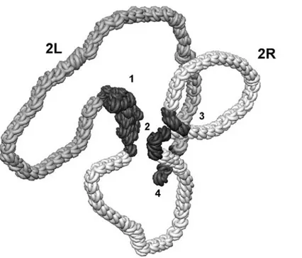
Figure 2. Example of 3D model of an elementary fiber of chromosome 2 in Drosophila sperm genome constructed with using of an
additional object in the form of a spiral-like structure along the spline and contrast coloration for the “hot” nodes.
Figure 3. Example of 3D model of an elementary fiber of chromosome 2 in Drosophila sperm genome constructed with using of
is not clearly consistent with the polar Rabl configura-tion of interphase chromosomes in animal or plant somatic cells. However, this 2D model does not give an idea what this fiber is arranged in space. To eluci-date this issue, 3D modeling of chromosome under study was performed.
3D model
The method of B splines is obviously the most accept-able for the construction of the three-dimensional model of the chromosome taking into account that the chromatide thread is a smooth curve without breaks. In such case, the screen is divided into four equal win-dows of projections in such a way that the lower right window shows the isometric perspective projection of the image and the lower left window shows the planar projection (the views from the right). It is favorable to show the planar frontal projection of the thread above the perspective projection.
During construction of the model, the distance of inversion DSBs interaction calculated (Table 1). They were used as the radii of embedded spheres. According to those data, concentric circles were ini-tially placed at the center of all projection planes. Then the smoothed adaptive spline of order n = 6 was con-structed on the frontal projection. Unlike the case of the construction of the two-dimensional model, the reference points were edited, in turn, by handles on all three projections inside the largest sphere with the radius ro = 70 nm.
At the next stage, it is necessary to color chromo-some arms. For this purpose it is suitable to use two approaches by using the powerful mechanism of “stringing” additional three-dimensional objects onto the constructed spline with quality, initial position or an interobject distance control which is called Spacing Tools. In the simplest case, the chromosome arm is set as the auxiliary three-dimensional object. The color of the arm after their superposition onto the spline is changed manually and their position on the planar pro-jections is edited for a more accurate edge matching.
The method for the construction of the additional three-dimensional object strongly twisted into a spiral, in the form of the Torus Knot, turned out to be most pictorial for coloring sections of the three-dimensional model. A rather large number of these “knots” are located on the spline, but repeat all its curls more smoothly. For this reason it is then required just to change the color of the knots. It is performed easily using the operation of the group selection of objects
called SelectbyNAME. Then, it is usually necessary to correct the illumination of the picture using different light sources, to rotate the thread for better view, and to add section markers if required (Chumachenko, 2004). It is very convenient to use the mechanism of the temporary removal of selected objects from the picture for the separate visualization of heterochro-matin, or a particular arm of the chromosome thread (Zaikin at al., 2005).
The examples of the construction of 3D model of Drosophila chromosome 2 in sperm genome using the additional objects and operations are pictured in Fig. 2 and 3. It is significant that the chromosome regions without relevant inversion breakpoints were modeled in the relaxed state as in the case of 2D model. More complex and more realistic 2D and 3D models con-structed with consideration of independently arising inversions in the chromosome 2 of the “point” b, cn and vg mutants as well as of inversions with an invari-ant break at the b gene (middle of 2L arm, section 34D) of the same chromosome will be presented at the next paper.
References
Agard DA, Hiraoka Y, Shaw P and Sedat UW, Fluorescence microscopy in three dimensions. Meth Cell Biol. 30: 353-377, 1989.
Alexandrov ID, Alexandrova MV, Korablinova SV and Korovina LN. Spatial arrangement of animal male germ cell genome: 1. Non-random pattern of radiation-induced inversions involving the vestigial region in autosome 2 of Drosophila melanogaster. Advances in Molecular Biology. 1: 23-29, 2007. Ananiev EV and Barsky VE. Elementary structures in
polytene chromosomes of Drosophila melanogaster. Chromosoma. 93:104-112, 1985 Boveri T. Die Blastomerenkerne von Ascaris
megalocephala und die theorie der cromosomenindividualität. Archiv für Zellforschung. 3: 181-268, 1909.
Brenner DJ. On the probability of interaction between elementary radiation-induced chromosomal injuries. Radiat Environt Biophys. 27: 189-199, 1988.
Chumachenko IN. 3Ds max. Moscow, DMK Press, 416, 2004
Cremer T and Cremer C. Chromosome territories, nuclear architecture and gene regulation in Spatial arrangement of male-cell genome
Alexandrov et al.
mammalian cells. Nat Rev Genet. 2(4) : 292-301, 2001.
Croft JA, Bridger JM, Boyle S, Perry P, Teague P and Bickmore WA. Differences in the localization and morphology of chromosomes in human nucleus. JCB 145 (6): 1119-1131, 1999.
Edelmann P, Bornfleth H, Zink D, Cremer T and Cremer C. Morphology and dynamics of chromosome territories in living cells. Biochimica and Biophysica Acta-Reviews on Cancer. 1551 (1): M29-M40, 2001.
Habermann FA, Cremer M, Walter JV. Hase J, Wienberg U, Cremer C and Cremer T. Arrangements of macro- and microchromosomes in chicken cells. Chromosome Res. 9 (7): 569-584, 2001.
Kimmins S and Sassone-Corsi P. Chromatin remodelling and epigenetic features of germ cells. Nature. 31: 434 (7033): 583-589, 2005
Kreth G, Finsterle J, von Hase J, Cremer M and Cremer C. Radial arrangement of chromosome territories in human cell nuclei: a computer model approach based on gene density indicates a probabilistic global positioning code. Biophys J. 86(5): 2803-2812, 2004.
Lukasova E, Kozubek S, Kozubek M, Falk M, Amrichova J. The 3D structure of human chromosomes in cell nuclei. Chromosome Res. 10 (7): 535-548, 2002.
Manders EM, Visser AE, Koppen A et al., Four-dimensional imaging of chromatin dynamics during the assembly of the interphase nucleus. Chromosome Res. 11 (5): 537-547, 2003.
Ostashevsky JY. Higher-order structure of interphase chromosomes and radiation-induced chromosomal exchange aberrations. Int J Radiat Biol. 76(9): 1179-1187, 2000.
Rabl C. Über Zelltheilung. Morphologisches jahrbuch. 10: 214-330, 1885.
Sachs RK, Awa A, Kodama Y et al., Ratios of radiation-produced chromosome aberrations as indicators of large-scale DNA geometry during interphase. Radiation Res. 133: 345-350, 1993.
Sachs RK, Van den Engh G, Trask B, Yokata H and Hearst JE. A random-walk giant-loop model for interphase chromosomes. Proc Natl Acad Sci USA. 92(7): 2710-2714, 1995.
Sassone-Corsi P. Unique chromatin remodeling and transcriptional regulation in spermatogenesis. Science. 296: 2176–2178, 2002.
Savage JR. The production of chromosome structural changes by radiation, Experientia. 45: 52–59, 1989. Skalnikova M, Kozubek S, Lukasova E, Bartova E, Jirsava P, Cafourkava A, Koutra I and Kozubek M. Spatial arrangement of genes, centromeres and chromosomes in human blood cell nuclei and its changes during the cell cycle, differentiation and after irradiation. Chromosome Res. 8(6): 487-499, 2000.
Solovei I, Cavallo A, Schermelleh L, Jaunin F, Scasselati C, Cmarko D, Cremer C, Fakan S and Cremer T. Spatial preservation of nuclear chromatin architecture during three-dimensional fluorescence in situ hybridization (3D-FISH). Exp Cell Res. 276(1): 10-23, 2002.
Tanabe H, Habermann F, Solovei I, Cremer M and Cremer T. Non-random radial arrangements of interphase chromosome territories: Evolutionary considerations and functional implications. Mutat Res. 504: 37-45, 2002.
Trask BJ, Allen S, Massa H, Fertitta A, Sachs R, vanden Engh G, wund WUM. Studies of metaphase and interphase chromosomes using fluorescence in situ hybridization. Cold Spring Harb Symp Quant Biol. 58: 767-775, 1993. Zaikin NS, Pervushova OV, Stepanenko VA,
Alexandrov ID. Genome macroarchitecture: elaboration of approaches to 3D modeling of structure with it radiation-induced alterations. Annual Report 2003, Joint Inst. Nucl.Res., Lab. Inform. Technol. (Eds. Gh.Adam, VV Ivanov, TA Strizh), Dubna, Russia, p.59-65, 2004.
Zaikin NS, Korenkov VV and Stepanenko VA. 3D modeling and visualization of subjects on the base of GMAX program. Annual Report 2005, Joint Inst. Nucl.Res., Lab. Inform. Technol. (Eds. Gh.Adam, VV Ivanov, TA Strizh), Dubna, Russia, p.160-162, 2005
Zhimulev IF. Heterochromatin and position effect of the gene. Nauka, Novosibirsk, Russia, 491, 1993.