1
Case Study
Treatment of Multiple Gingival Recessions by Tunnel Technique and Acellular Dermal Matrix Graft: A Case Report
İlknur Özenci1*, Şebnem Dirikan İpçi1, Gökser Çakar1, Selçuk Yılmaz2
1Department of Periodontology, Faculty of Dentistry, Altınbas University, Istanbul, Turkey. 2Private Practice, Taksim, Istanbul, Turkey
Submitted: April 3, 2018; Accepted: July 30, 2018
Abstract: Successful clinical outcomes have been achieved with various root coverage procedures for the treatment of Miller Class I and II multiple gingival recessions. Regarding Miller Class III recessions, unpredictable success rate was reported due to the loss of interproximal tissue support. Recently, it has been demonstrated that complete root coverage is possible in Miller Class III defects, when the defects present mild interdental attachment loss. This case report describes the application of acellular dermal matrix graft (ADM) and tunnel technique (TUN) combination, for the treatment of Miller Class III multiple gingival recession defects. At 12-month, complete root coverage on teeth #41 and partial root coverage on teeth #31 with mean root coverage of 83.3% were achieved. Root coverage esthetic score was 7 with successful esthetic results. Besides significant defect coverage increase in keratinized tissue height and gingival thickness were obtained. According to the present clinical case, Miller Class III multiple gingival recessions can be predictably treated with TUN+ADM, if the attachment loss is not too severe.
Key Words: Acellular dermal matrix graft, Multiple gingival recessions, Tunnel technique, Miller Class III recessions.
Address of Correspondence: İlknur Özenci - [email protected]
Department of Periodontology, Faculty of Dentistry, Altınbas University, Kartaltepe Mahallesi, Incirli Caddesi No: 11-A, 34144 Bakırköy/Istanbul, Turkey
Introduction
Gingival recession is a common condition in general population and defined as an apical shift of the gingival margin from its physiologic position, 1-2 mm coronal to the cemento-enamel junction (American Academy of Periodontology, 1996). Traumatic tooth brushing, prominence of teeth and thin gingival biotype are predisposing factors for soft tissue recession (Serino et al., 1994). The recession of the gingiva results in attachment loss and root exposure and may cause various clinical problems. The main objective of root coverage procedures is to achieve complete coverage of exposed roots, reduction of
2
root sensitivity without any residual periodontal pocket and aesthetic blending of the surrounding soft tissues (Cortellini & Pini Prato, 2012).
Several reviews reported that periodontal plastic surgical procedures may predictably determine complete root coverage (CRC) in multiple Miller Class I and II recessions (Graziani et al., 2014; Cairo, 2017; Stefanini et al., 2018). Classifications of gingival recessions indicate unpredictable success rate for Miller’s Class III and IV recessions, due to loss of interproximal bone and soft tissues, reduced periosteal bed and, limited blood supply to the graft (Miller 1985).
The treatment of multiple recessions are challenging because of broader area of root exposure, wider avascular surface, limited blood supply, differences in defect depth and tooth anatomic position. Different procedures have been suggested to overcome these difficulties and to improve aesthetic results. Successful outcomes have been accomplished in the treatment of multiple recessions and review of the literature showed that modified coronally advanced flap (CAF) and tunnel approaches (TUN) produced the highest percentages of CRC (Cairo, 2017).
The TUN is accepted as minimally invasive, safe and predictable technique. In this technique, the dental papillae is kept intact which may accelerate the initial wound healing and cause less scarring and minimal trauma (Zabalegui et al., 1999). Recent reports have demonstrated that the TUN in combination with subepithelial connective tissue grafts (CTG) is an excellent technique for predictable coverage of Miller Class I, II, and even III single and multiple gingival recessions (Aroca et al., 2010a; Sculean et al., 2017). Although CTG grafting is gold-standard, its harvesting is often associated with increased patient morbidity, prolonged surgical time, limited amount availability and possible postoperative complications (Zucchelli et al., 2014).
Recent systematic review showed that the probability of being the best treatment in terms of recession reduction is associated with the combined use of CAF and a grafting procedure, such as acellular dermal matrix (ADM) graft in multiple recessions (Graziani et al., 2014). ADM allograft has been successfully used as a soft tissue graft which provides a matrix consisting of type I collagen, elastin, vascular channels, and proteins that support revascularization, cell repopulation and tissue remodeling (Tal, 1999).
Successful clinical outcomes were obtained with ADM and TUN combination, in the treatment of Miller Class I, II and III recessions (Modaressi & Wang, 2009; Mahn, 2010; Ozenci et al., 2015). In the light of the available data derived from the literature, TUN+ADM might represent a successful approach in multiple gingival recession treatment. The aim of this case report was to evaluate the clinical outcomes of ADM in combination with TUN in the treatment of Miller Class III multiple gingival recession defects.
Description of the Case
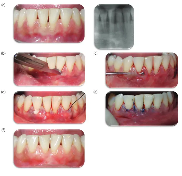
A 38-year-old systemically healthy female patient was referred to our clinic with chief complaints of hypersensitivity and poor aesthetics due to gingival recessions. Clinical and radiographical examination revealed buccal gingival recession defects affecting the mandibular central incisors with slight interdental attachment and bone loss (Figure 1.a). The defects were classified as Miller Class III type recessions. At
3 baseline periodontal examination, mild plaque accumulation and minimal bleeding on probing were present. Tooth positions and thin tissue biotype are considered possible predisposing factors for gingival recessions.
Figure 1. (a) Pre-operative clinical and radiographical views of recession defects. (b) Intra-sulcular insicions. (c) Flap
elevation and tunnel preparation with special instruments. (d) ADM placed into the tunnel. (e) Suturing. (f) 12-month postoperative view of recession defects
(a)
(b) (c)
(d) (e)
(f)
As an initial periodontal therapy, supragingival scaling and polishing were performed. Detailed oral hygiene instructions were given and the patient was informed to perform a non-traumatic (roll) brushing technique with a soft toothbrush.
The patient was re-evaluated 8 weeks after initial therapy and clinical measurements were taken with a calibrated periodontal probe (PCP 15 UNC, Hu-Friedy, USA) by using individual acrylic occlusal stent for proper probe positioning. Gingival thickness (GT) was measured with a digital caliper (Stainless Steel
4
Digital Caliper 75 mm, Shan, China) by using an endodontic spreader. Recession height (RH) of mandibular left central incisor (#31) and right central incisor (#41) was 3 and 2 mm, respectively, with buccal probing depths (PD) of 1 mm. The keratinized tissue height (KT) at the buccal aspect of the involved teeth was 2 and 3 mm, respectively (Table 1). The patient presented a thin gingival biotype, and GT measured as 0.8 mm at each.
For the treatment of gingival recessions at tooth #31 and #41, all the treatment alternatives explained to the patient. Since, the patient didn’t want a second surgical site at the palate; therefore, TUN in combination with ADM procedure was decided. After all risks and benefits were explained to the patient, oral and written consent was obtained.
Surgical treatment was based on TUN technique, in which papillae keep intact for better blood supply (Aroca et al., 2013) with the placement of ADM graft inside the tunnel. Following local anesthesia, root planing of denuded root surfaces was performed. The tunnel is prepared with intrasulcular incisions at each area of recession by using 15C blade (Figure 1.b). Care is taken not to damage the interdental papillae. Mucoperiosteal flap dissection was made with a tunnel elevator till the mucogingival junction and extended under each papillae, then papillae were gently raised. The tissue undermined beyond the mucogingival junction in order to obtain a tension-free tunnel (Figure 1.c). The ADM graft is inserted into the tunnel by applying a suture to the graft. This suture is inserted through the most distal site of #31 and then the graft is gently slided under the tunnel (Figure 1.d), positioned at the CEJ level and secured to the flap. The flap margin was positioned coronal to the CEJ to completely cover the defect and ADM and sutured with double sling suture technique with 5-0 non-resorbable polypropylene sutures (Figure 1.e).
Post-surgically, systemic antibiotic for 5 days (2x1000 mg, amoxicillin clavulanate, Augmentin BID; GlaxoSmithKline Pharmaceuticals, Turkey), oral analgesics as necessary (2x550 mg, naproxen sodium, Apranax Forte; Abdi Ibrahim, Turkey) and oral rinse twice a day for 4 weeks (0,2% chlorhexidine gluconate, Klorhex Oral Rinse; Drogsan Pharmaceuticals, Turkey) were prescribed. The patient was informed not to brush or floss her teeth in the operated areas for 14 days.
After 2 weeks, sutures were removed and patient was seen weekly for 1 month. One month after surgery, patient was reinstructed for mechanical tooth cleaning with a soft toothbrush and a roll technique. Recall sessions scheduled once a month for supragingival plaque removal, polishing and oral hygiene reinforcement in order to support healing process. At 12 months, all clinical measurements were repeated (Table 1). Post-operative healing was uneventful. She had no complaints about dentinal hypersensitivity. CRC was achieved at #41 and partial root coverage on #31 with mean root coverage (MRC) of 83.3% was obtained (Figure 1.f). RH reduction at #31 and #41 was 2.5 mm and 2 mm, respectively. GT increase was 0.6 mm for both teeth and KT gain was 1 mm and 1.5 mm, respectively. Patient satisfaction was assessed by using a three-point rating scale (Mahajan et al., 2007) and patient was questioned with regard to root coverage attained, relief from dentinal hypersensitivity, colour of gums, shape and contour of gums, surgical procedure, post surgical phase and cost effectiveness. This score was detected as 18.
5
Table 1. Measurements at baseline and at 12 months
Baseline 12 months PD (mm) #31#41 11 11 CAL (mm) #31#41 4 1.5 3 1 RH (mm) #31#41 3 0.5 2 0 RW (mm) #31 3 1 #41 2 0 GT (mm) #31#41 0.80.8 1.41.4 KT (mm) #31#41 2 3 3 3.5
Probing depth (PD), clinical attachment level (CAL), recession height (RH), recession width (RW), gingival thickness (GT), keratinized tissue height (KT)
Aesthetic outcomes of the procedure were also evaluated by using a scoring system called root coverage aesthetic score (RES) (Cairo et al., 2009). Gingival margin level, marginal tissue contour, soft tissue texture, mucogingival junction alignment and gingival colour were evaluated. Zero, 3 and 6 points were used for the evaluation of the position of the gingival margin, whereas a score 0 or 1 point was used for each of the other variables. Patient’s RES was found 7 which means that the aesthetic outcome was satisfying.
Discussion
The aim of this case report was to present a predictable procedure by using ADM along with TUN for the treatment of multiple Miller Class III recessions. Within the limit of this case, significant defect coverage, aesthetical results, KT and GT increase was achieved with TUN+ADM combination.
In the literature, limited data is available about the clinical results obtained with TUN+ADM in Miller Class III multiple gingival recessions. With regard to Miller Class III recessions, data from the literature stated that the accomplishment of CRC was possible in defects presenting mild interdental attachment loss of 1 to 3 mm (Esteibar et al., 2011) and similar treatment techniques of Miller Class I and II recessions can also be proposed for Class III recession defects (Sculean et al., 2016).
Aroca et al. (2010b) evaluated the efficacy of the TUN in combination with CTG for the treatment of multiple class III gingival recessions, and 1-year results showed 82% MRC and 38% CRC. In another study comparing the outcomes of Miller Class III gingival recessions treated with TUN and CAF with ADM, it has been reported that the similar CRC and MRC results can be obtained with both technique (Moussa & Bissada, 2017). In this case report comparable CRC results were obtained.
Successful treatment of recession defects is based on the selection of proper surgical technique, and that depends on many factors, such as tooth related and surgery related factors. Previous studies
6
indicate that releasing incisions are an advantage when coronal positioning is needed (Mahn, 2010; Papageorgakopoulos et al., 2008). However, vertical releasing incisions can disturb the vascularity of tissues and lead to aesthetical problems as a result of scar tissue formation (Zucchelli et al., 2009). It has been reported that the main blood supply of the gingiva is directed from vestibule to gingival margin providing more blood vessels, nutrients and source of cells which in turn leads to better integration of the ADM into the host’s tissues (Felipe et al., 2007; Zuhr et al., 2018). Furthermore, it has been suggested that additional grafting can provide a scaffold to support wound healing with increasing the thickness of the wound area (Baldi et al., 1999). Systematic reviews reported variable results with the use of ADM as an alternative to the gold standard CTG (Graziani et al., 2014). But, number of studies reported that ADM is an effective alternative to CTG for the treatment of multiple recessions, which support the gingival margin and change the gingival biotype (Thombre et al., 2013; Ahmedbeyli et al., 2014). In the present case, GT was increased from 0.8 mm to 1.4 mm.
Patient was questioned about her satisfaction with regard to the patient-centered criteria (Mahajan et al., 2007) and she reported no significant postoperative complications and minimal discomfort with the used approach. Reports in the literature also assessed pain, aesthetics and postoperative discomfort after TUN+ADM procedure (Mounssif et al., 2018) and results showed that patients had minimal discomfort and healed uneventfully with this approach by increasing its thickness as observed in our case report. In spite of the fact that the main goal of all root coverage procedures is complete coverage of exposed roots, aesthetic outcomes also have the great importance. Aesthetic outcomes of the procedure was assessed by using RES (Cairo et al., 2009). According to RES, gingival margin level contributes 60% of the total score. Due to partial coverage at tooth #31, RES value was found 7 in this case, 12 months after TUN+ADM procedure. This finding can be attributed to tooth related factors present in this case, such as thin tissue biotype and tooth positions, which will lead to difficulties to achieve CRC.
Within the limits of the present case report and based on previous studies, procedures involving ADM graft in combination with TUN technique may be a predictable treatment alternative for the treatment of Miller Class III multiple gingival recessions with successful root coverage and aesthetic outcomes.
Conflict of Interests
Authors declare no conflict of interests
References
Ahmedbeyli, C., Ipçi, Ş. D., Cakar, G., Kuru, B. E., & Yılmaz, S. (2014). Clinical evaluation of coronally advanced flap with or without acellular dermal matrix graft on complete defect coverage for the treatment of multiple gingival recessions with thin tissue biotype. Journal of Clinical Periodontology, 41(3), 303–310. American Academy of Periodontology. (1996). Consensus report on mucogingival therapy. Proceedings of the World Workshop in Periodontics. Annals of Periodontology, 1(1), 702–706.
7 Aroca, S., Keglevich, T., Nikolidakis, D., Gera, I., Nagy, K., Azzi, R., & Etienne, D. (2010a). Treatment of class III
multiple gingival recessions: a randomized-clinical trial. Journal of Clinical Periodontology, 37(1), 88–97. Aroca, S., Keglevich, T., Nikolidakis, D., Gera, I., Nagy, K., Azzi, R., & Etienne, D. (2010b). Treatment of class III multiple gingival recessions: a randomized-clinical trial. Journal of Clinical Periodontology, 37(1), 88–97. Aroca, S., Molnár, B., Windisch, P., Gera, I., Salvi, G. E., Nikolidakis, D., & Sculean, A. (2013). Treatment of multiple adjacent Miller class I and II gingival recessions with a Modified Coronally Advanced Tunnel (MCAT) technique and a collagen matrix or palatal connective tissue graft: a randomized, controlled clinical trial. Journal of Clinical Periodontology, 40(7), 713–720.
Baldi, C., Pini-Prato, G., Pagliaro, U., Nieri, M., Saletta, D., Muzzi, L., & Cortellini, P. (1999). Coronally advanced flap procedure for root coverage. Is flap thickness a relevant predictor to achieve root coverage? A 19-case series. Journal of Periodontology, 70(9), 1077–1084.
Cairo, F. (2017). Periodontal plastic surgery of gingival recessions at single and multiple teeth. Periodontology 2000, 75(1), 296–316.
Cairo, F., Rotundo, R., Miller, P. D., & Pini Prato, G. P. (2009). Root coverage esthetic score: a system to evaluate the esthetic outcome of the treatment of gingival recession through evaluation of clinical cases. Journal of Periodontology, 80(4), 705–710.
Cortellini, P., & Pini Prato, G. (2012). Coronally advanced flap and combination therapy for root coverage. Clinical strategies based on scientific evidence and clinical experience. Periodontology 2000, 59(1), 158–184. Esteibar, J. R. V., Zorzano, L. A. A., Cundín, E. E., Blanco, J. D. M., & Medina, J. R. O. de G. (2011). Complete root coverage of Miller Class III recessions. The International Journal of Periodontics & Restorative Dentistry, 31(4), e1-7.
Felipe, M. E. M. C., Andrade, P. F., Grisi, M. F. M., Souza, S. L. S., Taba, M., Palioto, D. B., & Novaes, A. B. (2007). Comparison of two surgical procedures for use of the acellular dermal matrix graft in the treatment of gingival recessions: a randomized controlled clinical study. Journal of Periodontology, 78(7), 1209–1217. Graziani, F., Gennai, S., Roldán, S., Discepoli, N., Buti, J., Madianos, P., & Herrera, D. (2014). Efficacy of periodontal plastic procedures in the treatment of multiple gingival recessions. Journal of Periodontology, 41(Suppl 15), 63-79.
Harris, R. J. (2004). A short-term and long-term comparison of root coverage with an acellular dermal matrix and a subepithelial graft. Journal of Periodontology, 75(5), 734–743.
Mahajan, A., Dixit, J., & Verma, U. P. (2007). A patient-centered clinical evaluation of acellular dermal matrix graft in the treatment of gingival recession defects. Journal of Periodontology, 78(12), 2348–2355. Mahn, D. H. (2010). Use of the tunnel technique and an acellular dermal matrix in the treatment of multiple adjacent teeth with gingival recession in the esthetic zone. The International Journal of Periodontics & Restorative Dentistry, 30(6), 593–599.
8
Miller, P. D. (1985). A classification of marginal tissue recession. The International Journal of Periodontics & Restorative Dentistry, 5(2), 8–13.
Modaressi, M., & Wang, H.-L. (2009). Tunneling procedure for root coverage using acellular dermal matrix: a case series. The International Journal of Periodontics & Restorative Dentistry, 29(4), 395–403.
Moslemi, N., Mousavi Jazi, M., Haghighati, F., Morovati, S. P., & Jamali, R. (2011). Acellular dermal matrix allograft versus subepithelial connective tissue graft in treatment of gingival recessions: a 5-year randomized clinical study. Journal of Clinical Periodontology, 38(12), 1122–112d9.
Mounssif, I., Stefanini, M., Mazzotti, C., Marzadori, M., Sangiorgi, M., & Zucchelli, G. (2018). Esthetic evaluation and patient-centered outcomes in root-coverage procedures. Periodontology 2000, 77(1) 1-35.
Moussa, E. W., Bissada, N.F. (2017) Comperative outcomes of Miller Class III gingival recession treatment using the tunnel or coronally advanced flap approach: a case report. Clinical Advances in Periodontics, 7(1), 35-41.
Novaes, A. B., Grisi, D. C., Molina, G. O., Souza, S. L., Taba, M., & Grisi, M. F. (2001). Comparative 6-month clinical study of a subepithelial connective tissue graft and acellular dermal matrix graft for the treatment of gingival recession. Journal of Periodontology, 72(11), 1477–14dddd84.
Ozenci, I., Ipci, S. D., Cakar, G., & Yilmaz, S. (2015). Tunnel technique versus coronally advanced flap with acellular dermal matrix graft in the treatment of multiple gingival recessions. Journal of Clinical Periodontology, 42(12), 1135–1142.
Papageorgakopoulos, G., Greenwell, H., Hill, M., Vidal, R., & Scheetz, J. P. (2008). Root coverage using acellular dermal matrix and comparing a coronally positioned tunnel to a coronally positioned flap approach. Journal of Periodontology, 79(6), 1022–1030.
Sculean, A., Cosgarea, R., Katsaros, C., Arweiler, N. B., Miron, R. J., & Deppe, H. (2017). Treatment of single and multiple Miller Class I and III gingival recessions at crown-restored teeth in maxillary esthetic areas. Quintessence International (Berlin, Germany: 1985), 48(10), 777–782.
Sculean, A., Cosgarea, R., Stähli, A., Katsaros, C., Arweiler, N. B., Miron, R. J., & Deppe, H. (2016). Treatment of multiple adjacent maxillary Miller Class I, II, and III gingival recessions with the modified coronally advanced tunnel, enamel matrix derivative, and subepithelial connective tissue graft: A report of 12 cases. Quintessence International (Berlin, Germany: 1985), 47(8), 653-659.
Serino, G., Wennström, J. L., Lindhe, J., & Eneroth, L. (1994). The prevalence and distribution of gingival recession in subjects with a high standard of oral hygiene. Journal of Clinical Periodontology, 21(1), 57-63. Stefanini, M., Marzadori, M., Aroca, S., Felice, P., Sangiorgi, M., Zucchelli, G. (2018). Decision making in root-coverage procedures for the esthetic outcome. Periodontology 2000, 77(1), 1-11.
Tal, H. (1999). Subgingival acellular dermal matrix allograft for the treatment of gingival recession: a case report. Journal of Periodontology, 70(9), 1118–1124.
9 Thombre, V., Koudale, S. B., & Bhongade, M. L. (2013). Comparative evaluation of the effectiveness of coronally
positioned flap with or without acellular dermal matrix allograft in the treatment of multiple marginal gingival recession defects. International Journal of Periodontics and Restorative Dentistry, 33(3), e88-94. Zabalegui, I., Sicilia, a, Cambra, J., Gil, J., & Sanz, M. (1999). Treatment of multiple adjacent gingival recessions with the tunnel subepithelial connective tissue graft: a clinical report. The International Journal of Periodontics & Restorative Dentistry, 19(2), 199–206.
Zucchelli, G., Mele, M., Mazzotti, C., Marzadori, M., Montebugnoli, L., & De Sanctis, M. (2009). Coronally advanced flap with and without vertical releasing incisions for the treatment of multiple gingival recessions: a comparative controlled randomized clinical trial. Journal of Periodontology, 80(7), 1083–1094.
Zucchelli, G., Mounssif, I., Mazzotti, C., Montebugnoli, L., Sangiorgi, M., Mele, M., & Stefanini, M. (2014). Does the dimension of the graft influence patient morbidity and root coverage outcomes? A randomized controlled clinical trial. Journal of Clinical Periodontology, 41(7), 708–716.
Zuhr, O., Rebele, S. F., Cheung, S. L., Hürzeler, M. B., & Research Group on Oral Soft Tissue Biology and Wound Healing. (2018). Surgery without papilla incision: tunneling flap procedures in plastic periodontal and implant surgery. Periodontology 2000, 77(1), 1-27.