Abstract
IntroductIon
Speech and language disorders can occur due to a host of reasons related to many factors such as etiology, associated anatomic disorders, and affected components of the language. Such diverse problems demand different modes of assessment. For instance, disorders, like cleft palate, that develop because of an anatomical impairment, and together with multiple problems, should be extensively evaluated. To that end, a number of studies are conducted in Europe and in the United States to standardize clinical applications.[1] The Great Ormond
Street Speech Assessment and The Cleft Audit Protocol for Speech-A developed under the Eurocleft project can be named as examples.[2,3]
In Turkey, there is no standard assessment form or protocol that can be used for evaluating speech and language in individuals with cleft lip and palate (CLP). The same tools that are used for evaluating various disorders are also used for this purpose, whereas assessment of individuals with CLP should target feeding, respiration, hearing, language, articulation, resonance,
cognition, dental, skeletal (occlusion), esthetic, psychosocial, and academic problems. Furthermore, assessments should allow for discriminating between syndromic and nonsyndromic cases.
Assessment in speech and language therapy is in fact a decision making process for referrals as well as for the mode of therapy.[4] To help making the right decisions, the tools used in
assessment should be able to both define the externally visible problems and identify the underlying causes which these problems possibly arise from.[5] Therefore, any information
possibly related to the underlying causes should be considered in the assessment of speech and language disorders associated with CLP.
Objective: The objective of the study is to identify the medical histories, oral-peripheral characteristics and speech problems of children with
cleft lip and palate (CLP) or craniofacial anomalies, and eventually create an assessment form that highlights the factors that should be taken into consideration in the examination of those children. Materials and Methods: An assessment form was developed and used to assess with
a descriptive method the medical history, oral peripheral, speech, and resonance characteristics of children with CLP. The study included 56 (21 females and 35 males) children with CLP. The results are presented as frequency or percentage. Results: About 20% of the CLP patients
were offspring of consanguineous marriage and about 30% had positive family history of CLP. The major difficulty families experienced was feeding. Hearing impairment at various degrees was reported in 23% of the participants. The presence of cardiovascular, renal, neurological and endocrinological problems, and symptoms addressed in peripheral examination were identified as critical in discriminating syndromic cases. Assessment of the speech skills showed backing to be the most prevalent error among the participants. Conclusion: The form that was
developed in scope of this study was observed to successfully define the medical history, oral-peripheral characteristics and speech problems of individuals with CLP. The form, however, needs further testing in larger populations and comparison to non-CLP populations.
Keywords: Assessment, cleft lip and palate, speech disorders
Address for correspondence: Dr. Özlem Ünal‑Logacev, Department of Speech and Language Therapy, Istanbul Medipol University, School of Health Sciences, Istanbul, Turkey. E‑mail: [email protected]
Access this article online
Quick Response Code:
Website:
http://www.turkjplastsurg.org
DOI:
10.4103/tjps.tjps_27_18
This is an open access journal, and articles are distributed under the terms of the Creative Commons Attribution‑NonCommercial‑ShareAlike 4.0 License, which allows others to remix, tweak, and build upon the work non‑commercially, as long as appropriate credit is given and the new creations are licensed under the identical terms.
For reprints contact: [email protected]
How to cite this article: Ünal-Logacev Ö, Kazanoğlu D, Balo E, Nemutlu A. Cleft lip and palate assessment form: Medical history, oral-peripheral characteristics, speech problems. Turk J Plast Surg 2018;26:156-68.
Cleft Lip and Palate Assessment Form: Medical History,
Oral‑peripheral Characteristics, Speech Problems
Özlem Ünal-Logacev, Deniz Kazanoğlu1, Eren Balo1, Ayşe Nemutlu1
Department of Speech and Language Therapy, Istanbul Medipol University, School of Health Sciences, Istanbul, 1Department of Language and Speech Therapy,
The purpose of this study is to develop a form – as described later in the methods section – that can be used in the assessment of individuals with CLP and craniofacial anomalies, and to use this form to identify the medical history, oral-peripheral characteristics, as well as speech and resonance characteristics in these individuals. The form is designed to help address the following areas:
1. Discriminating between syndromic and nonsyndromic cases
2. Identifying the underlying anatomic and physiologic causes of speech problems
3. Facilitating the diagnosis of speech disorder and contributing to the planning of the therapy
4. Providing accurate information and guidance.
MaterIals and Methods
This study aims at identifying the medical history, oral-peripheral characteristics and speech problems of children with CLP or craniofacial anomalies. To that end, a descriptive study was designed to assess and define the current state of patients. Participants
Participants of the study are the individuals who presented with cleft lip ± palate, submucous cleft, occult submucous cleft, or velopharyngeal insufficiency (VPI) to the Research Centre for Speech and Language Disorders Center (DİLKOM) of Anadolu University, and Faculty of Medicine of Kocaeli University in 2015 and 2016. Individuals with syndromes accompanied by cleft palate or VPI were also included in the study.
All participants were examined by plastic surgeons, otorhinolaryngologist, orthodondist, and evaluated by an audiologist. Parents of all participants gave their informed consent and signed the voluntary participation form.
Of the 56 participants included in the study, 21 are female and 35 are male and were aged 0–18 years at the time of the study. Twenty-one participants were ineligible and/or nor suitable to perform the sentence repetition task, therefore excluded from the speech and resonance assessment which was eventually conducted with 35 participants. Distribution of participants by age and gender is given in Table 1. Distribution of participants by cleft type is given in Table 2.
Thirty-two of the participants received speech and language therapy: Eight participants continued the therapy for <6 months, six continued for 6–12 months, and 18 continued for more than 12 months.
Forms and implementation
The CLP Assessment form designed in this study consists of three subforms, namely (i) the cleft lip palate family interview form, (ii) the cleft lip palate oral peripheral assessment form, (iii) the cleft lip palate speech and resonance assessment form.
Cleft lip palate family interview form
This form serves to explore the possible genetic and
environmental factors underlying the CLP, and to obtain information about the child’s prenatal, perinatal, postnatal history, as well as overall health status and development. The form also addresses the various problems which can arise independently and in multiple systems due to the syndrome [Annexure 1]. The form includes questions that address previous surgeries, the possible causes of CLP development, the symptoms associated with the differential diagnosis of syndromic and nonsyndromic CLP, developmental delays, feeding and swallowing problems, airway problems, and hearing problems.
The above information includes only those obtained from the families. This content certainly does not suffice for evaluating or diagnosing the specific area. Areas such as swallowing or hearing, for instance, need thorough assessment. It should be noted that the information obtained from the families serve merely as preliminary information for further considerations. Cleft lip palate oral peripheral assessment form
The peripheral examination part of this form addresses anomalies that are often seen in craniofacial syndromes, such as those of the skull, finger/toe, ear, nose, lip, and eyes. This section intends to draw the attention of speech therapists to the issues that should be considered in their cases and are particularly associated with syndromic individuals [Annexure 2].
Although there is no direct correlation between CLP-related speech problems and the size of the anatomic defect, the size of the defect varies between syndromic and nonsyndromic cases. Secondary clefts are more common among syndromic
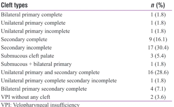
Table 2: Cleft types of participants
Cleft types n (%)
Bilateral primary complete 1 (1.8)
Unilateral primary complete 1 (1.8)
Unilateral primary incomplete 1 (1.8)
Secondary complete 9 (16.1)
Secondary incomplete 17 (30.4)
Submucous cleft palate 3 (5.4)
Submucous + bilateral primary 1 (1.8)
Unilateral primary and secondary complete 16 (28.6) Unilateral primary complete secondary incomplete 1 (1.8) Bilateral primary secondary complete 4 (7.1)
VPI without any cleft 2 (3.6)
VPI: Velopharyngeal insufficiency
Table 1: Gender and age range of participants
Gender Age range
0‑3,
n (%) n (%)4‑7, n (%)8‑12, 13‑18, n (%) n (%)Total,
Female 6 (10.7) 9 (16.1) 5 (8.9) 1 (1.8) 21 (37.5) Male 9 (16.1) 13 (23.2) 9 (16.1) 4 (7.1) 35 (62.5) Total 15 (26.8) 22 (39.3) 14 (25) 5 (8.9) 56
cases.[6,7] The presence of fistulae, VPI, or occlusion problems
have direct and severe influence on speech performance. Therefore, occlusion and dental problems, anomalies of the tongue that can directly influence speech, localization and size of a possible fistula, and any anatomic defects of the velum, uvula, and oropharynx should be examined prior to speech assessment. The second part of the oral peripheral assessment form is specifically designed to help correlate these anatomic issues with speech disorders [Annexure 2].
Cleft lip palate speech and resonance assessment form The forms are designed to take into account only those areas that are required in the assessment of individuals with CLP. As there are some widely used, valid, reliable and standardized language and articulation tests in Turkey, the areas that are already covered by these are not included in our forms with the thought that they can be used in combination. Instead, we added fields for commenting on the results of language tests, single-word naming articulation tests, and nasometric assessment tools.[8-13]
Given its practicality and its capacity to demonstrate the individual’s connected speech skills, sentence repetition test is included in this form [Annexure 3]. In the sentence repetition section of this form, one target sentence was included for each of the consonants in the Turkish alphabet (including the allophones of “k” and “g” and excluding “ğ”) for the purposes of identifying the individual’s articulation errors. One consonant was targeted in each sentence and sentences were constructed by placing the targeted consonant within the words. Nasal consonants (“m, n”) are not included in the sentences testing oral consonants (all consonants except for “m” and “n” are classified as oral consonants) – so that the sentences can also be used in nasoendoscopic examination. In normal speech, the velopharyngeal sphincter is completely closed when oral consonants are articulated, and open when nasal sounds are articulated. When nasoendoscopy is performed using words that contain nasal sounds, this will give the impression that there is no velopharyngeal closure.
Another aspect that is considered in this form is to avoid placing “s, z, ş, j, ç, c” in sentences that are used for testing other sound groups. The purpose is to allow for the testing of phoneme-specific nasal emission (PSNE), a disorder that will be discussed later in this article. PSNE is not the result of a structural disorder, but of mislearning where some of the “s, z, ş, j, ç, c” sounds are produced nasally and can be corrected only by speech therapy. PSNE should be considered if the individual sounds hypernasal when articulating the sentences that contain only these phonemes and sounds normal in those sentences that do not contain them.
In practice, sentences are read out loud by the examiner and repeated by the examined person. Correct articulation of target sound is marked in the True column, and wrong articulation is marked in the False column on the form. This section also includes a field for the phonetic transcription of the sentences according to the International Phonetic Alphabet (IPA) (The
IPA is a system of phonetic symbols designed by linguists to uniquely and accurately represent each of the wide variety of sounds [phones or phonemes] that are used in spoken human language).
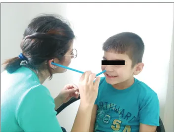
Resonance assessment, as part of this form, is measured with a simple and perceptual test using a straw (in this test, one end of the straw is placed in the nostril of the child with CLP, and the other end is placed near the ear of the examiner and the child is asked to repeat high-pressure phonemes. One of the below described resonance disorders should be considered if air passes through the straw when the individual with CLP articulates these sounds) [Figure 1].
These assessments should be measured with instruments (e.g., nasometer and nasoendoscopy) and verified before diagnosis. Below is a description of how resonance disorders are assessed:
• Hypernasality: Hypernasality can be suspected if airflow is identified in stimuli that contain oral + vowel combinations (papapa, tatata, kakaka, sasasa, şaşaşa) in the straw test. To distinguish hypernasality from nasal emission and turbulence, the nose opens and closes during prolonged articulation of “i” (i.e., “iiii”). Any variances in the open-close process are observed. Speech is marked as hypernasal in case of variances. As a final check, articulation should be observed for any similarities between the phonemes “b” and “m” and also “d” and “n” • Nasal emission: The straw test is used for testing
nasal emission in the same way it is used for testing hypernasality. The presence of airflow in the articulation of oral + vowel combinations (papapa, tatata, kakaka, sasasa, şaşaşa) is checked during the straw test. Vowel production is verified to discriminate from hypernasal speech. Nasal emission box is marked if vowels are not considerably affected
• Nasal rustle/turbulence: This box is marked if nasal leakage which resembles snoring is heard during the production of fricatives (s, z, ş, j). Then, a straw test can be performed as described above. The turbulence originates
from a very small opening in the velopharyngeal region. Surgical intervention may not be always necessary • Hyponasality: This is tested with speech stimuli using
words and nasal + vowel combinations (e.g., ma and na) that contain the phonemes “m, n.” The person is asked to vocalize a prolonged “m” sound (“mmm”) both with open and plugged nose. Any variances in the production process are observed. Hyponasality box is marked in the presence of minor variances. The accuracy of the markings is verified by checking whether the sounds of “m” and “n” are articulated in a way similar to the sounds “b” and “d”
• Mixed resonance: This is marked if resonance quality changes during sentence repetition. This is to say that resonance is sometimes perceived as hyponasal and sometimes hypernasal
• PSNE: In sentence repetition test, the PSNE box is marked if nasality is perceived in sentences which “s, z, ş, j, ç, c” are used frequently, but no nasality is perceived in sentences that do not contain these phonemes • Cul-de-sac resonance: This type of resonance disorder
occurs when airflow is trapped in the vocal tract because of conditions such as large tongue, large tonsils, or hypertrophy. The speech is perceived as muffled
• Nasal grimace: This box is marked if the child wrinkles his/her nose during sentence repetition.
Data analysis
Data analysis was performed in Microsoft Excel and given the descriptive nature of the study; results are presented as frequency and percentage. Fit index (FI) was used to estimate interrater reliability in the speech and resonance characteristics assessment section. The video recordings of all participants were reviewed by the three researchers of the study and scored based on the presence or absence of any errors (eg. backing, lateralization, weak consonant) in the production of the phonemes. FI value was calculated at 98.1% according to the below FI formula:
FI = ([Total number of Fits]/[Total number of Assessments]) × 100.[14]
results
The below given are the highlights of the results obtained in the study.
Results from cleft lip palate family interview form
Prenatal history
According to the information provided by their primary caretakers, 19.6% of the children are offspring of consanguineous marriage. About 30.4% have one or more case/s of CLP in their family. Regarding the medical history of their mothers, 14.3% had advanced maternal age and 23.2% had either a miscarriage or an abortion in the past. When mothers were asked about the substances they were exposed to in the first trimester of their pregnancy, 12.5%
indicated to have smoked, and 10.7% indicated exposure to radiation. While 21.4% indicated to have used medication, 51.8% indicated severe stress during pregnancy [Figure 2].
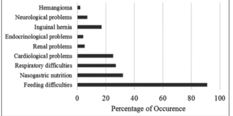
Postnatal history
According to the information provided by their primary caretakers, 91.1% of the participants experienced feeding difficulties, and 32.1% was fed nasogastrically. 26.8% experienced respiratory difficulties. About 25% had cardiological problems, 5.4% had renal problems, 3.6% had endocrinological problems, and 7.1% had neurological problems. 17.9% suffered inguinal hernia, and hemangioma developed in 1.8% [Figure 3].
State of health at the time of assessment
At the time of assessment, 32.1% had chewing difficulties and 19.6% had swallowing difficulties. 32.1% had nasal regurgitation. 23.2% had hearing impairment at various levels [Figure 4].
Regarding otitis media – one of the causes of hearing impairment – 44.6% of the participants had a history of 1–3 occurrences, and 8.9% had more than 10 occurrences [Figure 5]. While 57.1% of the participants was followed up by a hospital clinic, 42.9% was not followed by any health institution. Majority of the participants had undergone a series of surgeries that are directly associated with CLP, including for cleft lip, cleft palate, nose, fistula, pharynx, alveolar bone grafting, maxillary advancement, and ventilation tube, or surgeries that are mostly associated with the syndromes involving the kidneys, heart, testicular, or hernia. Of the 56 participants, 12.5% had undergone one surgery, 19.6% two surgeries, 30.4% three, 5.4% four, 3.6% five, 1.8% six, and 7.1% seven surgeries in the past [Figure 6]. Ventilation tube was the most commonly used (28.6%) procedure after cleft lip or palate surgery.
Results from the cleft lip palate oral peripheral assessment form
Regarding the cranial characteristics of the participants, 11% had a triangular face structure, and 7% had flat zygomatic bones. Examination of the fingers as part of the peripheral assessment showed that 5% of the participants had hyperextension and elongated digits. Septal deviation was the most encountered condition in peripheral assessment (18%). About 16% of the participants had low nasal bridge. Regarding the lips, participants with open mouth and asymmetrical lips were seen to be equal in number (11%). Findings related to the eyes showed narrow palpebral fissure in 13%, and hypertelorism in 7% [Figure 7].
The most distinct issues observed in oral examination were malocclusion, occlusion defects, dental problems, fistulae, and problems in the velar area [Figure 8]. Of the individuals with CLP included in the study, 29% had Type III malocclusion, 23% had crossbite, 9% had anterior crossbite, and 14% had posterior crossbite. Regarding dental problems, 59% of the
participants had missing teeth. Participants with anterior and posterior fistulae were equal in number (13%). As for velar area problems, 29% had short velum and 25% had minimal velum movement.
Results from the cleft lip palate speech and resonance assessment form
The results of speech problems consist of the information from 35 participants. These are presented under two headings,
Figure 2: Prenatal history of participants Figure 3: Postnatal history of participants
Figure 4: Current state of health during the assessments Figure 5: Occurrence of otitis media
Figure 6: Number of surgeries that participants underwent Figure 7: Percent of problems observed during peripheral examination
populations.[15,16] Our findings also show a high rate of CLP
history in the families of the participants (30%). Given these data, families should be referred to the genetics department before further pregnancies.
The major difficulty families experienced after birth was feeding. In fact, 32% of the participants were fed through the nasogastric route (for more than 24 h in 44% of the cases). This shows that families should be informed in detail about feeding, and that bottles that facilitate feeding should be made more accessible to families.
Findings related to hearing are another significant aspect of the study. Hearing impairment at various degrees was reported in 23% of the participants. Most of these are mild and likely to have been caused by recurrent otitis media. That almost 35% of the families reported no past occurrences of otitis media in their children is another finding worth noting since some of these families come from rural areas and do not have the means for routine audiological examinations. This increases the likelihood that otitis media remained unnoticed in these children.
The section of the cleft lip palate oral peripheral assessment form which addresses craniofacial characteristics is deemed to provide important clues for discriminating those with the syndromes based on the information collected from those who have or are suspected to have syndromes.
The results of this study should be regarded not as defining prevalence, but rather as highlighting the major problems that develop with the condition. Some of these major problems – all of which trigger speech disorders – are missing teeth, carious deciduous teeth that families leave untreated thinking that they will fall off anyway, and Type III malocclusion which is not orthodontically treated and monitored (61.5%).
More than half of the participants were seen to have short velum or insufficient velum movement. This has direct impact on resonance and articulation skills. Supporting these findings with nasometric and nasoendoscopic data in further studies and examining them in terms of surgical approach and technique will enhance the effectiveness of surgical procedures.
The assessment of speech skills of participants showed backing to be the most prevalent issue, whereas backing is reported to be rare in normal populations.[11] This finding of our study matches
those reported in the Hardin-Jones and Jones (2005) study.[17]
Backing is a compensatory articulation error that children with CLP develop due to insufficient intraoral pressure secondary to a fistula or VPI. Phonemes that are normally produced in the alveolar area (e.g., “t, d, s, z”) are produced in the velar or its posterior area. The most common acts after backing are also associated with VPI, namely nasalization, weak production of sounds, and double articulation. Another articulation error caused by the presence of fistulae is lateralization. Here, the child covers the fistula with his/her tongue and allows the air to flow along the sides of the tongue instead of the midline.[18] This Figure 10: Percent of resonance problems that participants experienced
namely articulation and resonance.
Articulation problems
As shown in Figure 9, the most common type of error made by the participants was backing (22%). Backing was followed by nasalization, weak production of sound, and double articulation errors (10%). Fronting and differentiation of glides, two of the most common types of error seen in normally developing children, were seen to be less common among the participants with CLP. Bilabialization, lateralization, changes in voice characteristics, and active nasal fricative sound production errors were each seen at a rate of 6%.
Resonance problems
As shown in Figure 10, while 38% of the participants had no resonance problems, hypernasality was observed in 9%, and hypernasality plus nasal emission was observed in 9%. Emission and nasal turbulence was identified in 17% of the participants.
Speech and language disorders mostly develop from a specific cause. It is not possible to provide adequate information and administer effective therapies without a complete understanding of the causes of the disorder. In the presence of a complex anomaly such as cleft palate, which also brings about problems in numerous areas, the underlying causes that can lead to language, articulation or resonance disorders, and all associated structures should be evaluated to identify the correlation between these structures and the existing speech disorders. The forms developed as part of this study include the recommended aspects that speech and language therapists should consider in a thorough examination, also provide the content in a practical format that can be used in reporting and therapy planning. The information obtained through these forms in the assessment of individuals with CLP will highlight many critical data about the individual.
dIscussIon
According to the data obtained from the cleft lip palate family interview form, about 20% of the individuals with CLP are offspring of consanguineous marriage, and about 23% of the mothers had prior miscarriage. These findings are comparable with the results of the studies that report a prevalence of 22% for consanguineous marriages and miscarriages in normal
act affects the articulation of “s, z, ş, j” and is difficult to correct in the presence of a fistula. Finally, Type III malocclusion is seen to cause some articulation errors. For instance, children with overjet where the incisors do not touch the lower lip were seen to bilabilize “f” and “v.”
Another finding worth noting in the study is that only 38% of the participants had no resonance disorders. Resonance disorders at different levels and forms were identified in the remaining 62%. Nasal emission and nasal turbulence were the most common disorders. This rate is considerably higher than the rates reported in the literature.[17,19-21] These results
obtained through perceptual assessment using low technology should be compared to nasometric and nasoendoscopic data to demonstrate the similarities and differences between objective and subjective findings.
conclusIon
This study presents the data from the first pilot of the form which is developed to be used in the assessment of individuals with CLP. Given the low number of participants, as well as the absence of instrumental measurements, the results should not be deemed to pertain to prevalence. However, the use of this form in other clinics can provide further detailed information on both the language and speech disorders and their underlying causes in children with CLP and allow for statistical analyses that provide a basis for instrumental measurements. Furthermore, we believe that this form will help close a significant gap by providing surgeons practicing in cities where there are no speech therapists with the means to conduct simple and practical speech assessment. Nevertheless, this pilot study needs to be used and assessed in higher numbers of participants to take its final form.
Declaration of patient consent
The authors certify that they have obtained all appropriate patient consent forms. In the form the patient(s) has/have given his/her/their consent for his/her/their images and other clinical information to be reported in the journal. The patients understand that their names and initials will not be published and due efforts will be made to conceal their identity, but anonymity cannot be guaranteed.
Acknowledgement
We would like to thank our participants for participating to our study; Tufan Bakır for designing the forms; Prof. Dr. Cengiz Çetin, Prof. Armağan İncesulu, Dr. İlhan Metin Dağsuyu, Dr. Nurdan Cankuvvet Aykut for taking parts in the cleft palate and craniofacial team work.
Financial support and sponsorship Nil.
Conflicts of interest
There are no conflicts of interest.
references
1. Shaw WC, Semb G, Nelson P, Brattström V, Mølsted K, Prahl-Andersen B, et al. The Eurocleft project 1996-2000: Overview. J Craniomaxillofac Surg 2001;29:131-40.
2. Sell D, Harding A, Grunwell P. GOS.SP.ASS.’98: An assessment for speech disorders associated with cleft palate and/or velopharyngeal dysfunction (revised). Int J Lang Commun Disord 1999;34:17-33. 3. John A, Sell D, Sweeney T, Harding-Bell A, Williams A. The cleft audit
protocol for speech-augmented: A validated and reliable measure for auditing cleft speech. Cleft Palate Craniofac J 2006;43:272-88. 4. Tylor AA, Tolbert LC. Speech-language assessment in the clinical
setting. Am J Speech Lang Pathol 2002;11:215-20.
5. Bleile K. Evaluating articulation and phonological disorders when the clock is running. Am J Speech Lang Pathol 2002;11:243-9.
6. Dixon MJ, Marazita ML, Beaty TH, Murray JC. Cleft lip and palate: Understanding genetic and environmental influences. Nat Rev Genet 2011;12:167-78.
7. Saal HM. Classification and description of nonsyndromic clefts. In: Wyszynski DF, editor. Cleft Lip and Palate: From Origin to Treatment. Oxford: Oxford University Press; 2002.
8. Güven S, Topbaş S. Adaptation of the test of early language development-(TELD-3) into Turkish: Reliability and validity study. Int J Early Child Spec Educ 2014;6:151-76.
9. Topbaş S, Güven S. Turkish Test of Language Development-Primary: Fourth Edition (TOLD-P:4). Ankara: Detay Yayıncılık: 2017.
10. Berument SK, Güven AG. Turkish Expressive and Receptive Language Test: I. Standardization, Reliability and Validity Study of the Receptive Vocabulary Sub-Scale. Turk Psikiyatri Dergisi 2013;24:192-201. 11. Topbaş SS. Turkish Articulation and Phonology Test. Ankara: Ministry
of National Education Publishing: 2005.
12. Ege P, Acarlar F, Turan F. Ankara Articulation Test-AAT. Research Projects Publishing, Ankara University. Ankara: Key Design; 2004. 13. Ünal Ö. Nasometric evaluation of resonance disorders: A norm study
with 4-18 years old Turkish speaking individuals. (Phd Dissertation). Eskisehir: Anadolu University; 2011.
14. Şencan H. Reliability and Validity in Social and Behavioral Measurements/Scaling. Ankara: Seçkin Publishing; 2005.
15. Koc I. Prevalence and sociodemographic correlates of consanguineous marriages in Turkey. J Biosoc Sci 2008;40:137-48.
16. Tomatir AG, Demirhan H, Sorkun HC, Köksal A, Ozerdem F, Cilengir N, et al. Major congenital anomalies: A five-year retrospective regional study in Turkey. Genet Mol Res 2009;8:19-27.
17. Hardin-Jones MA, Jones DL. Speech production of preschoolers with cleft palate. Cleft Palate Craniofac J 2005;42:7-13.
18. Kummer AW. Cleft Palate Craniofacial Anomalies: Effects on Speech and Resonance. 2nd ed. New York: Delmar; 2008.
19. Grunwell P, Sell DA. Speech and Cleft Palate/Velopharyngeal Anomalies. In: Watson AC, Sell DA, Grunwell P, editors. Management of Cleft Lip and Palate. London: Whurr; 2001.
20. Kummer AW. Cleft Palate Craniofacial Anomalies: Effects on Speech and Resonance. 3rd ed. New York: Delmar; 2014.
21. Prathanee B, Pumnum T, Seepuaham C, Jaiyong P. Five-year speech and language outcomes in children with cleft lip-palate. J Craniomaxillofac Surg 2016;44:1553-60.
annexures
Annexure 2: Cleft Lip Palate Oral Peripheral Assessment Form
Annexure 3: Cleft Lip Palate Speech and Resonance Assessment Form