Department of Cardiology, Kartal Koşuyolu High Specialization Training and Research Hospital, İstanbul, Turkey
7Department of Cardiology, Ümraniye Training and Research Hospital, İstanbul, Turkey 8Division of Health Sciences, Ardahan University, Ardahan, Turkey
Objective: Prosthetic valve thrombosis (PVT) is a serious complication among patients with prosthetic heart valves. Thrombolytic therapy (TT) is now widely used as first-line treat-ment for PVT. Endothelial dysfunction has previously been reported in patients with PVT. The aim of this study was to in-vestigate the changes in endothelial function soon after TT in PVT patients.
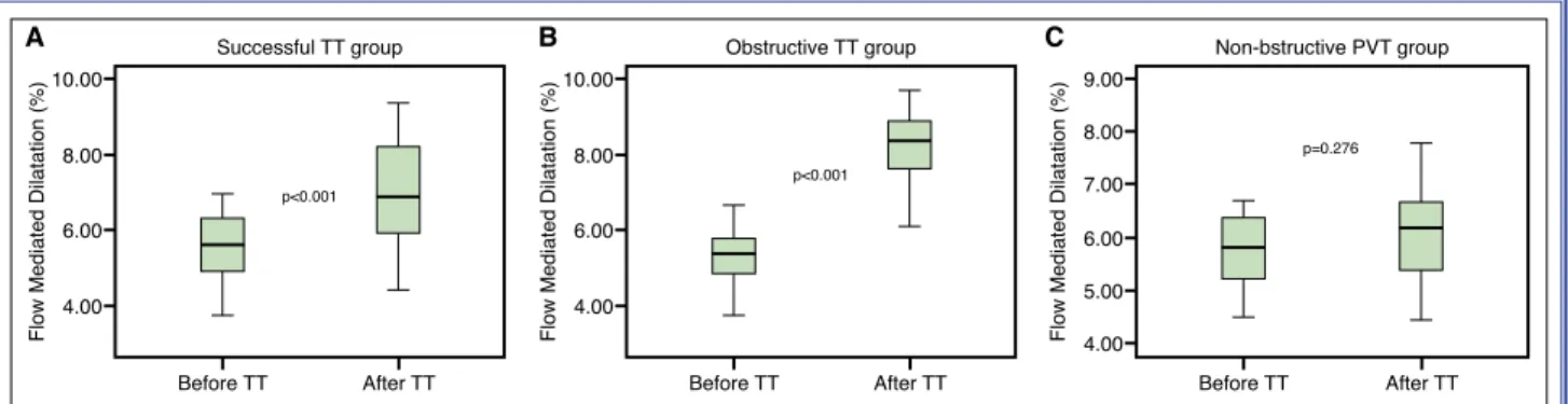
Methods: The study group included 85 patients with PVT [fe-male: 53 (62.3%); age: 48.7±13.9 years] who were evaluated prospectively before and shortly after TT. All of the patients were evaluated using transthoracic and transesophageal echocar-diography. TT was administered in all cases with a low-dose, ultra-slow infusion regimen. Endothelial function was evaluated using a noninvasive measurement of flow-mediated dilatation (FMD) of the brachial artery during reactive hyperemia. Results: The study population included 38 (44.7%) obstruc-tive and 47 (55.3%) non-obstrucobstruc-tive PVT patients. The ob-structive PVT patients had lower baseline FMD values than the non-obstructive PVT group (5.31±0.76% vs. 5.87±0.84%; p=0.003). TT was successful in 79 patients (92.9%). FMD was significantly increased in the successfully thrombolyzed patients after TT (5.65±0.86% vs. 7.13±1.26%; p<0.001). There was no significant difference in the FMD values after TT in patients who were unresponsive to TT (5.07±0.61%
vs. 5.38±0.95%; p=0.371). There was a significant increase
in FMD values after TT in patients with obstructive PVT (5.31±0.76% vs. 8.22±1.15%; p<0.001). However, this differ-ence was not statistically significant for patients with non-ob-structive PVT (5.87±0.84% vs. 6.11±0.95%; p=0.276). Conclusion: This study demonstrated that successful TT may contribute to improvement of impaired endothelial func-tion in patients with obstructive PVT.
Amaç: Protez kapak trombozu (PKT) gelişimi protez kalp kapaklı hastalarda ciddi bir komplikasyondur. Son zaman-larda trombolitik tedavi (TT) PKT tedavisinde ilk tercih olarak yaygın olarak kullanılmaktadır. Daha önceki çalışmalarda PKT hastalarında endotel disfonksiyonunun varlığı bildirilmiştir. Bu çalışmada, PKT hastalarında TT sonrasında endotel fonksiyonlarında olan değişiklikleri araştırmayı amaçladık. Yöntemler: Bu çalışmaya TT öncesi ve sonrası prospektif olarak takip edilen 85 PKT hastası [kadın: 53 (%62,3), orta-lama yaş: 48,7±13,9 yıl] dahil edildi. Tüm hastalar transtora-sik ve transözofajiyal ekokardiyografi ile değerlendirildi. Tüm hastalarda düşük doz ultra yavaş infüzyon rejimine göre TT uygulandı. Endotel fonksiyonları reaktif hipereminin neden olduğu akım aracılı genişleme (Flow Mediated Dilation, FMD) ölçülmesi ile değerlendirildi.
Bulgular: Çalışmaya 38 (%44,7) tıkayıcı ve 47 (%55,3) tıkayıcı olmayan PKT hastası alındı. Tıkayıcı PKT hastaları tıkayıcı olmayan PKT hastalarına göre daha düşük bazal FMD değerlerine sahiplerdi (%5,31±0,76 ve %5,87±0,84; p=0,003). TT 79 (%92,9) PKT hastasında başarılı idi. Ortama FMD değerleri başarılı TT grubunda TT sonrasında anlamlı olarak yükseldi (%5,65±0,86 ve %7,13±1,26; p<0,001). Başarısız TT grubunda TT sonrası FMD değerlerinde anlamlı değişiklik izlenmedi (%5,07±0,61 ve %5,38±0,95; p=0,371). Tıkayıcı PKT olan hastalarda TT sonrasında FMD değerleri anlamlı olarak yükseldi (%5,31±0,76 ve %8,22±1,15; p<0,001). Fakat, tıkayıcı olmayan PKT hastalarında TT sonrası FMD değerlerinde anlamlı değişiklik izlenmedi (%5,87±0,84 ve %6,11±0,95; p=0,276).
Sonuç: Bu çalışmada, tıkayıcı PKT hastalarında başarılı TT’nin bozulan endotel fonksiyonlarının düzelmesine katkı sunabildiği gösterilmiştir.
Received:February 18, 2020 Accepted:April 14, 2020
Correspondence: Dr. Macit Kalçık. Hitit Üniversitesi Tıp Fakültesi, Kardiyoloji Anabilim Dalı, Çorum, Turkey. Tel: +90 364 - 222 11 00 e-mail: [email protected]
© 2020 Turkish Society of Cardiology
T
he development of prosthetic valve thrombo-sis (PVT) represents one of the most important causes of morbidity and mortality in patients with prosthetic heart valves.[1] The annual incidence of left-sided PVT may vary from 0.5% to 8% per pa-tient[2] and even reach 20% for mechanical prostheses in the tricuspid position.[3] The most common cause of PVT is inadequate anticoagulant therapy.[4] The presenting clinical picture ranges from the absence of symptoms to cardiogenic shock. The traditional treatment of this complication has been emergency surgery, but thrombolytic therapy (TT), which has been available for many years, is considered first-line treatment in the current guidefirst-lines.[5] There have been reports from several trials regarding the safety and efficacy of TT regimens with a low-dose and slow infusion of tissue-type plasminogen activator (t-PA) in PVT patients, including pregnant patients. [6,7] More recently, results of the PROMETEE trial (PROsthetic MEchanical valve Thrombosis and the prEdictors of outcomE) demonstrated that an ultra-slow (25 hours) infusion of low-dose (25 mg) t-PA without a bolus appears to be associated with con-siderably lower rates of non-fatal complications and mortality for PVT patients without a loss of effec-tiveness.[8]Vascular endothelium secretes numerous factors that regulate cell growth, vascular tone, platelet and leukocyte interactions, and thrombogenicity. It has been recognized that endothelium takes part in some pathological processes during inflammation. The in-cremental role of inflammatory processes in PVT development was highlighted in a study reporting increased inflammatory parameters in patients with PVT.[9]
Endothelial dysfunction has been associated with various cardiovascular disorders.[10] Endothelial func-tion had previously been investigated in patients with PVT, and it was reported that patients with PVT had endothelial dysfunction, which might contribute to the development of PVT.[11] However, an association between endothelial dysfunction and PVT does not necessarily indicate causality. It is unclear whether endothelial dysfunction causes PVT or vice versa. Hence, this study was planned to investigate the re-versibility of endothelial dysfunction in patients with PVT after successful TT.
METHODS
Study population
A total of 85 PVT patients [female: 53 (62.3%); mean age: 48.7±13.9 years] who underwent TT with a reg-imen of an ultra-slow infusion of low-dose t-PA were enrolled in this single-center study. Patients with end-stage liver or renal disorders, excessive alcohol consumption,
ac-tive infection, acute coronary syndrome, renal insufficiency, pregnancy, chronic inflammatory dis-ease, deep vein thrombosis, coag-ulopathy, or
malig-nancies were excluded. A complete blood count and blood chemistry panel were completed for all of the patients at the time of admission. All of the patients provided written, informed consent and the study pro-tocol was approved by the local ethics committee of Kartal Kosuyolu Training and Research Hospital, Is-tanbul, Turkey on October 20, 2014 (no: 2014.3/15). The research was conducted in accordance with the Declaration of Helsinki and The Guideline of Good Clinical Practice.
Echocardiography
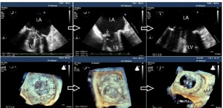
Transthoracic echocardiography (TTE) and 2-dimen-sional (2D) and real-time 3-dimen2-dimen-sional (RT 3D) transesophageal echocardiography (TEE) were per-formed for each patient using an X7-2t transducer on an iE33 ultrasound machine (Philips Healthcare, Inc., Andover, MA, USA). Parasternal long-axis and short-axis views and the apical 5-chamber view were used during the TTE evaluation. Transmitral gradients and the effective orifice area were measured with 2D TTE according to the current guidelines.[12] A TEE study was scheduled when there was an echocardiographic and/or clinical suspicion of PVT. A thrombus was recognized as a homogeneous, mobile, or fixed mass with similar echo density to the myocardium located at the valve occluder and/or valve struts and was visu-alized in all of the patients with PVT using echocar-diography[13] (Fig. 1). PVT was classified according to the identification of an obstructive or non-obstructive thrombus. The presence of an obstruction was defined on the basis of Doppler echocardiographic
measure-Abbreviations:
2D Two-dimensional FMD Flow-mediated dilatation PVT Prosthetic valve thrombosis RT 3D Real-time three-dimensional TEE Transesophageal echocardiography t-PA Tissue-type plasminogen activator TT Thrombolytic therapy
ments (peak velocity, mean gradient, effective orifice area, dimensionless index, and acceleration time, as appropriate). The cut-off values for these Doppler pa-rameters were defined based on recent recommenda-tions.[14]
The largest thrombus area was measured with 2D TEE between 0° and 180° where there was less inter-ference from acoustic shadowing. In the presence of a single mass, the thrombus was traced. In cases of multiple thrombi, each was traced separately and the thrombus areas were summed.[6–8]
Rationale for thrombolytic therapy
Based on previous reports regarding the safety and efficacy of a low-dose, ultra-slow infusion TT proto-col[4,8,15,16] in the absence of contraindications, a low-dose and ultra-slow TT regimen with a 25-hour in-fusion of 25 mg t-PA without a bolus (repeat up to 8 times if needed, maximum total dose of 200 mg) was administered as first-line therapy to all of the patients with obstructive PVT and those with non-obstructive PVT with a thrombus diameter of ≥10 mm accord-ing to the protocol described previously.[17] Anticoag-ulation with intravenous unfractionated heparin was withheld during the t-PA infusion due to the increased risk of bleeding. All of the patients underwent serial
TTE and TEE examination between each TT session. (A) Doppler documentation of complete improve-ment in valve hemodynamics and complete normaliza-tion of leaflet mobility, (B) reducnormaliza-tion in major diameter and/or area of the thrombus by 75%, and (C) sympto-matic improvement were considered the major criteria for TT success in patients with obstructive PVT in the absence of fatal and non-fatal major adverse events. For the patients with non-obstructive PVT, the only criterion for TT success was the complete lysis of the mobile component of the thrombus and an overall re-duction in thrombus burden of >50%.[8]
Vascular assessment of flow-mediated dilatation
All of the patients were assessed at least 12 hours af-ter their most recent meal, according to standard pro-tocol.[18] Patients were placed in the supine position for 10 minutes at rest before measurement. Right arm was fixed in an extended, relaxed position to provide correct analysis of the brachial artery 2–5 cm above the antecubital fossa. Next, the brachial artery was monitored longitudinally via a 17–5 MHz linear array ultrasound transducer (Vivid 5; GE Healthcare, Inc. Chicago, IL, USA). The brachial artery was scanned in the longitudinal section, the focus zone was set to optimize images of the lumen-arterial wall interface, Figure 1. Serial 2-dimensional and real-time 3-dimensional transesophageal echocardiography images of obstructive prosthetic mitral valve thrombosis that was successfully treated with a low-dose ultra-slow infusion of tissue-type plasminogen activator. After 2 sessions of thrombolytic therapy, the thrombus burden was remarkably reduced. LA: Left atrium; LAA: Left atrium ap-pendage; LV: Left ventricle.
aortic+mitral, and 3 tricuspid valve patients. The ma-jority of the patients (69.4%) had subtherapeutic anti-coagulation within the previous 3 months. The mean thrombus area was 0.9 cm2 (range: 0.6–1.6 cm2) and the mean t-PA dose was 50 mg (range: 25–90 mg). TT was successful in 79 patients (92.9%) and failed in 6 patients. Two of those 6 patients underwent redo and the machine operating parameters were not
al-tered for the remainder of the study. Measurements were taken from anterior to posterior M line at end-di-astole, incident with the R wave on the electrocardio-graphy. Three cardiac cycles (7 cardiac cycles in pa-tients with atrial fibrillation) were analyzed for each scan, and the measurements were averaged. A base-line brachial artery diameter was evaluated and a cuff placed around the forearm distal to the scanned artery segment was inflated to about 30 mmHg above sys-temic systolic arterial pressure for 5 minutes. A max-imal brachial artery diameter was established from 6 enrollments taken every minute after the cuff release. Percentage FMD was calculated using the formula of FMD (%) = (maximum diameter − baseline diame-ter)/baseline diameter × 100). Assessment of FMD values in the PVT patients was performed just before TT and repeated 6 hours after TT.
Statistical analysis
Statistical analyses were performed using IBM SPSS Statistics for Windows, Version 19.0 (IBM Corp., Ar-monk, NY, USA). Descriptive statistics were reported as mean±SD for continuous variables with normal dis-tribution and median (25th-75th percentiles) values for continuous variables without normal distribution. Fre-quency with percentage was used for the categorical variables. The Shapiro-Wilk test was used to test the normality of the distribution of continuous variables. Since the data were from the same patients and paired before and after TT, the Wilcoxon signed rank or a paired sample t-test was used to analyze these results, as appropriate. Continuous variables for independent samples were compared between groups using the Student’s t-test or the Mann-Whitney U test, as appro-priate. Categorical variables were compared using a chi-square test. Correlational analyses were performed using Pearson, Kendall’s tau or Spearmen’s correla-tion tests, as appropriate. The significance level was accepted as p<0.05 in all of the statistical analyses.
RESULTS
Baseline demographic, clinical, and echocardio-graphic parameters of the study population are pre-sented in Table 1. Laboratory parameters are included in Table 2. Study population was composed of 38 (44.7%) obstructive and 47 (55.3%) non-obstructive PVT patients. There were 22 aortic, 49 mitral, 11
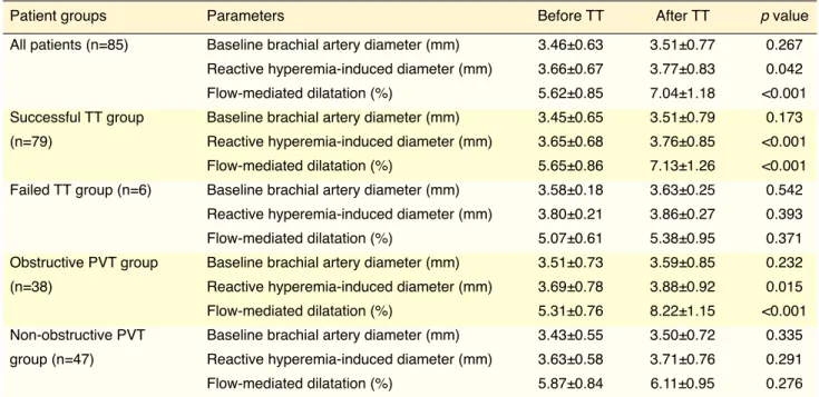
Table 1. Baseline demographic, clinical, and echocardiographic characteristics of the study population
PVT patients (n=85)
Age, (years) 48.7±13.9
Gender, n (%) Male: 32 (37.7)
Female: 53 (62.3) Prosthetic valve position, Mitral: 49 (57.7)
n (%) Aortic: 22 (25.9)
Aortic+mitral: 11 (12.9) Tricuspid: 3 (3.5)
Leaflet status, n (%) Monoleaflet: 12 (14.1)
Bileaflet: 73 (84.9)
Valve type, n (%) St. Jude Medical: 29 (34.1)
Carbomedics: 21 (24.7) Sorin-Bicarbon: 7 (8.2) Medtronic Hall: 12 (14.1)
ATS Medical: 14 (16.5) Omnicarbon: 2 (2.4)
Elapsed time since valve 61 (28–122)
surgery, (months)
Atrial fibrillation, n (%) 27 (36)
Left ventricular ejection 51.8±5.3
fraction, (%)
Left atrial diameter, (mm) 42.6±5.1
New York Heart Association I-II: 69 (81.1)
Functional Class, n (%) III-IV: 16 (18.9)
Recent subtherapeutic 59 (69.4)
anticoagulation, n (%)
Thrombus type, n (%) OT: 38 (44.7)
NOT: 47 (55.3)
Thrombus area, (cm2) 1.3 (0.9–1.7)
Thrombolytic therapy Successful: 79 (92.9)
outcome, n (%) Failed: 6 (7.1)
Tissue type plasminogen 50 (25–90)
activator dose, (mg)
NOT: Non-obstructive thrombus; OT: Obstructive thrombus; PVT: Prosthetic valve thrombosis.
nificant difference between FMD values before and after TT in patients who were unresponsive to TT (5.07±0.61% vs. 5.38±0.95%; p=0.371). There was a significant increase in FMD values after TT in patients with obstructive PVT (5.31±0.76% vs. 8.22±1.15%; p<0.001) (Fig. 3b). However, the difference was not significant in the non-obstructive PVT patients (5.87±0.84% vs. 6.11±0.95%; p=0.276) (Fig. 3c).
Correlation analysis yielded a statistically sig-nificant moderate and negative correlation between thrombus area and baseline FMD values in patients with PVT (r=-0.573; p<0.001) (Fig. 4a). Furthermore, there was a moderate positive correlation between the magnitude of change in FMD values and thrombus area (Fig. 4b).
DISCUSSION
In this single-center study, we focused on the revers-ibility of the endothelial dysfunction after TT in
pa-Table 3. Comparison of baseline and reactive hyperemia-induced parameters between patients with obstructive and non-obstructive PVT
Parameters Obstructive PVT Non-obstructive PVT p value
(n=38) (n=47)
Before TT Baseline brachial artery diameter (mm) 3.51±0.73 3.44±0.55 0.661
Reactive hyperemia-induced diameter (mm) 3.69±0.78 3.64±0.58 0.742
Flow-mediated dilatation (%) 5.31±0.76 5.87±0.84 0.003
After TT Baseline brachial artery diameter (mm) 3.56±0.84 3.48±0.71 0.658
Reactive hyperemia-induced diameter (mm) 3.85±0.91 3.69±0.76 0.408
Flow-mediated dilatation (%) 8.22±1.15 6.11±0.95 <0.001
PVT: Prosthetic valve thrombosis; TT: Thrombolytic therapy.
Platelet (x10/mL) 214 (185–324)
Glucose (mg/dL) 102.5 (89–116)
Urea (mg/dL) 31.5 (27–41)
Creatinine (mg/dL) 0.8 (0.7–1.1)
Uric acid (mg/dL) 6.4±1.5
Aspartate aminotransferase (U/L) 28 (20–39)
Alanine aminotransferase (U/L) 17 (13–29)
Sodium (mEq/L) 138 (134–141)
Potasium (mEq/L) 4.2 (4.1–4.4)
Total bilirubin (mg/dL) 0.9 (0.5–1.2)
Erthyrocyte sedimentation rate (mm/h) 29 (8.3–42)
C-reactive protein (mg/dL) 0.8 (0.3–1.7)
International normalized ratio 1.8 (1.4–2.1)
on admission
Triglyceride (mg/dL) 135 (94–178)
Total cholesterol (mg/dL) 212 (174–235)
High-density lipoprotein (mg/dL) 46 (43–52)
Low-density lipoprotein (mg/dL) 124 (104–153)
ty and morbidity.[19] TTE, 2D and RT-3D TEE, and multi-detector cardiac computed tomography play an important role in the diagnosis of PVT.[20–22] Despite the technological advancements, prosthetic valves are still thrombogenic as a result of foreign body reaction and endothelial damage. Effective anticoagulation is crucial for preventing PVT and its complications. Although subtherapeutic anticoagulation is the most tients with PVT. TT with a low-dose and ultra-slow
infusion of t-PA was performed with a considerable success rate in patients with PVT. Endothelium-de-pendent, reactive hyperemia-induced FMD in the bra-chial arteries significantly increased after successful TT in patients with obstructive PVT.
PVT is a rare complication of valvular replace-ment surgery that is associated with high
mortali-Flow Mediated Dilatation (%)
6.00 7.00 p=0.003 5.00 4.00 3.00 Obstructive PVT Non-obstructive PVT Before TT
Flow Mediated Dilatation (%)
9.00 10.00 p<0.001 5.00 6.00 7.00 8.00 4.00 Obstructive PVT Non-obstructive PVT After TT
Figure 2. Box-plot graphs comparing the percentage of flow-mediated dilatation between patients with obstructive and non-ob-structive prosthetic valve thrombosis (PVT) (A) before and (B) after thrombolytic therapy (TT).
A B
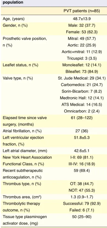
Table 4. Comparison of baseline and reactive hyperemia-induced endothelial parameters before and after thrombolytic therapy in PVT subgroups
Patient groups Parameters Before TT After TT p value
All patients (n=85) Baseline brachial artery diameter (mm) 3.46±0.63 3.51±0.77 0.267
Reactive hyperemia-induced diameter (mm) 3.66±0.67 3.77±0.83 0.042
Flow-mediated dilatation (%) 5.62±0.85 7.04±1.18 <0.001
Successful TT group Baseline brachial artery diameter (mm) 3.45±0.65 3.51±0.79 0.173
(n=79) Reactive hyperemia-induced diameter (mm) 3.65±0.68 3.76±0.85 <0.001
Flow-mediated dilatation (%) 5.65±0.86 7.13±1.26 <0.001
Failed TT group (n=6) Baseline brachial artery diameter (mm) 3.58±0.18 3.63±0.25 0.542
Reactive hyperemia-induced diameter (mm) 3.80±0.21 3.86±0.27 0.393
Flow-mediated dilatation (%) 5.07±0.61 5.38±0.95 0.371
Obstructive PVT group Baseline brachial artery diameter (mm) 3.51±0.73 3.59±0.85 0.232
(n=38) Reactive hyperemia-induced diameter (mm) 3.69±0.78 3.88±0.92 0.015
Flow-mediated dilatation (%) 5.31±0.76 8.22±1.15 <0.001
Non-obstructive PVT Baseline brachial artery diameter (mm) 3.43±0.55 3.50±0.72 0.335
group (n=47) Reactive hyperemia-induced diameter (mm) 3.63±0.58 3.71±0.76 0.291
Flow-mediated dilatation (%) 5.87±0.84 6.11±0.95 0.276
out compromising success rates with low-dose and slow-infusion protocols.[6–8] Following recent studies, the 2017 American College of Cardiology/American Heart Association valve disease guideline was revised and TT has been recommended in PVT therapy with an indication of class 1b and, alternatively, equivalent to emergency surgery.[5]
The endothelium is a large paracrine organ that se-cretes a variety of factors for regulation of vascular tone, cell growth, platelet and leukocyte interactions, and thrombogenicity.[10] The endothelium can sense and respond to numerous internal and external stimuli via cell membrane receptors and signal transduction mechanisms, leading to the synthesis and release of prominent cause of PVT development, several
eso-teric causes, such as genetic mutations,[23] elevated fi-brinogen,[24] anti-cardiolipin antibodies,[25] anti-tissue plasminogen activator antibodies,[26] increased hepa-ranase levels,[27] and AB0 blood groups,[28] may play a role in the aggravation of thrombus formation despite effective anticoagulation.
Treatment options for PVT include intensified anti-coagulation, TT, and redo valve surgery. Until recent-ly, surgery was recommended as first-line treatment for left-sided PVT, despite high morbidity and mor-tality rates.[4,19,29] TT is now increasingly performed in the treatment of PVT. Complications like hemor-rhage and thromboembolism have been reduced
with-A B
Figure 4. Scatter plot graphs illustrating (A) the moderate negative correlation between thrombus area and the percentage of baseline flow-mediated dilatation values and (B) the moderate positive correlation between the magnitude of change in flow-me-diated dilatation values and thrombus area in patients with prosthetic valve thrombosis.
Flow Mediated Dilatation (%)
Thrombus Area (cm2) 7.00 6.00 5.00 4.00 3.00 0.00 1.00 2.00 3.00 4.00 p=0.573; p<0.001 Dif
ference in Flow Mediated Dilatation after
TT (%) Thrombus Area (cm2) 4.00 3.00 2.00 1.00 0.00 -1.00 0.00 1.00 2.00 3.00 4.00 p=0.456; p<0.001
reported that acute arterial thrombosis caused endo-thelial dysfunction without causing endoendo-thelial cell loss and that supplementing TT with urokinase ame-liorated the endothelial dysfunction seen after acute thrombosis. Blum et al.[35] compared the FMD values of patients with sickle cell anemia during and after sickle cell crises characterized by increased adher-ence of sickle cell erythrocytes to vascular endothe-lial cells. The FMD values were significantly lower during the crises. Peng et al.[36] investigated the effects of TT and anticoagulation on the functions of vascu-lar endothelial cells and observed that TT and antico-agulation therapies were beneficial in protecting the functions of vascular endothelial cells in patients with a pulmonary thromboembolism. In the current study, we aimed to investigate the reversibility of endotheli-al dysfunction after TT in patients with PVT. Success-ful TT was found to improve endothelial function in PVT patients.
Conclusion
This study demonstrated that endothelial dysfunction was reversible in patients with PVT. Successful TT may contribute to the improvement of impaired en-dothelial function in patients with obstructive PVT proportional to the thrombus burden. TT with a low-dose and ultra-slow infusion of t-PA may be per-formed with considerable safety and efficacy in pa-tients with PVT.
Financial disclosure: This research received no specific
grant from any funding agency, commercial or not-for-profit sectors.
Ethical statement: The study protocol was approved by
the local ethics committee of Kartal Kosuyolu Training and Research Hospital, Istanbul, Turkey on October 20, 2014 (no: 2014.3/15).
Peer-review: Externally peer-reviewed. Conflict-of-interest: None.
Authorship contributions: Concept: B.Ç., M.K., M.Ö.;
Design: B.Ç., M.K., A.G.; Supervision: M.Ö., S.G.; Mate-rials: M.Y., E.B., M.O.G.; Data: A.G., E.B., S.K.; Analysis: M.K., S.K.; Literature search: A.G., M.Y., S.G.; Writing: B.Ç., M.K., A.G., M.O.G.; Critical revision: M.Ö.
REFERENCES
1. Cáceres-Lóriga FM, Pérez-López H, Santos-Gracia J, Mor-lans-Hernandez K. Prosthetic heart valve thrombosis: patho-genesis, diagnosis and management. Int J Cardiol 2006;110:1–
various vasoactive, thromboregulatory, and growth factor substances. A noninvasive technique has been widely used to evaluate flow-mediated vasodilation, an endothelium-dependent function, in the brachial artery.[18] In this technique, a reactive hyperemia-in-duced nitric oxide release is provoked, and the sub-sequent vasodilation is imaged and quantitated as an index of endothelial function.
Endothelial dysfunction plays an important role in the development of cardiovascular diseases, including atherosclerosis, hypertension, and heart failure.[10,30,31] In a previous study, it was demonstrated that patients with PVT had significantly lower levels of FMD com-pared with an age- and sex-matched control group of patients with normally functioning prosthetic valves. [11] However, the study could not determine whether endothelial dysfunction is the cause or the effect. In the current study, we focused on the reversibility of endothelial dysfunction after TT in patients with PVT. Persistence of the thrombus burden in the failed TT group resulted in no significant change in FMD val-ues. However, significantly increased FMD values af-ter successful TT in patients with PVT have revealed that endothelial dysfunction was predominantly the consequence of the presence of PVT.
It has been recognized that the endothelium partic-ipates in some pathological processes during inflam-mation and that inflaminflam-mation may affect endotheli-al function.[32] The incremental role of inflammatory processes in PVT development was highlighted in a previous study reporting increased inflammatory pa-rameters in patients with PVT. The difference in in-flammatory parameters was more significant in cases of obstructive PVT, indicating more serious inflam-mation in these patients.[9] Consistent with the results of earlier studies, our research also indicated that en-dothelial dysfunction resolved more significantly in patients with obstructive PVT. This may be explained by the association between the thrombus burden, in-flammation, and endothelial functions.
It was reported in an experimental animal study that endothelial exposure to thrombus formation significantly decreased endothelium-dependent va-sorelaxation. Furthermore, TT with urokinase was superior to thrombectomy in preserving endothelial functions.[33] In another study, Kashyap et al.[34] stud-ied rats with an infrarenal aortic occlusion created with a clip ligature to induce arterial thrombosis. They
with prosthetic valve thrombosis. J Thromb Thrombolysis 2018;46:399–402. [CrossRef]
18. Corretti MC, Anderson TJ, Benjamin EJ, Celermajer D, Char-bonneau F, Creager MA, et al. Guidelines for the ultrasound assessment of endothelial-dependent flow-mediated vasodi-lation of the brachial artery: a report of the International Brachial Artery Reactivity Task Force. J Am Coll Cardiol 2002;39:257–65. [CrossRef]
19. Castilho FM, De Sousa MR, Mendonça AL, Ribeiro AL, Cáceres-Lóriga FM. Thrombolytic therapy or surgery for valve prosthesis thrombosis: systematic review and meta-analysis. J Thromb Haemost 2014;12:1218–28. [CrossRef] 20. Gürsoy OM, Karakoyun S, Kalçık M, Özkan M. The
incre-mental value of RT three-dimensional TEE in the evaluation of prosthetic mitral valve ring thrombosis complicated with thromboembolism. Echocardiography 2013;30:E198–E201. 21. Ozkan M, Gürsoy OM, Astarcıoğlu MA, Gündüz S, Cakal
B, Karakoyun S, et al. Real-time three-dimensional trans-esophageal echocardiography in the assessment of mechan-ical prosthetic mitral valve ring thrombosis. Am J Cardiol 2013;112:977–83. [CrossRef]
22. Gündüz S, Özkan M, Kalçik M, Gürsoy OM, Astarcioğlu MA, Karakoyun S, et al. Sixty-Four-Section Cardiac Com-puted Tomography in Mechanical Prosthetic Heart Valve Dysfunction: Thrombus or Pannus. Circ Cardiovasc Imaging 2015;8:e003246. [CrossRef]
23. Kalcik M, Gursoy MO, Karakoyun S, Yesin M, Astarcioglu MA, Ozkan M. Potential inherited causes of recurrent pros-thetic mitral valve thrombosis in a pregnant patient suffering from recurrent miscarriage. Korean Circ J 2014;44:268–70. 24. Aykan AC, Gökdeniz T, Gündüz S, Astarcioğlu MA, Gürsoy
OM, Ertürk E, et al. Value of serum fibrinogen levels in the as-sessment of mechanical prosthetic valve thrombosis. J Heart Valve Dis 2014;23:222–7.
25. Aykan AÇ, Gökdeniz T, Kalçık M, Astarcıoğlu MA, Gündüz S, Karakoyun S, et al. Role of anticardiolipin antibodies in the pathogenesis of prosthetic valve thrombosis: An observa-2017;135:e1159–e95. [CrossRef]
6. Özkan M, Gündüz S, Biteker M, Astarcioglu MA, Çevik C, Kaynak E, et al. Comparison of different TEE-guided throm-bolytic regimens for prosthetic valve thrombosis: the TROIA trial. JACC Cardiovasc Imaging 2013;6:206–16. [CrossRef] 7. Özkan M, Çakal B, Karakoyun S, Gürsoy OM, Çevik C,
Kalçık M, et al. Thrombolytic therapy for the treatment of prosthetic heart valve thrombosis in pregnancy with low-dose, slow infusion of tissue-type plasminogen activator. Cir-culation 2013;128:532–40. [CrossRef]
8. Özkan M, Gündüz S, Gürsoy OM, Karakoyun S, Astarcıoğlu MA, Kalçık M, et al. Ultraslow thrombolytic therapy: A novel strategy in the management of PROsthetic MEchanical valve Thrombosis and the prEdictors of outcomE: The Ultra-slow PROMETEE trial. Am Heart J 2015;170:409–18. [CrossRef] 9. Gürsoy OM, Karakoyun S, Kalçik M, Gökdeniz T, Yesin M,
Gündüz S, et al. Usefulness of novel hematologic inflamma-tory parameters to predict prosthetic mitral valve thrombosis. Am J Cardiol 2014;113:860–4. [CrossRef]
10. Verma S, Anderson TJ. Fundamentals of endothelial function for the clinical cardiologist. Circulation 2002;105:546–9. 11. Kaya H, Ozkan M, Yildiz M. Relationship between
endo-thelial dysfunction and prosthetic heart valve thrombosis: a preliminary investigation. Eur Rev Med Pharmacol Sci 2013;17:1594–8.
12. Zoghbi WA. New recommendations for evaluation of pros-thetic valves with echocardiography and doppler ultrasound. Methodist Debakey Cardiovasc J 2010;6:20–6. [CrossRef] 13. Ozkan M, Kaymaz C, Kirma C, Sönmez K, Ozdemir N,
Balkanay M, et al. Intravenous thrombolytic treatment of mechanical prosthetic valve thrombosis: a study using se-rial transesophageal echocardiography. J Am Coll Cardiol 2000;35:1881–9. [CrossRef]
14. Lancellotti P, Pibarot P, Chambers J, Edvardsen T, Delgado V, Dulgheru R, et al. Recommendations for the imaging as-sessment of prosthetic heart valves: a report from the Euro-pean Association of Cardiovascular Imaging endorsed by the
32. Stenvinkel P. Endothelial dysfunction and inflammation-is there a link?. Nephrol Dial Transplant 2001;16:1968–71. 33. Reil TD, Moore WS, Kashyap VS, Nene SS, Gelabert HA,
Quinones-Baldrich WJ. The effects of thrombus, thrombec-tomy and thrombolysis on endothelial function. Eur J Vasc Endovasc Surg 2000;19:162–8. [CrossRef]
34. Kashyap VS, Reil TD, Moore WS, Hoang TX, Gelabert HA, Byrns RE, et al. Acute arterial thrombosis causes endothelial dysfunction: a new paradigm for thrombolytic therapy. J Vasc Surg 2001;34:323–9. [CrossRef]
35. Blum A, Yeganeh S, Peleg A, Vigder F, Kryuger K, Khatib A, et al. Endothelial function in patients with sickle cell anemia during and after sickle cell crises. J Thromb Thrombolysis 2005;19:83–6. [CrossRef]
36. Peng K, Wang C, Pang BS, Yang YH. Effects of thrombolysis and anticoagulation on the functions of vascular endothelial cells and coagulation and fibrinolysis in patients with pul-monary thromboembolism. [Article in Chinese] Zhonghua Jie He He Hu Xi Za Zhi 2005;28:596–9.
tional study. Herz 2015;40:528–33. [CrossRef]
26. Özkan M, Kalçık M, Gürsoy MO, Öcal L, Griffini S, Karakoyun S, et al. Assessment of Anti-Tissue Type Plas-minogen Activator Antibodies in Patients With Prosthetic Heart Valve Thrombosis: The ATA Trial. J Cardiovasc Phar-macol Ther 2016;21:372–80. [CrossRef]
27. Bayam E, Kalçık M, Gürbüz AS, Yesin M, Güner A, Gündüz S, et al. The relationship between heparanase levels, thrombus burden and thromboembolism in patients receiving unfrac-tionated heparin treatment for prosthetic valve thrombosis. Thromb Res 2018;171:103–10. [CrossRef]
28. Astarcıoğlu MA, Kalçık M, Yesin M, Gürsoy MO, Şen T, Karakoyun S, et al. AB0 blood types: impact on development of prosthetic mechanical valve thrombosis. Anatol J Cardiol 2016;16:820–3. [CrossRef]
29. Lengyel M, Horstkotte D, Völler H, Mistiaen WP; Working Group Infection, Thrombosis, Embolism and Bleeding of the Society for Heart Valve Disease. Recommendations for the management of prosthetic valve thrombosis. J Heart Valve Dis 2005;14:567–75.
30. Laurent S, Lacolley P, Brunel P, Laloux B, Pannier B, Safar M. Flow-dependent vasodilation of brachial artery in essential hypertension. Am J Physiol 1990;258:H1004–H11. [CrossRef] 31. Celermajer DS, Sorensen KE, Gooch VM, Spiegelhalter DJ,
Miller OI, Sullivan ID, et al. Non-invasive detection of en-dothelial dysfunction in children and adults at risk of athero-sclerosis. Lancet 1992;340:1111–5. [CrossRef]
Keywords: Echocardiography; endothelial functions; flow-mediated
dilatation; prosthetic valve thrombosis; transesophageal echocar-diography.
Anahtar sözcükler: Ekokardiyografi; endotel fonksiyonları; akım
aracılı dilatasyon; protez kapak trombozu; transözofajiyal ekokar-diyografi.