Türk Kardiyol Dern Arş - Arch Turk Soc Cardiol 2015;43(5):481-483 doi: 10.5543/tkda.2015.25295
Conservative management of a left ventricle cardiac fibroma
in an asymptomatic child patient
Semptomsuz sol ventrikül kalp fibroması olan çocukda konservatif yaklaşım
Department of Pediatric Cardiology, Zekai Tahir Burak Maternity Teaching Hospital, Ankara#Department of Pediatric Cardiology, Ufuk University Faculty of Medicine, Ankara
Handan Ünsal, M.D., Enver Ekici, M.D.#
Özet– Çocuk ve infantlarda nadir görülen primer kalp tümör-leri çoğunlukla iyi huyludur. Rabdomiyomdan sonra en sık görülen iyi huylu kalp tümörü fibromaların insidansı olduk-ça düşüktür. Fibromalar çıkım yolu obstrüksiyonu, ventrikül fonksiyon bozukluğu ve ciddi aritmi oluşturması durumunda cerrahi müdahale gerektirirler. Bu yazıda dokuz yaşında er-kek çocukta saptanan sol ventrikülde yerleşik dev fibroma, nadir görülmesi yanında semptomsuz seyretmesi ve kon-servatif yaklaşımla izlenebilmesi nedeniyle sunuldu.
Summary– Primary cardiac tumors in infancy and child-hood are rare and usually benign. Fibroma is the second most common bening cardiac tumor after rhabdomyo-ma. Surgery is required when fibromas cause ventricu-lar outflow tract obstruction, ventricuventricu-lar dysfunction and life-threatening arrhythmia. This case report describes a 9-year-old asymptomatic male presenting with a giant left ventricular cardiac fibroma who was followed up using con-servative treatment.
481
P
rimary cardiac tumors in infancy and childhoodare rare and usually benign. Fibromas are gen-erally reported as the second most common benign cardiac tumor after rhabdomyoma in the pediatric age group. Clinical signs and symptoms vary depending
on tumor size and location.[1,2] The tumor may occur
with cardiomegaly, heart failure, arrhythmias, cya-nosis and chest pain, or it may be a cause of sudden
death.[3] In rare cases, a tumor of gigantic size may
also be asymptomatic.[4–6]
The case here was thought to be valuable for pre-sentation because of its rarity among diseases and the tumor’s being asymptomatic despite its large size.
CASE REPORT
A 9-year-old boy was admitted for evaluation of a cardiac murmur which had been detected on day 15 in the newborn period. A detailed cardiac examina-tion (physical examinaexamina-tion, electrocardiography and echocardiography) was done and he was diagnosed with a primary cardiac tumor after computed
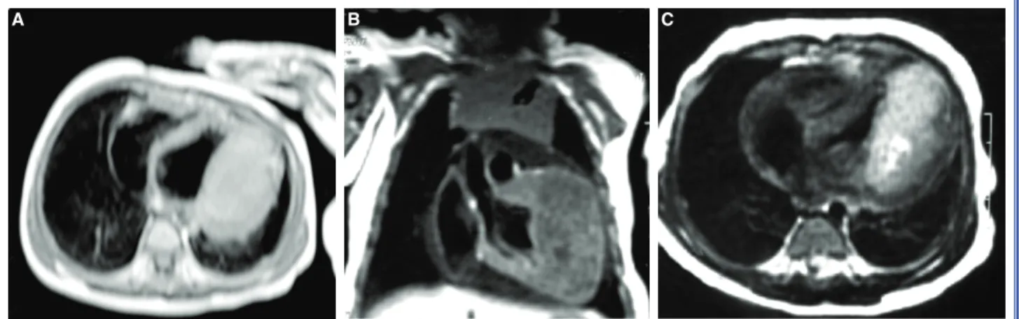
tomog-raphy (CT) and magnetic resonance imaging (MRI) (Figure 1a). The cardiac MRI demonstrated a large
(4x6.5x6.1 cm) homogeneous mass extending from the apex to basal segment and covering the lateral, anterolateral and inferolateral walls (Figure 1b, c). A biopsy was done from the left ventricle apex. At the histopathological exam, the material showed the char-acteristics of a fibroma, thus confirming the diagnosis. At the age of 3 years, when the patient was admit-ted to our clinic for the first time, he had already been put on the heart transplant list in another center. At that visit, the overall physical examination of patient, who was asymptomatic, was normal, except for the pres-ence of the soft systolic heart murmur. Electrocardiog-raphy showed ventricular extrasystoles and negative T waves on the anterolateral derivations. Echocardiogra-py showed a large (8.2x3.1 cm) mass containing cal-cific and cystic areas, appended to the left ventricular free wall (Figure 2a, b). The mass did not influence
Received:January 20, 2015 Accepted:April 09, 2015
Correspondence: Dr. Handan Ünsal. Dr. Zekai Tahir Burak Kadın Hastalıkları Eğitim Hastanesi, Pediatrik Kardiyoloji Bölümü, 06230 Ankara, Turkey.
Tel: +90 312 - 306 52 70 e-mail: [email protected]
© 2015 Turkish Society of Cardiology
Abbreviations:
CT Computed tomography MRI Magnetic resonance imaging
left ventricular functions or lead to left ventricular outflow tract obstruction. As electrocardiography and 24-hour Holter monitoring revealed ventricular extra-systoles, beta blocker therapy was started. No signifi-cant growth in tumor size was seen during the clinical follow-up. Currently, the patient is still asymptomatic and conservative follow-up continues at our clinic.
DISCUSSION
Primary cardiac tumors are rare, with a prevalence of lower than 0.03% according to postmortem studies. Approximately 90% are benign, and the majority of
them are diagnosed before the age of 1 year.[1,2]
Car-diac fibromas, which normally arise from heart fibro-blasts, are solitary and white in appearance, and have
no capsule to help ensure separation from the sur-rounding tissue. They are located primarily in the left ventricular free wall or septum, and less frequently in the right ventricular or atrial free wall. They tend to
grow slowly and show no spontaneous regression.[5–7]
In the present case, the diagnosis was made before the age of 1 year, and no significant regression was seen during follow-up.
Clinical presentation varies, depending on size and
location of the tumor.[3] Affected children may develop
left ventricular outflow obstruction, valve dysfunction, heart failure—depending on the ventricular dysfunc-tion—or life-threatening arrhythmias and even sudden
death (10%). They may may also be asymptomatic.[4,5,7]
Although echocardiography is the mainstay of
Türk Kardiyol Dern Arş 482
Figure 1. (A) Axial Computed Tomography showing the intraventricular mass. (B) Coronal T1 Weighted image of the intraven-tricular mass isointense to the myocardium. (C) Axial Gradient magnetic resonance image showing the mass isointense to the myocardium.
A B C
Figure 2. (A) Echocardiographic image of the tumor in the left ventricule. (B) Image of calcific and cystic areas in the tumor.
Conservative management of a left ventricle cardiac fibroma in an asymptomatic child patient 483
non-invasive diagnostic tools for initial evaluation of cardiac fibromas, imaging techniques such as CT or MRI are valuable in their detection. MRI espe-cially, provides the identification, location, surround-ing structures and the hemodynamic effects of the tumor. Cardiac fibromas appear as regular, limited, and mildly hyperechogenic solid lesions. They can be distinguished from other tumors because of their solitary, regular and limited nature. In large fibromas, hypoechogenic or calcific and cystic areas can be seen
according to the ischemia in the center of the tumor.[9]
In the initial radiological examination of our patient, the tumor was demonstrated as a regular limited solid lession, with cystic and calcific areas appearing over time during follow-up. Although biopsy and histo-pathological examination are needed for definitive di-agnosis, recent improvements in imaging technologies eliminates the necessity for this.[10] In this case,
diag-nosis was confirmed by histopathological evaluation of material taken from the apex of the left ventricle.
Location, size and clinical presentation of the tu-mor define the treatment strategies. Surgical interven-tion is warranted for symptomatic patients, and the type of intervention, such as a total or subtotal resec-tion, depends on the location of the tumor. Symp-tomatic cases where surgical resection is not possible may need heart transplantation. In surgical resections, even if it is subtotal, short and long-term results are
reported to be very good.[11]
The approach is controversial in asymptomatic tu-mors. As outcomes of even partial resections are fine, and the tumors create increased risk of sudden death due to fatal arrhythmia, some authors suggest
surgi-cal intervention for asymptomatic patients.[12] On the
other hand, careful long-term follow-up adopting the conservative approach may be preferred because of
the risk of surgery.[13] In our case, although the tumor
mass was large, the patient was asymptomatic. We decided to do follow-up with conservative modali-ties, and in the 6 years of this process it has continued without any problems. However, we think that in time surgery may be needed due to changes in the growth process of the patient.
Consequently the authors consider that in asymp-tomatic patients with a cardiac fibroma, careful long-term follow-up using the conservative approach may be an alternative treatment, even when the tumor mass is large.
Conflict-of-interest issues regarding the authorship or article: None declared.
REFERENCES
1. Bruce CJ. Cardiac tumours: diagnosis and management. Heart 2011;97:151–60. CrossRef 2. Kır M, Çatalyürek H, Karadaş U, Ünal N, Saylam G. A prena-tally diagnosed newborn with an intracardiac rhabdomyoma obstructing the left ventricular outflow tract. Turkish Journal of Thoracic and Cardiovascular Surgery 2011;19:623–6. CrossRef 3. Burke AP, Rosado-de-Christenson M, Templeton PA, Virmani R. Cardiac fibroma: clinicopathologic correlates and surgical treatment. J Thorac Cardiovasc Surg 1994;108:862–70. 4. Becker AE. Primary heart tumors in the pediatric age group: a review of salient pathologic features relevant for clinicians. Pediatr Cardiol 2000;21:317–23. CrossRef 5. Becker AE. Tumours of the heart and pericardium. In: Fletch- er CDM, editor. Diagnostic histopathology of tumours. Edin-burgh, UK: Churchill Livingstone; 1995. p. 7–41. 6. Demir F, Akın A, Bilici M, Aktar F, Uluca Ü, Turan Mİ, et al. An infant with a giant right ventricular fibroma. Dicle Medi-cal Journal 2014;41:608–10. CrossRef 7. Feldman PS, Meyer MW. Fibroelastic hamartoma (fibroma) of the heart. Cancer 1976;38:314–23. CrossRef 8. Stratemann S, Dzurik Y, Fish F, Parra D. Left ventricular car- diac fibroma in a child presenting with ventricular tachycar-dia. Pediatr Cardiol 2008;29:223–6. CrossRef 9. Kiaffas MG, Powell AJ, Geva T. Magnetic resonance imag-ing evaluation of cardiac tumor characteristics in infants and children. Am J Cardiol 2002;89:1229–33. CrossRef
10. De Cobelli F, Esposito A, Mellone R, Papa M, Varisco T, Besana R, et al. Images in cardiovascular medicine. Late en-hancement of a left ventricular cardiac fibroma assessed with gadolinium-enhanced cardiovascular magnetic resonance. Circulation 2005;112:242–3. CrossRef
11. Cho JM, Danielson GK, Puga FJ, Dearani JA, McGregor CG, Tazelaar HD, et al. Surgical resection of ventricular cardiac fibromas: early and late results. Ann Thorac Surg 2003;76:1929–34. CrossRef
12. Nwachukwu H, Li A, Nair V, Nguyen E, David TE, Butany J. Cardiac fibroma in adults. Cardiovasc Pathol 2011;20:146– 52. CrossRef
13. Pozzi M, Deux JF, Kirsch M. Conservative management of left ventricle cardiac fibroma in an adult asymptomatic pa-tient. Int J Cardiol 2012;161:61–2. CrossRef
Key words: Child; echocardiography; fibroma/complications/diagno-sis; heart neoplasms.
Anahtar sözcükler: Çocuk; ekokardiyografi; fibroma/komplikasyon/ tanı; kalp neoplazileri.