Published online 2014 February 5. Research Article
Unusual Findings in Appendectomy Specimens of Adults: Retrospective
Analyses of 1466 Patients and a Review of Literature
Hakan Yabanoglu
1; Kenan Caliskan
1; Huseyin Ozgur Aytac
1; Emin Turk
1; Erdal Karagulle
1,*;
Fazilet Kayaselcuk
2; Mehmet Akin Tarim
11 Department of General Surgery, Faculty of Medicine, Baskent University, Ankara, Turkey 2 Department of Pathology, Faculty of Medicine, Baskent University, Ankara, Turkey
*Corresponding Author: Erdal Karagulle, Department of General Surgery, Faculty of Medicine, Baskent University, Ankara, Turkey. Tel: +90-03322570606, Fax: +90-03322570637, E-mail:
Received: June 14, 2013; Revised: July 29, 2013; Accepted: August 23, 2013
Background: Diseases and tumors of the appendix vermiformis are very rare, except acute appendicitis.
Objectives: This retrospective study was conducted to document the unusual findings in appendectomy specimens.
Patients and Methods: Data of 1466 adult patients were gathered retrospectively. Appendectomy was performed in 1169 and in 297
patients following a diagnosis of acute appendicitis and during other abdominal operations, respectively. The data of 57 (3.88 %) patients who were pathologically reported to have unusual appendix findings were retrospectively collected. The records were analyzed according to patients’ age, gender, clinical presentations, operative reports, pathological reports and follow up.
Results: Unusual pathologic examination findings were detected in the appendectomy specimens of 57 patients with a mean age of 48.34
± 19. Twenty-nine patients (50.8 %) were male and 28 (49.2 %) were female. Normal appendix tissues were observed in specimens of 26 (45.6 %) patients and inflamed appendix in 31 (54.3 %). The most common unusual finding was parasitic diseases of the intestine. Pathological diagnosis of malignancy and benign features were reported in specimens of 14 and 43 patients, respectively. Macroscopic evaluation of appendectomy specimens during surgery might result in negligence of the presence of unusual pathology.
Conclusions: Even if the macroscopic appearance of the specimen is normal or acute appendicitis, we suggest routine histopathological
examination.
Keywords:Appendectomy; Appendiceal Neoplasms; Enterobius; Entamoeba Histolytica
Implication for health policy/practice/research/medical education:
The most commonly performed surgery is appendectomy. Various pathologies are seen in patient’s undergone appandectomy. The article is important with regard to number of patients examined and the pathologic results reported. In other words, pathological examination of appendectomy specimens of each patient should be considered.
Copyright © 2014, Iranian Red Crescent Medical Journal; Published by Kowsar Corp. This is an open-access article distributed under the terms of the Creative Com-mons Attribution License, which permits unrestricted use, distribution, and reproduction in any medium, provided the original work is properly cited.
1. Background
Appendectomy is one of the most common surgical procedures done for acute appendicitis or during vari-ous abdominal operations. The lifetime incidence of acute appendicitis is 8.6% and 6.7 % for men and women, respectively (1). However, the incidence of appendectomy performed for various reasons is 12 % for men and 25 % for women (1). Most common cause of acute appendicitis is obstruction of the lumen. Although fecal impactions and lymphoid hyperplasia are the most frequent reasons of lumen obstruction, rare and unusual causes like intes-tinal parasites, radiocontrast agents, actinomycetes, en-dometriosis, tuberculosis, stromal tumors, carcinoid tu-mors, fruit pippins, adenomas, mucoceles, lymphomas, dysplastic changes, primary and secondary adenocarci-nomas, eosinophilic granulomas, and granulomatous diseases may be seen (2-8).
2. Objectives
This retrospective study was designed to document the unusual findings in appendectomy specimens.
3. Materials and Methods
The data of adult patients who underwent appendecto-my at Baskent University, Adana Teaching Hospital from January 1999 through February 2013 were analyzed retro-spectively. This study was a retrospective clinical research study. During a 14-year period, patients who underwent appendectomy in our hospital were included regardless of the preoperative diagnosis. Specimens from patients operated at different centers and those examined by pa-thology department of our hospital were excluded from the study. The collected records were patients’ age, gen-der, clinical presentation, operative reports, pathological reports, and up durations. The duration of follow-up was reported as months, beginning from the date of diagnosis until the last clinical information available up to February 2013. Histological reports were analyzed ac-cording to diagnosis and unusual findings were noted. The original pathology specimens with unusual findings were re-evaluated by an experienced pathologist (FK). This study was approved by Baskent University Institu-tional Review Board and supported by Baskent University
Research Fund (KA13/50). Data were collected on a com-puter media and analyzed by SPSS software (SPSS Inc, Chi-cago, Illinois, united states).
4. Results
Hospital records of 1466 patients who had underwent appendectomy (due to inflammatory or incidental rea-sons, either open or laparoscopic) were reviewed dur-ing the 14 years period beginndur-ing from January 1999 to February 2013. The data of 57 (3.88 %) patients who were pathologically reported to have unusual appendix find-ings were collected retrospectively. Of the 1466 patients who underwent appendectomy, 1169 were operated for acute appendicitis and the rest 297 were operated dur-ing other surgical procedures (gynecological cancers, laparotomies intending to acute abdomen, colon can-cers, mesenteric ischemias, Amyand’s hernias, and etc.). Among these patients, 730 (49.7 %) were males and 736 (50.3 %) were females. Mean age of the patients was 36 ± 12 years ranging from 16 to 83 years. Out of 1466 cases, path-ological evaluation of 328 (22 %) was normal. In 1138 cases (78 %), pathological reports were associated with acute inflammation showing changes of acute appendicitis (abscess, perforation, or gangrene). Unusual pathologic findings were detected in the appendectomy specimens of 57 patients with a mean age of 48 ± 19 years. Twenty-nine patients (50.8 %) were males and 28 (49.2 %) were fe-males. Of these 57 specimens with unusual pathology, 26 (45.6 %) were non-inflamed and 31 (54.3 %) were inflamed (phlegmonous, perforated, or acute) appendix tissues. Eighteen (31.5 %) of the patients having normal histo-pathology were females and 8 (14 %) were males. On the other hand, 10 (17.5 %) of the patients who were reported to have inflamed appendix tissues were females and 21 (36.3 %) were males. Amongst 57 patients having unusual histopathologic findings, 14 had malignant (primary mu-cinous adenocarcinoma, neuroendocrine -carcinoid- tu-mours, lymphomas, or pseudomyxoma peritonei) and 43 had benign disorders. Most of the benign disorders were due to intestinal parasites. Of the 15 patients (57.6 %) pre-senting intestinal parasitosis, 12 had Enterobious vermicu-laris and three had Entamoeba histolytica (amebiasis). The etiological causations of unusual histopathologic findings in appendectomy specimens of 57 cases are sum-marized in Table 1. Some images of specimens of benign and malignant disorders are demonstrated in Figure 1. The clinicopathological characteristics of five patients with appendix mucinous adenocarcinoma and seven pa-tients with neuroendocrine tumors (carcinoids) are sum-marized in Table 2 and Table 3, respectively.
In those with diagnosis of mucinous adenocarcinoma, immediate right-sided hemicolectomy was performed for three patients synchronously with appendectomy by perioperative pathological frozen section confirma-tion and for one patient at a following session. A
right-sided hemicolectomy at the second session was per-formed for another patient with neuroendocrine tumor invading the serosa. A patient for whom synchronous right-sided hemicolectomy was performed due to mu-cinous adenocarcinoma and another patient operated for rectum perforation secondary to metastasis of lung adenocarcinoma with a postoperative pathology diagno-sis of mucinous adenocarcinoma in appendix had died. A patient who was diagnosed with mucinous adenocar-cinoma and a previous metastatic colon caradenocar-cinoma had died three months later. Another patient who was di-agnosed with neuroendocrine tumor (carcinoid) with a previous history of cholangiocellular carcinoma had died two months later owing to multiple organ failure.
Table 1. Distribution of the 57 Cases Defined as ‘’Unusual
Find-ing’’ According to Etiological Causes
No. (%)
Total Patients 57 (3.88)
Mucinous cystadenoma ( + mucocele) 16 (1.09)
Enterobius vermicularis 15 (1.02) Neuroendocrine tumors 7 (0.47) Mucinous adenocarcinoma 5 (0.4) Amebiasis 4 (0.27) Tubular adenoma 2 (0.0 1) Lymphoma 1 (< 0.01) Endometriosis 1 (< 0.01) Actinomycosis 1 (< 0.01) Pseudomyxoma peritonei 1 (< 0.01)
Foreign body granuloma 1 (< 0.01)
Focal severe dysplasia 1 (< 0.01)
Serrated adenoma 1 (< 0.01)
Hyperplastic poly 1 (< 0.01)
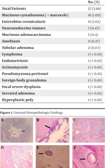
Figure 1. Unusual Histopathologic Findings
A: Enterobius vermicularis image within the lumen of the appendix (black arrow), (HE, x40); B: High grade mucinous appendicial neoplasia (black arrow), (HE, x200); C: CD 79 a (+) in appendix, low grade lymphoma (HE, x200); D: Presence of mucine in wall of appendix (HE, x200); E: Acti-nomyces colonies in the appendix (black arrow), (HE, x200); F: Endometri-al glands and stroma in the muscle layer of appendix wEndometri-all (black arrow), (HE, x200).
Table 2. Clinicopathological Characteristics of the Five Patients With Primary Appendicular Mucinous Adenocarcinoma
Age (y) Sex Tumor size (cm) Location Treatment Parietal Spread Follow-up (Month)
61 F 4 Distal Appendectomy + Right hemicolectomy Mucosa 13
47 M 2 Base Appendectomy + Right hemicolectomy Mucosa 11
71 F < 1 Distal Appendectomy + Right hemicolectomy Mucosa 12
60 F < 1 Distal Appendectomy Serosa 3 (exitus)
39 F 2 Distal Appendectomy + Right hemicolectomy Serosa 4
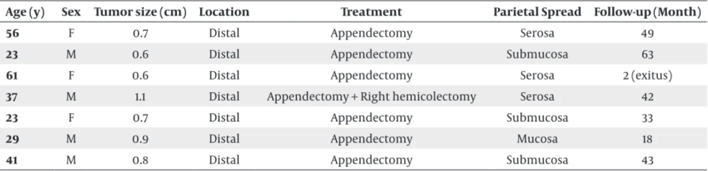
Table 3. Clinicopathological Characteristics of the Seven Patients With Primary Appendicular Neuroendocrine Tumors (Carcinoid)
Age (y) Sex Tumor size (cm) Location Treatment Parietal Spread Follow-up (Month)
56 F 0.7 Distal Appendectomy Serosa 49
23 M 0.6 Distal Appendectomy Submucosa 63
61 F 0.6 Distal Appendectomy Serosa 2 (exitus)
37 M 1.1 Distal Appendectomy + Right hemicolectomy Serosa 42
23 F 0.7 Distal Appendectomy Submucosa 33
29 M 0.9 Distal Appendectomy Mucosa 18
41 M 0.8 Distal Appendectomy Submucosa 43
Neither recurrence nor mortality had been seen after the mean follow up of 30 (4-63) months of the 12 patients that were reported to have primary malignant disease of the appendix. All patients with malignant tumors were diagnosed clinically with acute appendicitis, and none of them had symptoms of carcinoid syndrome or were preoperatively diagnosed with an appendicular tumors. After pathological confirmation of the diagnosis, abdom-inal ultrasonography, computed tomography (CT) scan-ning, and 24-hours urinary 5-hydroxyindoleacetic acid level measurements were performed for staging. Then, all patients were followed-up at the outpatient clinic every 3 or 6 months for the first year. Oral medications were prescribed to the patients that were found to have parasitic diseases of the intestine according to histopath-ological studies.
5. Discussion
Appendectomy is one of the most common surgical operations (9). Incidence of acute appendicitis is rather proportional with lymphoid development. It makes peak by the end of puberty and third decade of life. The gender ratio in acute appendicitis is about 1:1 prior to puberty. At puberty, male to female ratio becomes 2:1 (10). The most important causative factor for acute appendicitis is lu-minal obstruction. With the pathophysiological changes occurring due to luminal obstruction like continued mu-cus secretion, inflammatory exudation increasing intra-luminal pressure, which obstructs lymphatic drainage and developing edema and mucosal ulceration, disten-sion of appendix increases and results in venous obstruc-tion. At the end of this process, ischemic necrosis occurs at the wall of appendix vermicularis (11). Fecaloids are the
major factors resulting in luminal obstruction of appen-dix. There are many other rare reasons except this (2-8). Most prevalent unusual pathologic findings seen in ap-pendectomy specimens after apap-pendectomy due to any reason are parasitic diseases of the intestine and benign or malignant tumors (10). Enterobious vermicularis is the most common helminthic infestation agent of gas-trointestinal tract in the world (12). The relation between Enterobious vermicularis and appendicitis was first described by Stil in the late 19th century (13). Although it is generally asymptomatic, its major symptom is pru-ritus ani. On the other hand, it may appear with serious complications like ileocolitis, enterocutaneous fistulas, urinary infections, mesenteric abscesses, salpingitis, and appendicitis. Incidence of Enterobious vermicularis existence in appendectomy specimens is between 0.6 % and 3.8 % (12, 14-19). Inflammation rate in appendectomy specimens that were infected with Enterobious vermicu-laris differs from 13 % to 37 % (20-22). Similarly, in our study 15 (1 %) patients had Enterobious vermicularis in their ap-pendectomy specimens and of these, 3 (20 %) had shown inflammation related with acute appendicitis.
Appendicitis due to existence of Entemoeba histolytica is very rare and few cases are reported in literature (23-28). The certain frequency of this atypical presentation of parasitic disease is not known (29). It differs between 0.5 % and 2.3% among the limited number of studies in lit-erature (15, 23, 30). In our study, Entemoeba histolytica was demonstrated in 4 (0.2 %) patients. Only in one (25 %) of these patients inflammation was shown histopathologi-cally. Ratio of inflammation is very low in appendectomy specimens of patients determined to have both Entemoe-ba histolytica and Enterobious vermicularis (20-22). For this reason, negative laparotomy is seen more frequently in
this group in contrast to all the other unusual pathologi-cal situations. Proper medications (oral metronidazole/ pyrantel pamoate) were started after surgery for all the patients in this group. Neoplasms of the appendix are very uncommon and usually diagnosed at operation or autopsy. Malignant tumors of the appendix include carcinoids, lymphomas, mucoceles, primary adenocar-cinomas, and mucinous cystadenocarcinomas. Benign tumors of the appendix consist of tubular adenomas, vil-lous adenomas, leiomyomas, neuromas, and lipomas (3, 31, 32). In a study of Collins, investigating appendectomy specimens of 71000 patients operated for various clinical conditions, 958 (1.35 %) malignant and 3271 (4.6 %) benign tumors were determined (33). in our patients, we identi-fied 30 (2 %) cases with malignant neoplasm in appendix specimens.
Neuroendocrine tumors (carcinoids) are the most com-mon malignant tumors of appendix vermiformis (34). They are typically yellow-brown, small, hard and having limited surface tumors. They are generally diagnosed af-ter appendectomies done for acute appendicitis or other surgical procedures with coincidence. In the series by Collins, carcinoids made up 51 % of the malignant tumors of the appendix (33). The reported incidence of appendix carcinoids in several studies ranges from 0.02 to 2.27 % of surgically removed appendixes (3, 9, 35-37). In Collins’s study, carcinoids were found in 0.7 % of all appendectomy specimens (33). Carcinoids were seen in 7 (0.47 %) patients in our study. All patients in our study had signs and symp-toms of acute appendicitis. None of the patients had symptoms of carcinoid syndrome (flushing, diarrhea, cardiac symptoms, and bronchospasm). A large female preponderance is reported in all series (2-3:1) (38, 39). In many studies diameter of carcinoid tumors were found to be shorter than 1 cm and dominantly located at the tip of the appendix (3, 34, 40-42). While tumor localization in our study was concordant with the literature, incidence seemed to vary with gender (Table 3). The calculated risk of metastasis from tumors of 1 cm or smaller in diame-ter is nearly zero and therefore, can be managed with a simple appendectomy. Metastasis risk increases up to 85 % when the tumor diameter exceeds 2 cm. An appendiceal carcinoid tumor larger than 2 cm should be managed with a formal right-sided hemicolectomy (4, 14, 31, 34, 36, 41, 43-46). In our study, a right-sided hemicolectomy was performed to a patient with a carcinoid tumor of 11 mm in diameter invading serosa at a second session. No recur-rence was observed after 42 months follow-up of.
Primary adenocarcinoma of the appendix is an extraor-dinarily rare tumor. It is defined in limited series in litera-ture with an incidence of 0.08 % (33). This tumor is seen most commonly in patients between the age 50 and 55 years. Adenocarcinomas behave aggressively in a fashion similar to colonic adenocarcinomas; therefore, they must also be treated with the same aggressive approach (39). In our study, five patients had adenocarcinomas.
Right-sided hemicolectomy were performed synchronous with appendectomy for three patients and for another patient after permanent pathological confirmation at a second session. No further surgical intervention was planned for one of the patients with metastatic disease who died at the third month of follow up due to multiple organ fail-ure. Four patients are still alive without any recurrence and are still being followed up.
Mucinous cystadenoma is a rare tumor of the appen-dix associated with cystic dilatation, to which the more general term of mucocele has been applied. Incidence of mucocele in appendectomy specimens is between 0.2 % and 0.3 % (3). This ratio was found to be higher (1.09 %) than literature in our study. Mucocele generally behaves asymptomatic and is determined coincidentally during other abdominal operations. Mucoceles were found in five of our patients who were surgically explored for ovar-ian masses. However, mucoceles might be recognized clinically from features of acute appendicitis. Appendec-tomy is the standard of care for mucinous cystadenoma, whereas a cystadenocarcinoma requires a right-sided hemicolectomy. Because of the high association of mu-cinous cystadenoma with colonic and ovarian malignan-cies, follow-up with CT-scans, ultrasonography, and colo-noscopic examinations must be performed during the postoperative period (3, 4, 31, 42, 47-49).
Endometriosis is defined as the presence of endome-trial tissue outside uterine cavity (50). It rarely settles in gastrointestinal system. Intestinal endometriosis is clas-sified as external endometriosis and occurs in only about 10 % of women with endometriosis. Appendiceal endo-metriosis is usually asymptomatic, but it occasionally causes appendicitis, perforation, and intussusceptions. The diagnosis of appendiceal endometriosis is based on the histological presence of endometrial tissue in the specimen. We found endometriosis in one of our pa-tients’ appendectomy specimens. Rare pathogens like ac-tinomycosis, lymphoma, and pseudomyxoma peritonei were found in very few ratios, which is in concordance with the reports in the medical literature. Our series have the highest frequency of unusual pathological findings in appendectomy specimens (3.88 %) among single center series in literature. Although there are some case reports in English medical literature, there are only few reports analyzing large series of patients (9, 51-54). However, the weaknesses of this study are its retrospective nature and including data only from a certain region of Turkey.
Although fecaloids and lymphatic hyperplasia are the foremost reasons of acute appendicitis, rare conditions must not be neglected. Otherwise, pure macroscopic as-sessments of the specimens without histopathologic con-firmation may cause overlooking unusual reasons that result in incomplete surgical and medical treatments. This situation, especially with the existence of malignant tumors, brings important medical, social, ethical, and le-gal problems. Hence, we conclude that all the
appendec-tomy specimens must be examined histopathologically independent from macroscopic aspect.
Acknowledgements
This study was approved by Baskent University Institu-tional Review Board and supported by Baskent University Research Fund (KA13/50).
Author Contributions
Study concept and design: Yabanoglu, Caliskan, Aytac, Turk and Kayaselcuk. Analysis and interpretation of data: Yabanoglu, Caliskan, Karagulle and Tarim. Drafting of the manuscript: Yabanoglu, Aytac, Turk, Karagulle, Kayasel-cuk and Tarim
Funding Support
The authors declare that they have no competing inter-ests and no funding statement.
Financial Disclosure
There is no financial disclosure.
References
1. Flum DR, Koepsell T. The clinical and economic correlates of misdiagnosed appendicitis: nationwide analysis. Arch Surg. 2002;137(7):799–804.
2. Karatepe O, Adas G, Tukenmez M, Battal M, Altiok M, Karahan S. Parasitic infestation as cause of acute appendicitis. G Chir. 2009;30(10):426–8.
3. Duzgun AP, Moran M, Uzun S, Ozmen MM, Ozer VM, Seckin S. Un-usual findings in appendectomy specimens: Evaluation of 2458 cases and review of the literature. Indian J Surg. 2004;66:221–6. 4. Jones AE, Phillips AW, Jarvis JR, Sargen K. The value of routine
his-topathological examination of appendicectomy specimens. BMC
Surg. 2007;7:17.
5. Terada T. Schistosomal appendicitis: incidence in Japan and a case report. World J Gastroenterol. 2009;15(13):1648–9.
6. AbdullGaffar B. Granulomatous diseases and granulomas of the appendix. Int J Surg Pathol. 2010;18(1):14–20.
7. Karagulle E, Turan H, Turk E, Kiyici H, Yildirim E, Moray G. Ab-dominal actinomycosis mimicking acute appendicitis. Can J
Surg. 2008;51(5):E109–10.
8. Astroza G, Faundes V, Nanjari R, Fleiderman M, Rodriguez C. Ap-pendiceal endometriosis differentially diagnosed from acute ap-pendicitis. Chin Med J (Engl). 2010;123(12):1610–1.
9. Akbulut S, Tas M, Sogutcu N, Arikanoglu Z, Basbug M, Ulku A, et al. Unusual histopathological findings in appendectomy speci-mens: a retrospective analysis and literature review. World J
Gas-troenterol. 2011;17(15):1961–70.
10. Kozar RA, Roslyn JJ. The appendix. In : Schwartz SI, eds. Principles of
Surgery.New York: McGraw-Hill; 1999.
11. Russell RC, Williams NS, Bulstrode CJ. The vermiform appendix. In: Russell RC, Williams NS, Bulstrode CJ editors. Baileyand Love’s
Short Practice of Surgery. 23 th ed. london: Arnold Publishers;
around 2000. p. 1076–92.
12. da Silva DF, da Silva RJ, da Silva MG, Sartorelli AC, Rodrigues MA. Parasitic infection of the appendix as a cause of acute appendici-tis. Parasitol Res. 2007;102(1):99–102.
13. Still GF. Observations on Oxyuris Vermicularis in Children. Br
Med J. 1899;1(1998):898–900.
14. Sah SP, Bhadani PP. Enterobius vermicularis causing symptoms of appendicitis in Nepal. Trop Doct. 2006;36(3):160–2.
15. Chamisa I. A clinicopathological review of 324 appendices
re-moved for acute appendicitis in Durban, South Africa: a retro-spective analysis. Ann R Coll Surg Engl. 2009;91(8):688–92. 16. Gialamas E, Papavramidis T, Michalopoulos N, Karayannopoulou
G, Cheva A, Vasilaki O, et al. Enterobius vermicularis: a rare cause of appendicitis. Turkiye Parazitol Derg. 2012;36(1):37–40.
17. Yildirim S, Nursal TZ, Tarim A, Kayaselcuk F, Noyan T. A rare cause of acute appendicitis: parasitic infection. Scand J Infect Dis. 2005;37(10):757–9.
18. Aydin O. Incidental parasitic infestations in surgically removed appendices: a retrospective analysis. Diagn Pathol. 2007;2:16. 19. Isik B, Yilmaz M, Karadag N, Kahraman L, Sogutlu G, Yilmaz S, et
al. Appendiceal Enterobius vermicularis infestation in adults. Int
Surg. 2007;92(4):221–5.
20. Dahlstrom JE, Macarthur EB. Enterobius vermicularis: a possible cause of symptoms resembling appendicitis. Australian New
Zea-land J Surg. 1994;64(10):692–4.
21. Wiebe BM. Appendicitis and Enterobius vermicularis. Scand J
Gastroenterol. 1991;26(3):336–8.
22. Budd JS, Armstrong C. Role of Enterobius vermicularis in the ae-tiology of appendicitis. British J Surg. 1987;74(8):748–9.
23. Gupta SC, Gupta AK, Keswani NK, Singh PA, Tripathi AK, Krishna V. Pathology of tropical appendicitis. J Clin Pathol. 1989;42(11):1169– 72.
24. Nadler S, Cappell MS, Bhatt B, Matano S, Kure K. Appendiceal in-fection by Entamoeba histolytica and Strongyloides stercoralis presenting like acute appendicitis. Dig Dis Sci. 1990;35(5):603–8. 25. Malik AK, Hanum N, Yip CH. Acute isolated amoebic
appendici-tis. Histopathology. 1994;24(1):87–8.
26. Gotohda N, Itano S, Okada Y, Horiki S, Endo A, Terada N, et al. Acute appendicitis caused by amebiasis. J Gastroenterol. 2000;35(11):861–3.
27. Zardawi IM, Kattampallil JS, Rode JW. Amoebic appendicitis. Med
J Aust. 2003;178(10):523–4.
28. Ciftci AO, Karnak I, Senocak ME, Kale G, Buyukpamukcu N. Spec-trum of complicated intestinal amebiasis through resected spec-imens: incidence and outcome. J Pediatr Surg. 1999;34(9):1369–73. 29. Sartorelli AC, da Silva MG, Rodrigues MA, da Silva RJ. Appendi-ceal taeniasis presenting like acute appendicitis. Parasitol Res. 2005;97(2):171–2.
30. Guzman-Valdivia G. Acute amebic appendicitis. World J Surg. 2006;30(6):1038–42.
31. Bucher P, Mathe Z, Demirag A, Morel P. Appendix tumors in the era of laparoscopic appendectomy. Surg Endosc. 2004;18(7):1063–6. 32. Ma KW, Chia NH, Yeung HW, Cheung MT. If not appendicitis, then
what else can it be? A retrospective review of 1492 appendecto-mies. Hong Kong Med J. 2010;16(1):12–7.
33. Collins DC. 71,000 Human Appendix Specimens. A Final Report, Summarizing Forty Years' Study. Am J Proctol. 1963;14:265–81. 34. Shapiro R, Eldar S, Sadot E, Venturero M, Papa MZ, Zippel DB. The
significance of occult carcinoids in the era of laparoscopic ap-pendectomies. Surg Endosc. 2010;24(9):2197–9.
35. Moertel CG, Weiland LH, Nagorney DM, Dockerty MB. Carcinoid tumor of the appendix: treatment and prognosis. N Engl J Med. 1987;317(27):1699–701.
36. Tchana-Sato V, Detry O, Polus M, Thiry A, Detroz B, Maweja S, et al. Carcinoid tumor of the appendix: a consecutive series from 1237 appendectomies. World J Gastroenterol. 2006;12(41):6699–701. 37. Goede AC, Caplin ME, Winslet MC. Carcinoid tumour of the
ap-pendix. Br J Surg. 2003;90(11):1317–22.
38. Pelizzo G, La Riccia A, Bouvier R, Chappuis JP, Franchella A. Car-cinoid tumors of the appendix in children. Pediatr Surg Int. 2001;17(5-6):399–402.
39. Bast RC, Kufe DW, Pollock RE, Weichselbaum RR, Holland JF, Frei III E, et al. Holland-Frei Cancer Medicine. In: Barquist E, Zinner M editors. Neoplasms of the Small Intestine, Vermiform Appendix, and
Peritoneum. Hamilton.: BC Decker; 2000.
40. Spallitta SI, Termine G, Stella M, Calistro V, Marozzi P. [Carcinoid of the appendix. A case report]. Minerva Chir. 2000;55(1-2):77–87. 41. Hatzipantelis E, Panagopoulou P, Sidi-Fragandrea V, Fragandrea
I, Koliouskas DE. Carcinoid tumors of the appendix in children: experience from a tertiary center in northern Greece. J Pediatr
42. Sieren LM, Collins JN, Weireter LJ, Britt RC, Reed SF, Novosel TJ, et al. The incidence of benign and malignant neoplasia presenting as acute appendicitis. Am Surg. 2010;76(8):808–11.
43. Guraya SY, Khairy GA, Ghallab A, Al-Saigh A. Carcinoid tumors of the appendix. Our experience in a university hospital. Saudi Med
J. 2005;26(3):434–7.
44. Bucher P, Gervaz P, Ris F, Oulhaci W, Inan I, Morel P. Laparoscop-ic versus open resection for appendix carcinoid. Surg Endosc. 2006;20(6):967–70.
45. Coskun H, Bostanci O, Dilege ME, Mihmanli M, Yilmaz B, Akgun I, et al. Carcinoid tumors of appendix: treatment and outcome.
Ulus Travma Acil Cerrahi Derg. 2006;12(2):150–4.
46. Gali BM, Nggada HA, Eni EU. Schistosomiasis of the appendix in Maiduguri. Trop Doct. 2006;36(3):162–3.
47. Graham RP, Williams NP, West KA. Primary epithelial tumours of the appendix in a black population: a review of cases. World J
Gas-troenterol. 2009;15(12):1472–4.
48. Caliskan K, Yildirim S, Bal N, Nursal TZ, Akdur AC, Moray G.
Muci-nous cystadenoma of the appendix: a rare cause of acute abdo-men. Ulus Travma Acil Cerrahi Derg. 2008;14(4):303–7.
49. Kiyak G, Celik A, Sarikaya SM. Mucocele of the appendix due to mucinous cystadenoma. J Pak Med Assoc. 2009;59(5):336. 50. Olive DL, Schwartz LB. Endometriosis. N Engl J Med.
1993;328(24):1759–69.
51. Ojo OS, Udeh SC, Odesanmi WO. Review of the histopathological findings in appendices removed for acute appendicitis in Nigeri-ans. J R Coll Surg Edinb. 1991;36(4):245–8.
52. Ross E, Ruiz ME. [Pathology of the cecal appendix in our country. An analysis of 936 surgical specimens of appendectomy]. G E N. 1995;49(2):140–4.
53. Blair NP, Bugis SP, Turner LJ, MacLeod MM. Review of the patho-logic diagnoses of 2,216 appendectomy specimens. Am J Surg. 1993;165(5):618–20.
54. Royes CA, DuQuesnay DR, Coard K, Fletcher PR. Appendicectomy at the University Hospital of the West Indies (1984-1988). A retro-spective review. West Indian Med J. 1991;40(4):159–62.