Case Report
22
DOI: 10.4274/tjem.2861 Turk J Endocrinol Metab 2016;20:22-25
Turkish Journal of Endocrinology and Metabolism, published by Galenos Publishing.
Address for Correspondence: Filiz Ekşi Haydardedeoğlu MD, Başkent University Faculty of Medicine, Department of Endocrinology and Metabolism, Adana, Turkey
Phone: +90 322 327 27 27 E-mail: [email protected] Received: 20/10/2014 Accepted: 27/09/2015
Filiz Ekşi Haydardedeoğlu, Ayşe Nur İzol Torun, Çağatay Andıç*, Okan Bakıner, Emre Bozkırlı, Fazilet Kayaselçuk**,
Melek Eda Ertörer
Başkent University Faculty of Medicine, Department of Endocrinology and Metabolism, Adana, Turkey *Başkent University Faculty of Medicine, Department of Radiology, Adana, Turkey **Başkent University Faculty of Medicine, Department of Pathology, Adana, Turkey
Postprandial Hypoglycemia: An Unusual Presentation for
Insulinoma
Postprandiyal Hipoglisemi: İnsülinoma için Sıradışı Bir Prezentasyon
Abs tract
Öz
Introduction
Insulinoma is the most common type of islet cell tumor of the pancreas and its incidence is estimated at four per one million persons per year (1). It may occur at any age and does not show gender predominance. Only 10% of insulinomas have been reported to be malignant, while the only criterion for malignancy is the presence of metastases (2). Although fasting hypoglycemia is the typical presentation, cases with postprandial hypoglycemia have rarely been reported (3,4). Surgical resection is the preferred treatment modality for insulinomas, therefore, accurate localization of the tumor is mandatory (5). Herein, we present a case of malignant insulinoma presenting with postprandial
hypoglycemia and discuss the differential diagnosis and possible mechanisms of postprandial hypoglycemia in insulinoma cases.
Case Report
A 48-year-old woman was admitted to our department for further evaluation of hypoglycemia. Her symptoms, such as fatigue, sweating and palpitation that started four years before admission, were highly suggestive of hypoglycemia. Detailed history indicated possible hypoglycemia which begun 1-2 hours after meals and improved following carbohydrate ingestion. She had been given a diet for reactive hypoglycemia after a negative 72-hours fasting test, which did not work, at another medical center.
Insulinoma is the most common type of islet cell tumor of the pancreas and its incidence is estimated at four per one million persons per year. Although fasting hypoglycemia is the typical presentation, cases with postprandial hypoglycemia have rarely been reported. A 48-year-old woman was admitted to our department for evaluation of hypoglycemia. Laboratory data suggested a state of postprandial endogenous hyperinsulinemic hypoglycemia. Abdominal computed tomography revealed a mass lesion measuring 20 mm at the distal pancreas. A decision was made to perform an arterial calcium-stimulated venous sampling for excluding nesidioblastosis coexisting with a pancreatic incidental mass. After that the patient was referred to surgery. Pathological examination revealed a low-grade well-differentiated neuroendocrine tumor with regional lymph node metastasis. Herein, we report a case of malignant insulinoma presenting with postprandial hypoglycemia and discuss the differential diagnosis and possible mechanisms of postprandial hypoglycemia in insulinoma cases.
Keywords: Insulinoma, hypoglycemia, postprandial hypoglycemia
İnsülinoma, pankreasın en sık görülen adacık hücre kaynaklı tümörüdür ve yıllık insidansı bir milyonda 4 kişi olarak tahmin edilmektedir. Her ne kadar tipik başvuru açlık hipoglisemisi ile olsada, literatürde postprandiyal hipoglisemi ile seyreden olgular da bulunmaktadır. Kırk sekiz yaşında kadın hasta hipoglisemi değerlendirilmesi amacıyla Başkent Üniversitesi Adana Hastanesi Endokrinoloji ve Metabolizma Hastalıkları Kliniği’ne kabul edildi. Laboratuvar verileri, postprandiyal dönemde ortaya çıkan endojen hiperinsülinemik hipoglisemiyi işaret etmekteydi. Abdominal kompüterize tomografi çektirildi ve pankreas distalinde 20 mm çapında kitle tespit edildi. Pankreatik kitle ile eş zamanlı saptanabilecek bir nesidiyoblastozisi ekarte etmek amacıyla arteryel kalsiyum uyarısı sonrası venöz örnekleme yapılmasına karar verildi. Venöz örnekleme sonrası hasta cerrahi bölümüne refere edildi. Patolojik incelemede, bölgesel lenf nodu metastazının eşlik ettiği düşük dereceli iyi diferansiye nöroendokrin tümör tespit edildi. Bu yazıda, postprandiyal hipoglisemi ile seyreden bir malin insülinoma olgusu takdim edilmiş olup, ayırıcı tanı ve insülinoma olgularında olası postprandiyal hipoglisemi mekanizmaları tartışılmıştır.
23
Ekşi Haydardedeoğlu et al.Insulinoma and Postprandial Hypoglycemia Turk J Endocrinol Metab
2016;20:22-25
She was not using any insulin secretagogue, and had no history of gastrointestinal surgery. Her family history was uneventful. Her physical examination was normal and her body mass index was 27 kg/m2. Laboratory studies demonstrated an euthyroid
state. Her morning basal serum cortisol level was 17.35 µg/dl with normal renal and liver function tests (Table 1). A 72-hour fasting test was performed and not induced hypoglycemia or symptoms suggestive of hypoglycemia. Then a mixed meal tolerance test was performed which resulted in symptomatic hypoglycemia with a glucose level of 19 mg/dl starting one hour following food ingestion. A simultaneous blood sample was withdrawn for further investigation, because the patient was neuroglycopenic, the test was ended with intravenous dextrose infusion. Her simultaneous insulin and c-peptide levels were 66.3 uIU/mL and 2.68 pmol/mL, respectively, while serum glucose level was found to be 19 mg/dl. These laboratory data suggested a state of postprandial endogenous hyperinsulinemic hypoglycemia. Further investigation was required.
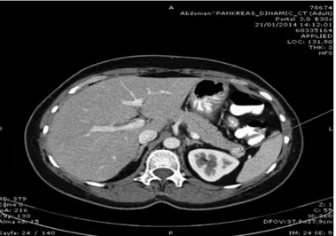
Abdominal computed tomography revealed a mass lesion measuring 20 mm in diameter at the distal pancreas which rapidly enhanced during arterial phase, showing similar imaging features with normal pancreas during portal phase (Figure 1). As the patient demonstrated postprandial hypoglycemia which was a rare presentation for insulinoma, a decision was made to perform arterial calcium-stimulated venous sampling for excluding nesidioblastosis coexisting with a pancreatic incidental
mass. The test was performed as it was described before by different centers (6). During ASVS, insulin concentration was found to increase 33-fold after calcium stimulation in the sample collected from proximal splenic artery (Figure 2). This finding suggested that the mass which we detected at pancreatic tail on abdominal CT was the source of hyperinsulinemia.
The patient underwent surgery including distal pancreatectomy and splenectomy. Splenic hilar lymph node dissection was performed due to the suspicion of nodal metastases. Intra-operative ultrasonography detected no other mass in the rest of the pancreas, thus, the surgery was limited to distal pancreas without further resection.
The patient was symptom-free after surgery. On pathological examination, the tumor cells and one of the four resected splenic hilar lymph nodes exhibited uniform atypical cell proliferation with positive immunostaining for chromogranin and synaptophysin (Figure 3). The Ki-67 index was <2%; necrosis and mitosis were not detected. The pathological findings suggested a low-grade well-differentiated neuroendocrine tumor with regional lymph node metastasis.
Discussion
Insulinoma is the one of the well-known causes of hyperinsulinemic hypoglycemia, which typically presents following prolonged fasting and, neuroglycopenic signs of hypoglycemia are its main clinical features (3). Nesidioblastosis, also known as noninsulinoma pancreatogenous hypoglycemia syndrome, in which hypoglycemia tends to occur after meals, is another rare cause of hyperinsulinemic hypoglycemia (7). Other
Table 1. Documentation of the laboratory analyses of the patient on admission
Findings Reference limits
Fasting glucose (mg/dl) 91 (70-110 mg/dl)
Insulin (uIU/ml) 5.6 (2.6-25 uIU/ml)
c-peptide (pmol/mL) 0.39 (0.37-1.47 pmol/mL)
Cortisol (µg/dl) 17.35 (6.20-19.4 µg/dl)
TSH (uIU/ml) 2.25 (0.4-4.67 uIU/ml)
TSH: Thyroid stimulating hormone
Figure 2. Schematic presentation of the results of Arterial Stimulation
Venous Sampling test with calcium
Figure 3. Microscopic examination of the resected tumor. Left panel:
Hematoxylin eosin staine section of the tumor (x100). Right panel: Synaptophysin stained section of the tumor (x100)
24
Ekşi Haydardedeoğlu et al.Insulinoma and Postprandial Hypoglycemia Turk J Endocrinol Metab2016;20:22-25
causes of postprandial hypoglycemia are alimentary (reactive) hypoglycemia and functional alimentary hypoglycemia (7). Here, we report a case of malignant insulinoma with postprandial hypoglycemia, which is an unusual presentation for insulinoma. Our case shared various clinical, imaging and laboratory aspects of these hypoglycemic disorders, as we mentioned above. Presentation of our case with postprandial hypoglycemia and accompanying negative 72-hour fasting test resulted in exclusion of the diagnosis of insulinoma at the former medical center, thus, she was diagnosed as having functional alimentary hypoglycemia. This is the most common cause of postprandial hypoglycemia in which patient’s history is enough for diagnosis and no further laboratory evaluation is suggested. Limiting simple carbohydrates intake and use of acarbose when needed usually resolve the postprandial symptoms in cases of reactive hypoglycemia. If symptoms do not improve with these interventions, the patient deserves further evaluation, as was in our case. Since a 72-hour fasting test was inconclusive, a mixed meal tolerance test was performed which induced symptomatic hypoglycemia after the first hour of food ingestion and endogenous hyperinsulinemic hypoglycemia was diagnosed in our center. Conditions associated with postprandial hypoglycemia, such as alimentary hypoglycemia (reactive hypoglycemia), late hypoglycemia of occult diabetes and nesidioblastosis should be kept in mind in the differential diagnosis of postprandial hypoglycemic states. Our patient’s hypoglycemic episode which happened one hour following meal ingestion was not usual for the disorders mentioned above and necessitated further investigation. Although postprandial hypoglycemia is usually not attributed to insulinoma, there are reports suggesting that insulinomas may rarely present with hypoglycemic symptoms after meals. It is not possible to determine the exact prevalence of insulinoma with postprandial hypoglycemia. In a retrospective analysis of 237 insulinoma patients, the symptoms of hypoglycemia have been reported in postprandial state in 6%, in whom 25% had a negative 72-hour fasting test (4). A negative 72-hour fasting test does exclude insulinoma, especially in cases presenting with symptoms highly suggestive of postprandial hypoglycemia. Patients with postprandial hypoglycemia, who do not improve after dietary therapy and acarbose treatment, may be selected for further evaluation. Objective demonstration of hypoglycemia is essential and insulinoma should be considered if postprandial hypoglycemia is proven.
There have been studies of the mechanism of hypoglycemia in patients with insulinoma. In contrast to normal pancreatic beta cells, insulinomas have been reported to express Glut-1 which is a peripheral cell dominant glucose transporter, and transports glucose into cells during basal low glucose milieu (8). This is why insulinomas cause fasting hypoglycemia. Pancreatic beta cells dominantly express Glut-2 which transports glucose into the cells at high glucose concentrations. On the basis of these findings, one of the possible explanations for insulinomas that present with postprandial hypoglycemia may be the predominance of Glut-2 expression of these tumors unlike the classical insulinomas (8). Another explanatory approach to postprandial hypoglycemia
in insulinomas may be through glucagon-like peptide-1 (GLP-1), which is a gastrointestinal peptide secreted in response to absorbed nutrients, such as glucose and triglycerides (9). It stimulates postprandial insulin release in response to food intake via glucagon-like peptide-1 receptor (GLP-1R). Benign insulinomas have been reported to over-express GLP-1R, and studies which attempt to use these receptors for localization and targeted nuclear therapies of insulinomas are promising (10). Malignant insulinomas have been reported to express GLP-1R rarely (11). The pathology of the case that we report resembled a low-grade malignant neuroendocrine tumor which might express GLP-1 like benign insulinomas. Based on these assumptions, postprandial hypoglycemia in this case may be either due to the presence of Glut-2 on tumor cells or aberrant expression of GLP-1R, which induced the secretion of insulin from abnormal insulinoma cells. Nevertheless, these possible mechanisms are only mechanistic theories in the absence of immunostaining for Glut-2 and/or GLP-1R for this case.
Conclusion
A negative 72-hour fasting test does not exclude the presence of insulinoma especially in patients with postprandial symptoms. Further evaluation is mandatory in patients whose symptoms do not improve after conventional treatment modalities.
Ethics
Informed Consent: Consent form was filled out by all participants, Peer-review: External and Internal peer-reviewed.
Authorship Contributions
Concept: Filiz Ekşi Haydardedeoğlu, Design: Filiz Ekşi Haydardedeoğlu, Ayşe Nur İzol Torun, Data Collection or Processing: Filiz Ekşi Haydardedeoğlu, Ayşe Nur İzol Torun, Okan Bakıner, Emre Bozkırlı, Analysis or Interpretation: Filiz Ekşi Haydardedeoğlu, Ayşe Nur İzol Torun, Melek Eda Ertörer, Literature Search: Filiz Ekşi Haydardedeoğlu, Ayşe Nur İzol Torun, Writing: Filiz Ekşi Haydardedeoğlu, Ayşe Nur İzol Torun, Melek Eda Ertörer, The pathological examination was performed: Fazilet Kayaselçuk, The Radiological procedure was performed: Çağatay Andıç, Conflict of Interest: No conflict of interest was declared by the authors, Financial Disclosure: The authors declared that this study has received no financial support.
References
1. Service FJ, Mc Mahon MM, O Brien PC, Ballard DJ. Functioning insulinoma-incidence-recurrence and long term survival of patients: A 60- year study. Mayo Clin Proc 1991:66:711-719.
2. Sotoudehmanesh R, Hedayat A, Shirazian N, Shahraeeni S, Ainechi S, Zeinali F, Kolahdoozan S. Endoscopic ultrasonography (EUS) in the localization of insulinoma. Endocrine 2007;31:238-241.
3. Service FJ. Diagnostic approach to adults with hypoglycemic disorders. Endocrinol Metab Clin North Am 1999;28:519-532.
4. Ploczkowski KA,Vella A,Thompson GB,Grant CS, Reading CC, Charboneau JW, Andrews JC, Lloyd RV, Service FJ. Secular trends in the presentation and management of functioning insulinoma at the Mayo clinic 1987-2007. J Clin Endocrinol Metab 2009;94:1069-1073.
25
Ekşi Haydardedeoğlu et al.Insulinoma and Postprandial Hypoglycemia Turk J Endocrinol Metab
2016;20:22-25
5. Okabayashi T, Shima Y, Sumiyoshi T, Kozuki A, Ito S, Ogawa Y, Kobayashi M, Hanazaki K. Diagnosis and management of insulinoma. World J Gastroenterol 2013;19:829-837.
6. Jackson JE. Angiography and arterial stimulation venous sampling in the localization of pancreatic neuroendocrine tumours. Best Pract Res Clin Endocrinol Metab 2005;19:229-239.
7. Gardner DG, Shoback D. Specific hypoglycemic disorders. Greenspan’s Basic and clinical endocrinology (9th ed). 2011:661-673.
8. Seino Y, Yamamoto T, Inoue K, Imamura M, Kadowaki S, Kojima H, Fujikawa J, Imura H. Abnormal facilitative glucose transporter gene expression in human islet cell tumours. J Clin Endocrinol Metab 1993;76:75-78.
9. Holst JJ. The physiology of glucagon-like peptide 1. Physiol Rev 2007;87:1409-1439.
10. Christ E, Wild D, Ederer S, Béhé M, Nicolas E, Caplin M, Brändle M, Clerici T, Fischli S, Stettler C, Ell P, Seufert J, Gloor B, Perren A, Reubi J, Forrer F. Glucagon-like peptide-1 receptor imaging for the localisation of insulinomas: a prospective multicentre imaging study. Lancet Diabetes Endocrinol 2013;115-122.
11. Eriksson O, Velikyan I, Selvaraju RK, Kandeel F, Johansson L, Antoni G, Eriksson B, Sörensen J, Korsgren O. Detection of metastatic insulinoma by positron emission tomography with [68ga] exendin-4-a case report. J Clin Endocrinol Metab 2014;99:1519-1524.