Restoration of pull-out strength of the failed pedicle
screw: biomechanical comparison of calcium
sulfate vs polymethylmethacrylate augmentation
Correspondence: Ümit Özgür Güler, MD. Ankara Omurga Merkezi, İran Caddesi, 45/2, Kavaklıdere, 06700 Ankara, Turkey.
Tel: +90 312 – 467 04 42 e-mail: [email protected] Submitted: February 16, 2013 Accepted: November 27, 2013
©2014 Turkish Association of Orthopaedics and Traumatology
doi: 10.3944/AOTT.2014.3193
Ümit Özgür GÜLER1, Alihan DERİNCEK2, Murat Ali HERSEKLİ2, Metin ÖZALAY2, Bekir Murat ÇINAR2, Emre ACAROĞLU1
1Department of Orthopedics and Traumatology, Ankara Spine Center, Ankara, Turkey;
2Department of Orthopedics and Traumatology, Adana Training and Research Hospital, Başkent University, Adana, Turkey
Objective: The aim of the present study was to compare calcium sulfate (CAS) and polymethylmeth-acrylate (PMMA) bone cements used for the augmentation of a failed pedicle screw with biomechani-cal pull-out strength (POS) testing.
Methods: Thirty lumbar vertebrae were harvested from 6 calves and bone mineral densities (BMD) were measured. Primary polyaxial pedicle screws were randomly inserted and pulled out and the POSs of the specimen were recorded. For revision, specimens were randomly assigned to the CAS-augment-ed pCAS-augment-edicle screws group (Group 1) or PMMA-augmentCAS-augment-ed pCAS-augment-edicle screw group (Group 2). Pull-out tests were repeated to compare both groups.
Results: Mean BMD of the specimens was 1.006±0.116 g/cm2. There were no statistically significant
differences between BMD results of the two groups (p=0.116). For Group 1, mean POS of primary screws was 2,441.3±936.4 N and was 2,499.5±1,425.1 N after CAS augmentation, demonstrat-ing no statistically significant difference (p=0.865). In Group 2, mean POS of the primary screws was 2,876.6±926.6 N and significantly increased to 3,745.5±1,299.2 N after PMMA augmentation (p=0.047). There was also a significant difference in mean POS between the CAS and PMMA groups (p=0.026).
Conclusion: Although CAS augmentation facilitates a revision screw POS as strong as that of pri-mary screws, it is not as strong as PMMA augmentation.
Key words: Calcium sulfate; pedicle screw; PMMA; pull-out strength.
The use of pedicle screws in the treatment of vertebral diseases has increased in the last two decades. The ad-vantages of pedicle screws include the stable fixation ca-pability until solid fusion is achieved and not requiring an intact lamina or spinous process. However, loosening,
pulling out and failure of pedicle screws are relatively frequent problems.[1] In such situations, revision of the
pedicle screws during primary or revision surgery may be unavoidable.
Enhancement of the fixation power of the
ped-Available online at www.aott.org.tr doi: 10.3944/AOTT.2014.3193 QR (Quick Response) Code
icle screws can be achieved using larger and/or longer pedicle screws or changing the direction of implemen-tation of pedicle trajectory in primary or revision pro-cedures. Pedicle trajectory can be sustained by augment-ing the defected pedicle with polymethylmethacrylate (PMMA), hydroxyapatite or calcium sulfate (CAS)/
calcium phosphate (CAP) bone cements.[1-10] PMMA
is the most frequently used and biomechanically sound method in revision surgery. On the other hand, PMMA may cause side effects such as heat and polymer release, bone necrosis and nerve damage.[11] Injectable calcium
salts are a good alternative to PMMA due to their non-exothermic nature and biological cohesion capabilities.
The aim of the present study was to compare CAS and PMMA bone cements used for failed pedicle screw augmentation with biomechanical pull-out strength (POS) testing.
Materials and methods
Approval of the Committee of Research Ethics was ob-tained for the present study. Thirty lumbar vertebrae (L1-L5) were obtained from 6 two-year-old calf cadav-ers. All specimens were cleaned of surrounding muscula-ture, ligaments and periosteum and separated into indi-vidual vertebrae. Anteroposterior and lateral radiographs were taken to exclude fractures and other pathologies.
Bone mineral density (BMD, g/cm2) of each vertebra
was measured in the anterior/posterior direction with dual-energy X-ray absorptiometry (DEXA-Hologic QDR 4500; Hologic, Inc., Waltham, MA, USA) to en-sure homogeneous study groups with similar BMD val-ues. Specimens were then wrapped with gauze, sealed in plastic bags, and stored frozen at -20°C in a deep freezer until the testing. Biomechanical tests were performed in two steps.
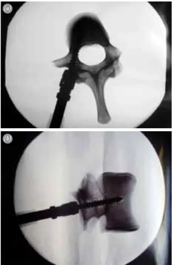
Prior to testing, all vertebrae were removed from the deep freezer and thawed to room temperature for 24 hours. Care was taken to keep the specimens moist throughout the experiment. Pedicle screw insertion points were identified with the help of the intersection method and screws inserted using the Roy-Camille tech-nique in left or right pedicular trajectories under fluoro-scope for each vertebra. Polyaxial, self-tapping, titanium pedicle screws (Cezmed Medical, Adana, Turkey) were placed into all vertebrae (Fig. 1). Screw diameter was 6.5 mm and length was 45 mm. No pedicle damage or an-terior wall penetration occurred in any of the vertebrae during screw replacement.
Following pedicle screw replacement, all vertebrae were embedded into cement (Amberok Model Stone) from the anterior side (anterior surface in a downward
direction and the pedicle entry point as the upper sur-face) using 30 aluminum embedding containers pre-pared in advance. Pull-out tests were performed using a material testing instrument (Instron Model No. 4505; Instron Corp., Canton, MA, USA). Each pedicle screw was tested for axial POS at 5 mm/min displacement un-til total screw pull-out (Fig. 2). Primary screw pull-out testing of the 30 calf vertebrae was completed without breakage in the cement, bending of the embedding con-tainer or screw breakage.
Vertebrae were then randomly divided into two equal groups receiving CAS (Group1) or PMMA (Group 2) cements. For the second step of the study, no drilling or tapping was used to insert the screws. Screws were used in the same specimens in both stages of the experiment.
In Group 1, 3 cc of CAS bone paste (Minimally Invasive Injectable Graft: MIIG X3; Wright Medical Technology, Inc., Arlington, TN, USA) was inserted into the damaged pedicle holes of each vertebra
with-Fig. 1. (a) Axial and (b) lateral view of the pedicle screw insertion under fluoroscopic imaging.
(a)
out any pressure. In Group 2, 3 cc of PMMA cement (SmartSet MV Endurance; DePuy International Ltd., Leeds, UK) was injected into the damaged pedicle holes without any pressure.
After augmentation of all damaged pedicle holes, ped-icle screws used in the first step of the study were placed in the same vertebra. All specimens were kept at room temperature for 24 hours to allow the CAS and PMMA cement to harden completely.[12,13] Vertebrae were placed
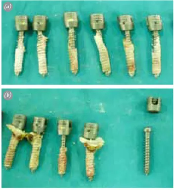
into the Instron machine again as in the primary screw pull-out tests and revision pull-out tests were success-fully concluded in 29 vertebrae (Fig. 3a). The polyaxial head of one PMMA-augmented screw detached from the screw body during the pull-out process at 6,415 N and the specimen was excluded from the study (Fig. 3b).
Data were analyzed using the SPSS v17.0 (SPSS Inc, Chicago, IL, USA) statistical package program. The Mann-Whitney U test was used in the comparison of primary and revision screw pull-out results. The Wil-coxon test was used for the evaluation of data obtained from changes in POS of PMMA and CAS revision groups and the Pearson’s correlation test for comparison of BMD values with the primary and revision pull-out test results. The level of statistical significance was deter-mined as p<0.05.
Results
Mean and standard deviation (SD) BMD of all segments was 1.006±0.116 g/cm2, suggesting low or osteoporotic
bone quality.[14] There was no statistically significant
dif-ference in BMD scores between the PMMA and CAS groups (p=0.116). When primary and revision pull-out BMD values were compared, a statistically positive cor-relation was observed between BMD and primary screw pull-out values (r=0.578; p<0.05).
No incidence of pedicle or lamina fractures was ob-served. One pedicle screw detached from the screw body in revision testing and its results were excluded from the study. In the CAS group, mean POS of primary screws was 2,441.3±936.4 N (range: 1,317 to 4,634 N) and 2,499.5±1,425.1 N (range: 760 to 5,336 N) for CAS-augmented screws. There was no statistical difference between primary and revision screws POS in Group 1 (p=0.865). Mean POS increased from 2,876.6±926.6 N (range: 1,964 to 4,612 N) for primary screws to 3,745.5±1,299.2 N (range: 1,246 to 4,903 N) for re-vision screws in the PMMA group. This increase was statistically significant (p=0.047). Additionally, there was a statistically significant difference in POS values between the CAS and PMMA groups after augmenta-tion (p=0.026) (Table 1).
Fig. 2. All vertebrae were embedded into cement from anterior sides and pull-out tests were performed using a material testing instrument. [Color figure can be viewed in the online issue, which is available at www.aott.org.tr]
Fig. 3. (a) View of the CAS-augmented screws after pull-out test. All the thread of the screw is coated with calcium sulfate cement. (b) View of the PMMA-augmented screws after pull-out test. Polyaxial head breaking off was observed dur-ing testdur-ing procedure in a screw. [Color figure can be viewed in the online issue, which is available at www.aott.org.tr]
(a)
Discussion
The majority of subjects with pedicle screw fixation do not require revision. However, intraoperative or post-operative augmentation is necessary in some cases. Loosened screws, pedicle breakage, inappropriate screw placement or osteoporotic bone generally result in loss of correction or nonunion. The relationship between bone and the metal of the pedicle screws, especially in osteoporotic bone, has been described as a weakness of the system.[15,16] Several recent studies have shown that
for revision procedures, the use of materials creating fa-vorable biomechanical properties can be used for pedicle screw augmentation.[6,7,17]
In general, the compounds of PMMA or hydroxy-apatite and CAS or CAP are used in revision surgery. In the literature, augmentation with PMMA was reported to provide the highest mechanical strength in both pri-mary and revision cases.[5,6,18,19] Although stability and
availability of PMMA and its efficiency in increasing POS have been ascertained, complications resulting from PMMA use create some limitations on its use in spinal surgery. PMMA is an exothermic polymer and may lead to bone necrosis, toxin release and/or neural injury.[8] However, bone graft materials made from CAS
or CAP are potential alternatives to PMMA. They have high biological incorporation and are not exothermic. Therefore, they do not have the potential risk of thermal damage to bones or nerves. Additionally, CAS bone graft requires a short period of preparation and long period of
hardening processes.[10] Still, both PMMA and
bioab-sorbable cement are widely used by spine surgeons for augmentation in revision procedures or in osteoporotic patients. The use of CAS bone graft materials is rela-tively new and has not been as well studied as PMMA.
Few articles comparing CAS and PMMA have been published in the literature.[6,17] Both processes were
ap-plied as primary pedicle screw augmentation techniques in our study, but not as a salvage process. Additionally, in the previous studies, BMD values were not measured, vertebrae were predicted to be osteoporotic before the process, and the effect of bone density on POS was not
discussed. In contrast with previous reports, a statisti-cally significant difference was found between CAS and PMMA in our study.
In the recent articles, axial POS was correlated with screw length, screw diameter, insertional torque and BMD.[20-22] They concluded that while the use of larger
diameter screws increased the insertional torque, creasing screw length did not have an effect on the in-sertional torque. Additionally, they reported that larger diameter and full-threaded screw insertion deep enough to fit into the anterior vertebral cortex provided the most secure fixation and that the incidence of non-union was
increased when BMD was below 0.674±1.04 g/cm2. In
the present study, the same CAS- and PMMA-augment-ed pPMMA-augment-edicle screws were usPMMA-augment-ed in both the primary and re-vision fixations in order to ensure that screw length and diameter did not affect screw POS. Additionally, BMD values were taken for all vertebrae and a statistically pos-itive correlation was detected between BMD and prima-ry pedicle screws while there was no correlation between the BMD and revision screw pull-out values. We, there-fore, concluded that BMD did not affect PMMA- and CAS-supported pedicle screw pull-out values.
This study had some limitations. First, we compared only two types of cements despite the availability on the market of other types of cements. Second, animal brae were used in the study while human cadaver verte-brae would have more clinical use.
In conclusion, PMMA, as a material of pedicle screw salvage, has been recognized as the golden standard due to its easy availability, low cost and superiority of aug-mentation. Although not as strong as PMMA augmen-tation, POS of revision screws with CAS-augmented bone cement is as strong as that of primary screws. In-jectable calcium salts give no exothermic reaction and have high biocompatibility and, consequently, a lower risk of complication. The use of these materials is prom-ising for the future.
Conflicts of Interest: No conflicts declared. Table 1. Statistical analysis of the primary and CAS- and PMMA-augmented revision screws’ pull-out strengths
(mean±SD).
Mean pull-out strength of Mean pull-out strength of p*
the primary screws (N) the revision screws (N)
CAS group 2,441.3±936.4 2,499.5±1,425.1 >0.05
PMMA group 2,876.6±926.6 3,745.5±1,299.2 <0.05
p† >0.05 <0.05
References
1. Lonstein JE, Denis F, Perra JH, Pinto MR, Smith MD, Winter RB. Complications associated with pedicle screws. J Bone Joint Surg Am 1999;81:1519-28.
2. Chang MC, Liu CL, Chen TH. Polymethylmethacrylate augmentation of pedicle screw for osteoporotic spinal sur-gery: a novel technique. Spine 2008;33:E317-24. CrossRef
3. Lotz JC, Hu SS, Chiu DF, Yu M, Colliou O, Poser RD. Carbonated apatite cement augmentation of pedicle screw fixation in the lumbar spine. Spine 1997;22:2716-23. CrossRef
4. Moore DC, Maitra RS, Farjo LA, Graziano GP, Goldstein SA. Restoration of pedicle screw fixation with an in situ setting calcium phosphate cement. Spine 1997;22:1696-705. CrossRef
5. Motzkin NE, Chao EY, An KN, Wikenheiser MA, Lewallen DG. Pull-out strength of screws from polymeth-ylmethacrylate cement. J Bone Joint Surg Br 1994;76:320-3.
6. Rohmiller MT, Schwalm D, Glattes RC, Elalayli TG, Spengler DM. Evaluation of calcium sulfate paste for aug-mentation of lumbar pedicle screw pullout strength. Spine J 2002;2:255-60. CrossRef
7. Spivak JM, Neuwirth MG, Labiak JJ, Kummer FJ, Ricci JL. Hydroxyapatite enhancement of posterior spinal in-strumentation fixation. Spine 1994;19:955-64. CrossRef
8. Wilkes RA, Mackinnon JG, Thomas WG. Neurological deterioration after cement injection into a vertebral body. J Bone Joint Surg Br 1994;76:155.
9. Yerby SA, Toh E, McLain RF. Revision of failed pedicle screws using hydroxyapatite cement. A biomechanical analysis. Spine 1998;23:1657-61. CrossRef
10. Bucholz RW. Nonallograft osteoconductive bone graft substitutes. Clin Orthop Relat Res 2002;395:44-52. CrossRef
11. Stürup J, Nimb L, Kramhøft M, Jensen JS. Effects of po-lymerization heat and monomers from acrylic cement on canine bone. Acta Orthop Scand 1994;65:20-3. CrossRef
12. Mitzner E, Albertus P, Pelt HM, Mueller C, Strohwig A, Mueller WD. Material properties and in vitro biocompat-ibility of a newly developed bone cement. Materials
Re-search 2009;12:447-54. CrossRef
13. Belkoff SM, Sanders JC, Jasper LE. The effect of the monomer-to-powder ratio on the material properties of acrylic bone cement. J Biomed Mater Res 2002;63:396-9. 14. Akbay A, Bozkurt G, Ilgaz O, Palaoglu S, Akalan N, Ben-zel EC. A demineralized calf vertebra model as an alter-native to classic osteoporotic vertebra models for pedicle screw pullout studies. Eur Spine J 2008;17:468-73. CrossRef
15. Esses SI, Sachs BL, Dreyzin V. Complications associated with the technique of pedicle screw fixation. A selected survey of ABS members. Spine 1993;18:2231-9. CrossRef
16. Okuyama K, Abe E, Suzuki T, Tamura Y, Chiba M, Sato K. Influence of bone mineral density on pedicle screw fixa-tion: a study of pedicle screw fixation augmenting poste-rior lumbar interbody fusion in elderly patients. Spine J 2001;1:402-7. CrossRef
17. Yi X, Wang Y, Lu H, Li C, Zhu T. Augmentation of pedi-cle screw fixation strength using an injectable calcium sul-fate cement: an in vivo study. Spine 2008;33:2503-9. CrossRef
18. Derincek A, Wu C, Mehbod A, Transfeldt EE. Biome-chanical comparison of anatomic trajectory pedicle screw versus injectable calcium sulfate graft-augmented pedicle screw for salvage in cadaveric thoracic bone. J Spinal Dis-ord Tech 2006;19:286-91. CrossRef
19. Wittenberg RH, Lee KS, Shea M, White AA 3rd, Hayes WC. Effect of screw diameter, insertion technique, and bone cement augmentation of pedicular screw fixation strength. Clin Orthop Relat Res 1993;296:278-87. 20. Fogel GR, Reitman CA, Liu W, Esses SI. Physical
char-acteristics of polyaxial-headed pedicle screws and bio-mechanical comparison of load with their failure. Spine 2003;28:470-3. CrossRef
21. Lehman RA Jr, Polly DW Jr, Kuklo TR, Cunningham B, Kirk KL, Belmont PJ Jr. Straight-forward versus anatomic trajectory technique of thoracic pedicle screw fixation: a biomechanical analysis. Spine 2003;28:2058-65. CrossRef
22. Yamagata M, Kitahara H, Minami S, Takahashi K, Isobe K, Moriya H, et al. Mechanical stability of the pedi-cle screw fixation systems for the lumbar spine. Spine 1992;17(3 Suppl):S51-4. CrossRef