Changes in selected haematological and biochemical parameters in
debeaked pheasant hens during the laying period
Petra HRABCAKOVA1, Eva VOSLAROVA1, Iveta BEDANOVA1, Vladimira PISTEKOVA1,
Jan CHLOUPEK2
1 Department of Veterinary Public Health and Animal Welfare; 2 Department of Animal Husbandry and Animal Hygiene, Faculty of Veterinary Hygiene and Ecology, University of Veterinary and Pharmaceutical Sciences Brno, Czech Republic.
Summary: Changes in selected haematological and biochemical parameters during the laying period were monitored in pheasant hens kept in a cage system. Thermal cauterization of one-third of the upper part of the beak was performed before the birds were placed in the cage at the beginning of the laying period. Randomly selected pheasant hens were sampled for haematological and biochemical analysis at the beginning of the laying period, and the same hens were subsequently sampled after 6 and 12 weeks of the laying period. The results showed significant changes in the monitored haematological and biochemical parameters during egg laying. At the time when laying capacity approached a maximum, a fall was seen (P<0.05) in haematocrit, haemoglobin, erythrocytes and plasma glucose values, whereas leukocytes, lymphocytes, basophils and plasma uric acid exhibited an increase (P<0.05). At the end of the laying period, a higher (P<0.05) count of leukocytes, lymphocytes, eosinophils, basophils, monocytes, plasma alanine aminotransferase, uric acid, lactate and calcium concentrations were seen, whereas for haematocrit, heterophil to lymphocyte ratio, plasma total protein, and glucose lower (P<0.05) values were recorded in comparison with the values of the given indicators at the beginning of the laying period. The results determined provide new information about the dynamics of selected haematological and biochemical indicators in clinically healthy common pheasant layers during the course of the laying period.
Key words: biochemistry, captive-rearing, egg laying, haematology, pheasant.
Introduction
The common pheasant (Phasianus colchicus) either lives in the wild or is kept in breeding facilities (pheasantries), from which it is released and subsequently hunted (13). Increasing numbers of pheasants are also raised commercially to be slaughtered for meat (19, 22). Captive-reared pheasants are frequently kept in large flocks in aviaries (13) or, particularly during the laying period, in cage systems (15, 58, 61).
Large pheasantries regularly order preventive health status examinations (13). The determination of haematological parameters and plasma metabolite levels may provide highly valuable information on the physiological state of the pheasants and forms the cornerstone of medical diagnosis. Although it is well known that plasma biochemistry, along with haematology, is important for medical diagnosis in bird species, limited information is available for pheasants (32). Numerous studies have reported the values of blood parameters in gallinaceous poultry (1, 16, 17, 30, 45, 52, 56), but only limited data on biochemical parameters in pheasants has been reported in the literature to date (42, 43, 54).
Our knowledge of haematological parameters in the common pheasant also remains incomplete. Some parameters of total leukocyte counts for phasianids were
reported by Prinzinger and Misovic (39). Selected parameters of the white blood cell count in the common pheasant were specified by Lucas and Jamroz (25) and Maxwell and Robertson (28, 29). Strakova et al. (49) investigated selected haematological indicators in six major species of feathered game including the common pheasant and evaluated the differences between individual species. Hauptmanova et al. (13) focused on the haematological parameters of the common pheasant, the dynamics of changes in blood count parameters with regard to the season of the year and reproduction status, and the effects of sex and body condition on haematological parameters of blood. The influence of age on haematological and biochemical parameters was investigated in juvenile ring-necked pheasants by Schmidt et al. (44), who also studied the connection with vaccination against Newcastle disease and the influence of sex and age on these parameters. Lloyd and Gibson (24) studied the haematology and biochemistry of healthy young pheasants and the effect of spironucleosis on these indicators. Kececi and Col (18) determined selected haematological and biochemical parameters of pheasants and compared these parameters among various age groups. Nazifi et al. (32) studied reference values for biochemical parameters in adult male and female
ring-necked pheasants. Suchy et al. (54) studied biochemical and mineral profiles in selected species of feathered game reared in the Czech Republic and Europe. Their work evaluates potential interspecies differences in the given values and compares their findings with the results reported for related domesticated species of birds. Blood biochemistry in pheasants has also been studied by Jerabek et al. (17), Muller et al. (31) and Strakova et al. (45, 46).
The main objective of this study was to determine values for selected haematological parameters and blood chemistries in debeaked pheasant hens kept in conventional cages during the laying period. It can be hypothesized that the laying period entails significant modifications of both biochemical and haematological blood indices in pheasants. Thus, to reliably interpret the results obtained by biochemical and haematological examinations of the blood of laying pheasant hens there is a need for knowledge of the dynamic changes in these parameters during egg laying. This study aims to provide such baseline data.
Materials and Methods
The birds and their treatment: The University of Veterinary and Pharmaceutical Sciences Brno Committee on Animal Care in Research gave approval of the experimental design through Protocol No. 15/2011.
The experiment was performed on approximate 1-year-old (average body weight 0.85 kg) common pheasant hens (Phasianus colchicus). In the rearing facility, the birds were housed in external aviaries prior to the experiment. The number of birds in the pheasantry was approximately 900 (parent flock). From the beginning of the laying period, pheasant hens were housed in a two-tiered cage battery. There was one breeding group in each cage, consisting of one male and five hens. Each laying cage was equipped with a wire floor, five automatic nipple drinkers and a trough feeder located at the front wall of the cage, with manual administration of feedings. The dimensions of the cage were as follows: 200 cm length, 85 cm depth, 58 cm back height and 70 cm front height. The floor was sloped towards the front wall to enable collection of laid eggs. The pheasants were fed with the pelleted feeding mixture BZN (ADW Agro a. s., Krahulov, Czech Republic). The cages were kept under natural light only (daylight), with no artificial lighting. Before being placed in the cage, thermal cauterization of one-third of the upper part of the beak was performed on the pheasants. All pheasants were ringed, i.e. an individually numbered plastic tag was attached to the leg of each bird.
At the beginning of the laying period 15 pheasant hens from 15 randomly selected cages (each hen from a different cage) were randomly selected and sampled for
biochemical and haematological blood analysis. Subsequently, the same hens were sampled after 6 and 12 weeks of the laying period.
Blood sampling: Blood samples (3 ml) for haematological and biochemical examination were taken from the vena basilica of the left wing, and collected using syringe-needle assemblies that had been flushed with heparin (15 of heparin for 3 ml of blood). The samples were collected within 1 min of capture to ensure that the levels of the monitored parameters were not affected by any stress induced by pre-sampling handling (5). Blood sampling was always performed at the same time of day (9:00 a.m.) and its duration did not exceed one hour. The heparinized blood was immediately centrifuged at 837 x g at 4 °C for 10 min., and plasma samples were stored at - 80 °C in Eppendorf test tubes until the analyses were performed. Samples for leukocyte profile examination were collected in tubes with EDTA and examined immediately.
Biochemical examinations: Selected plasma biochemical parameters (alanine aminotransferase, aspartate aminotransferase, total protein, cholesterol, glucose, uric acid, lactate dehydrogenase, lactate, calcium, phosphorus) were measured on the principle of photometric detection by a KONELAB 20i biochemical analyzer using commercial test kits (Biovendor -Laboratorni medicina a.s., Brno, Czech Republic).
Haematological examinations: The total erythrocyte and leukocyte counts were determined by means of the flask method of dilution and counting corpuscles using a Burker chamber. The diluent was Natt-Herick solution (33) and the dilution rate was 1:200 (a mixture of 25 ul blood and 4975 ul Natt-Herick solution was prepared). Haemoglobin levels were determined photometrically using a SPECOL-11 photometer and Drabkin's solution (6) at a wavelength of 540 nm, and haematocrit values were determined by means of the micro-haematocrit technique according to Janetzki (8). Proportions of individual leukocyte types (in 200 cells) were computed by classical histological methods using a light microscope with an immersion lens after staining blood smears according to Pappenheim with May-Grunwald and Giemsa- Romanowski solutions (35, 36). The type of blood cells was determined according to Lucas and Jamroz (25).
Statistical analysis: The results were analyzed using the statistical package UNISTAT 5.1 (Unistad Ltd., GB). The normality and homogeneity of variances were checked for all variables tested by means of a Shapiro-Wilk test and a Bartlett-Box test (62). In the case of non-normal data, logarithmic or square root transformations were used for analysis of variance, though actual mean values are presented in the tables. Because the same birds were measured on each day of sampling over the length of the experiment, the data was subjected to repeated
measures ANOVA using the general linear model procedure with a factor "Day of sampling" with 3 levels (Day 1, Day 42, Day 84) within the birds in a randomized block design. There were n = 15 pheasants (blocks) in the experiment. When the effect was statistically significant, the Tukey-HSD test was performed as a post hoc test for pairwise comparisons of means. A P-value of <0.05 was considered significant.
Results and Discussion
A haematological examination is one method that contributes to detection of some changes in health status which may not be apparent during physical examination, but which affect the fitness of the birds (2, 9, 20).
However, there is a lack of knowledge of the basic haematological parameters and dynamic changes in pheasants during the laying period. Consequently, the interpretation of results obtained by haematological research on birds is often difficult (47). Kececi and Col (18) observed that the red blood cell counts, haemoglobin amounts and haematocrit values increased with the advancement of age, being lowest in chicks and highest in adults. These age-related findings were substantiated for pheasants by reports by Pujman and Hanusova (40) and Schmidt et al. (43). In contrast, Schmidt et al. (43) reported no significant differences in packed cell volume, eosinophiles, monocytes and basophils among juvenile ring-necked pheasants of various ages. According to Pujman and Hanusova (40), Herbert et al. (14) and Schmidt et al. (42, 43) the total erythrocyte concentration and the PCV of birds are influenced not only by age, but also by other factors such as sex, hormones, hypoxia, environment and diseases (4). Our study was conducted over the course of 3 months during the laying period, and laying capacity can be considered the principal factor influencing the values of haematological parameters in pheasant hens during this period. The haematological
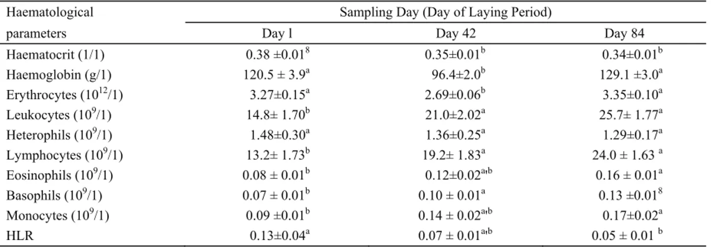
profile of pheasant hens during the laying period is given in Table 1.
Haematocrit values fell during the course of the laying period. A statistically significant fall in haematocrit values in pheasant hens was determined in week 6 (P=0.009) and week 12 (P=0.002) of the laying period in comparison with the haematocrit value at the beginning of the laying period.
Our results indicate that the red blood cell count and content of haemoglobin decreased with an increase in laying capacity. The lowest mean values of total erythrocyte count (2.69 ± 0.06 1012/1) and haemoglobin
content (96.4 ± 2.0 g/1) were determined in the 6th week of the laying period, which is roughly the time at which laying capacity in pheasant hens reaches a maximum, although the literary sources differ in relation to this figure. Gibes et al. (10) reported the highest egg production in pheasants in the 7th week of laying. Krystianiak et al. (21) recorded the peak of egg production in the 4th week of laying. Tserveni-Gousi and Yannakopoulos (55) reported the peak of laying in pheasants one week earlier, whereas Woodard and Snyder (59) reported it in week 5. The decrease in the total erythrocyte count and haemoglobin in the 6th week was followed by a highly significant increase in both indicators (P<0.001) in the 12th week. Like Strakova et al. (47) in laying hens, we assume that laying in pheasant hens represents an enormous metabolic stress that is manifested by decreasing erythropoiesis.
Chickens and domestic turkeys have a white blood cell distribution with lymphocytes as the most numerous leukocytes (1). This would also be expected in pheasants, and our results corroborate this. The leukocyte count has been found to be extremely variable in pheasants (3, 13). This variability is due to many factors, such as the season, age, individual properties of birds, hormones, stress, immune status, as well as the time of blood
Table 1. Haematological parameters in debeaked pheasant hens (n = 15/group) housed in a cage system during the laying period (means ± SEM).
Haematological Sampling Day (Day of Laying Period)
parameters Day l Day 42 Day 84
Haematocrit (1/1) 0.38 ±0.018 0.35±0.01b 0.34±0.01b Haemoglobin (g/1) 120.5 ± 3.9a 96.4±2.0b 129.1 ±3.0a Erythrocytes (1012/1) 3.27±0.15a 2.69±0.06b 3.35±0.10a Leukocytes (109/1) 14.8± 1.70b 21.0±2.02a 25.7± 1.77a Heterophils (109/1) 1.48±0.30a 1.36±0.25a 1.29±0.17a Lymphocytes (109/1) 13.2± 1.73b 19.2± 1.83a 24.0 ± 1.63 a Eosinophils (109/1) 0.08 ± 0.01b 0.12±0.02a'b 0.16 ± 0.01a Basophils (109/1) 0.07 ± 0.01b 0.10 ± 0.01a 0.13 ±0.018 Monocytes (109/1) 0.09 ±0.01b 0.14 ± 0.02a'b 0.17±0.02a HLR 0.13±0.04a 0.07 ± 0.01a'b 0.05 ± 0.01 b
l'b Means within a row with different superscripts differ (PO.05). HLR = heterophil to lymphocyte ratio.
sampling during the day (7, 23, 27, 29). In our study, pheasant hens exhibited an increase (P<0.01) in the number of leucocytes during the laying period. The number of heterophils, in contrast, tended to fall during the period from week 1 to week 12 of the laying period, although this fall was not statistically significant.
The heterophil/lymphocyte ratio in pheasant hens was largest at the beginning of the laying period before subsequently falling. It was significantly lower (P=0.011) in week 12 of the laying period than in week 1. Since the heterophil/lymphocyte ratio is an indicator of stress in birds (11), an increased value of the heterophil/ lymphocyte ratio at the beginning of the laying period can be explained by the stress caused by the caging of the pheasant hens and the beak cauterization performed as an anti-pecking measure before the laying period.
A significant increase was determined between week 1 and week 12 of the laying period for lymphocytes (P<0.001), eosinophils (P<0.001), basophils (P<0.001) and monocytes (P<0.001).
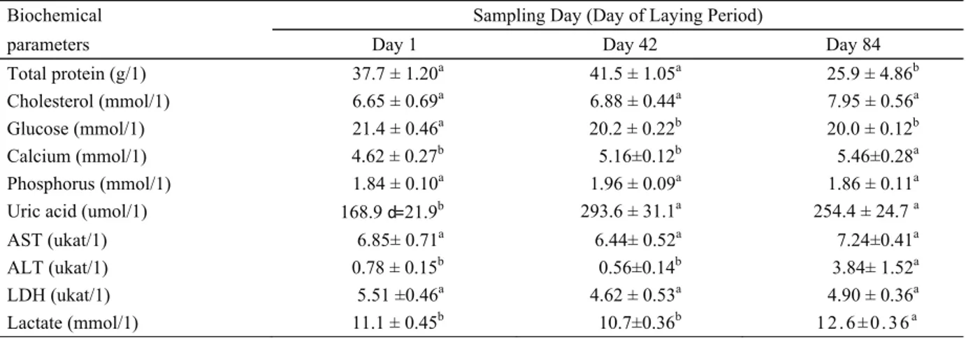
Our knowledge of chemistry in pheasants also still remains incomplete (32). Only a few studies on blood chemistry values in pheasants have been published so far, none of which has focused on changes in biochemical parameters during the laying period. Blood plasma mineral concentrations (37) and biochemical values generally can be influenced by many factors, such as laying rate and energy requirements (51). The relation between the phase of the laying period and the values of selected biochemical parameters in pheasant hens is presented in Table 2.
The values of plasma protein concentrations we determined correspond to the values stipulated by Lloyd and Gibson (24) in healthy, young, farmed pheasants. As
for Suchy et al. (51) in laying hens and Strakova et al. (46) in quails, we assume that the high protein concentrations in blood plasma are closely related to higher proteosynthesis, which is a prerequisite to high egg production. Our results confirm that plasma protein increases with an increase in laying capacity. Females of oviparous species demonstrate a marked increase in plasma total protein concentration just before egg production. This estrogen-induced hyperproteinemia is associated with an increase in vitellogenin and lipoproteins, which are necessary for yolk production. These proteins are produced in the liver, transported in the blood and incorporated into the oocytes of the ovary (26). Accordingly, a statistically significant fall in plasma protein (P=0.002) was discovered in pheasant hens at the end of the laying period.
No significant changes during the laying period were detected for plasma cholesterol concentrations ranging from 6.65 ± 0.69 mmol/1 to 7.95 ± 0.56 mmol/1. Suchy et al. (54) detected a cholesterol value of 3.880 mmol/1 in the common pheasant and, similarly, Nazifi et al. (32) found cholesterol values of 3.72 mmol/1 and 3.25 mmol/1 in pheasant males and females, respectively. Such values are supposed to be near the lower limit of the normal cholesterol range (3.55 - 10.25 mmol/1) for domestic fowl (48, 50) and common pheasants, respectively. The cholesterol metabolism in avian species is similar to that of mammals, but the plasma cholesterol level can significantly increase during vitellogenesis and egg formation in birds (12). It has also been reported that a wide variation of cholesterol levels among avian species may depend on circadian rhythms and the effect of diet (12, 34, 57).
Table 2. Selected biochemical parameters in debeaked pheasant hens (n = 15/group) housed in a cage system during the laying period (means ± SEM).
Biochemical Sampling Day (Day of Laying Period)
parameters Day 1 Day 42 Day 84
Total protein (g/1) 37.7 ± 1.20a 41.5 ± 1.05a 25.9 ± 4.86b
Cholesterol (mmol/1) 6.65 ± 0.69a 6.88 ± 0.44a 7.95 ± 0.56a
Glucose (mmol/1) 21.4 ± 0.46a 20.2 ± 0.22b 20.0 ± 0.12b
Calcium (mmol/1) 4.62 ± 0.27b 5.16±0.12b 5.46±0.28a
Phosphorus (mmol/1) 1.84 ± 0.10a 1.96 ± 0.09a 1.86 ± 0.11a
Uric acid (umol/1) 168.9 d= 21.9b 293.6 ± 31.1a 254.4 ± 24.7 a
AST (ukat/1) 6.85± 0.71a 6.44± 0.52a 7.24±0.41a
ALT (ukat/1) 0.78 ± 0.15b 0.56±0.14b 3.84± 1.52a
LDH (ukat/1) 5.51 ±0.46a 4.62 ± 0.53a 4.90 ± 0.36a
Lactate (mmol/1) 11.1 ± 0.45b 10.7±0.36b 1 2. 6 ± 0. 36a
h
' Means within a row with different superscripts differ (PO.05). ALT = alanine aminotransferase
AST = aspartate aminotransferase LDH = lactate dehydrogenase
The plasma glucose concentration in our study fell in pheasant hens during the laying period from 21.4 ± 0.46 mmol/1 to 20.0 ± 0.12 mmol/1. A statistically significant fall was found in week 6 (P=0.03) and in week 12 (P=0.007) of the laying period in comparison with the plasma glucose concentration at the beginning of the laying period.
The concentration of plasma calcium in pheasants during the laying period increased from 4.62 ± 0.27 mmol/1 in the 1st week of the laying period to 5.46 ± 0.12 mmol/1 in the 12th week (P=0.039). Significantly lower values of calcium in common pheasants were found by Lloyd and Gibson (24), Suchy et al. (54) and Nazifi et al. (32), who monitored them outside the laying period. In laying hens, Suchy et al. (51) characterized the concentration of calcium in blood plasma as a gradual increase from 5.35 mmol/1 in the early stages to 7.19 mmol/1 in the 40th week of the laying period, followed by a decrease to 5.92 mmol/1 in the 50th week. An increasing release of calcium into the blood in hens is closely related to its accumulation in the shell.
No significant changes in the plasma phosphorus concentration in pheasant hens were detected during the laying period. In the 1st week of the laying period the concentration was 1.84 ± 0.10 mmol/1, in the 6th week of the laying period 1.96 ± 0.09 mmol/1, and in the 12th week 1.86 ± 0.11 mmol/1. Similar values were discovered in common pheasants kept in aviaries outside the laying period by Suchy et al. (54).
Uric acid and urea are the main products of the nitrogen metabolism of birds, and uric acid is also the major nitrogenous waste product of birds (12, 38, 41). The plasma uric acid concentration increased statistically significantly, with the highest values being determined in the 6th week of the laying period, when laying capacity approaches its peak. The level of uric acid in the blood of pheasant hens during the laying period is, therefore, influenced not merely by species and diet (26), but in all probability also by the phases of the laying period.
Since plasma enzymes are characterised by an extremely wide range of activity, the interpretation of variations in these enzymes is difficult. Nevertheless, these enzymes may be adversely affected by factors such as muscular injury, organ rupture, nutritional status, physical activity, haemolysis, treatment, and the preservation of plasma samples, and their levels in blood can increase. These enzymes can be an important diagnostic tool in veterinary medicine (12, 38, 41). In our study, no significant changes were found in the plasma aspartate aminotransferase (AST) concentration during the laying period, whereas the mean plasma alanine aminotransferase (ALT) concentration in pheasant hens increased (P=0.002) from 0.56 ± 0.14 ukat/1 in the 6th week of the laying period to 3.84 ± 1.52 ukat/1 in the 12th week of the laying period.
There is little information available for AST, ALT and lactate dehydrogenase (LDH) values in pheasants to compare with our results. In our study, the plasma concentrations of LDH in pheasant hens ranged from 4.62 ± 0.53 to 5.51 ± 0.46 ukat/1. No significant changes were found during the laying period. The LDH levels in pheasants appear to be higher than those of chickens (60), and no differences have been found between the sexes (32).
Data is also lacking for plasma lactate dynamics in pheasants. The published data is limited to research on acute stress (5, 53). In our study, the plasma lactate concentration showed no changes in the first half of the laying period, but subsequently increased (P=0.004) from 10.7 ± 0.36 in the 6th week to 12.6 ± 0.36 mmol/1 in the 12th week of the laying period.
Conclusion
The present study described changes in selected haematological and biochemical parameters in debeaked pheasant hens housed in a cage system during the laying period. Similarly as for poultry, significant changes take place in the pheasant organism during the laying period. At the time when laying capacity approached a maximum, a fall was seen in haematocrit, haemoglobin, erythrocytes and plasma glucose values, whereas leukocytes, lymphocytes, basophils and plasma uric acid exhibited an increase. At the end of the laying period, a higher count of leukocytes, lymphocytes, eosinophils, basophils, monocytes, plasma alanine aminotransferase, uric acid, lactate and calcium concentrations were seen, whereas for haematocrit, heterophil to lymphocyte ratio, plasma total protein, and glucose lower values were recorded in comparison with the values of the given indicators at the beginning of the laying period.
Acknowledgements
This study was supported by Research Project no. MSM6215712402 Veterinary Aspects of Food Safety and Quality.
References
1. Bounous DI, Wyatt RD, Gibbs PS, Gilburn JV, Quist CF (2000): Normal haematologic and serum biochemical
reference for juvenile wild turkeys. J Wildl Dis, 36, 393-396.
2. Bradley LW, Threlfall W (1974): Blood cell indices of
five species of auk (Alcidae) from Newfoundland. J Zool,
174, 377-385.
3. Campbell TW (1994): Hematology. 176-198. In: Ritchie BW, Harrison GJ, Harrison LR (Ed), Avian Medicine: Principles and Application. Wingers Publishing, Inc., Lake Worth.
4. Campbell TW (2004): Hematology of birds. 225-258. In: Thrall MA (Ed.), Veterinary Hematology and Clinical Chemistry. Lippincott Williams and Wilkins, Philadelphia.
5. Chloupek P, Voslarova E, Suchy P Jr, Bedanova I, Pistekova V, Vitula F, Chloupek J, Vecerek V (2009):
Influence of Pre-Sampling Handling Duration on Selected Biochemical Indices in the Common Pheasant (Phasianus colchicus). Acta Vet Brno, 78, 23-28.
6. Drâbkin DR (1945): Crystallographic and optical
properties of human hemoglobin. A proposal for the standartization of hemoglobin. Am J Med Sci, 209, 268-270.
7. Fudge AM (2000): Laboratory Medicine: Avian and
Exotic Pets. 1. Ed., W.B. Saunders Company, Philadelphia.
8. Gaertner H, Pazdro Z (1969): Relationship between the
concentration of erythrocytes or hemoglobin and the hematocrit value as determined in the microhematocrit centrifuge of Janetzki. Wiadomosci lekarskie, 22,
1225-1231.
9. Gavett AP, Wakeley JS (1986): Blood constituents and
their relationship to diet in urban and rural House Sparrows. Condor, 88, 279-284.
10. Gibes C, Wasilewski M, £ukasiewicz M (1974):
[Performance estimation of pheasant flock bred at the state farm Malczewo]. Zesz Nauk AR Warszawa, 10,
181-191. [In Polish]
11. Gross WB, Siegel HS (1983): Evaluation of the heterophil:
lymphocyte ratio as a measure of stress in chickens. Avian
Dis, 27, 972-979.
12. Harr KE (2002): Clinical chemistry of companion avian
species: A Review. Vet Clin Pathol, 31, 140-151.
13. Hauptmanova K, Maly M, Literak I (2006): Changes of
haematological parameters in common pheasant throughout the year. Vet Med, 51, 29-34.
14. Herbert R, Nanney J, Spano JS, Pedersoli WM, Krista LM (1989): Erythrocyte distribution in ducks. Am J Vet Res, 50, 958-960.
15. Hrabcakova P, Bedanova I, Voslarova E, Pistekova V, Vecerek V (2012): Evaluation of tonic immobility in
common pheasant hens kept in different housing systems during laying period. Arch Tierz, 55, 626-632.
16. Itoh N, Moritsu Y, Ichikawa S (1995): Comparison of
blood chemical values of Japanese quail, White Leghorns and broiler chickens. J Vet Med Jpn, 48, 97-101.
17. Jerabek S, Suchy P, Illek J, Strakova E, Zelenka J (1993): Haematological and biochemical parameters of
the blood of hens with damaged and integral shells. Zivoc
Vyr, 38, 145-151.
18. Kececi T, Col R (2011): Haematological and biochemical
values of the blood of pheasants (Phasianus colchicus) of different ages. Turk J Vet Anim Sci, 35, 149-156.
19. Kokoszynski D, Bernacki Z, Cisowska A (2011): Growth
and development of young game pheasants (Phasianus colchicus). Arch Tierz, 54, 83-92.
20. Kronfeld DS, Medway W (1969): Blood chemistry. 522. In: Medway W, Prier JE, Wilkinson JS (Ed.), Textbook of Veterinary Clinical Pathology. Williams & Wilkins, Baltimore. 21. Krystianiak S, Kontecka H, Nowaczewski S, Rosinski A (2007): Laying Characteristics of One- and Two-year Old
Pheasants (Phasianus colchicus, L.). Folia biol Krakow,
55, 65-72.
22. Kuzniacka J, Adamski M (2010): Growth rate of body
weight and measurements in pheasants reared up to the 24th week of life. Arch Tierz, 53, 360-367.
23. Latimer KS, Bienzle D (2000): Determination and
interpretation of the avian leukogram. 417^32. In:
Feldman BF, Zinkl JG, Jain NC (Ed.), Schalm's Veterinary Hematology. Lippincott Williams and Wilikins, Philadelphia. 24. Lloyd S and Gibson JS (2006): Haematology and
biochemistry in healthy young pheasants and redlegged partridges and effects of spironucleosis on these parameters.
Avian Pathol, 35, 335-340.
25. Lucas AM, Jamroz C (1961): Atlas of Avian Hematology. Agriculture Monography 25. United States Department of Agriculture, Washington, USA, 200.
26. Lumeij JT (1997): Avian Clinical Biochemistry. 857-883. In: Kaneko JJ, Harvey JW, Bruss ML (Ed.), Clinical Biochemistry of Domestic Animals. Academic Press, San Diego.
27. Maxwell MH (1993): Avian blood leukocyte responses to
stress. Worlds Poult Sci J, 49, 34-43.
28. Maxwell MH, Robertson GW (1995): The avian basophil
leucocyte: a review. Worlds Poult Sci J, 51, 307-325.
29. Maxwell MH, Robertson GW (1998): The avian heterophil
leucocyte: a review. Worlds Poult Sci J, 54, 155-178.
30. Meluzzi A, Promiceri G, Giordani R, Fabrik G (1992):
Determination of blood constituents reference values in broilers. Poult Sci, 71, 337-345.
31. Muller H, Korber R (1993): Normal values for calcium,
phosphorus and alkaline phosphates in the blood serum of pheasants. Monat Vet- Med, 48, 357— 379.
32. Nazifi S, Mosleh N, Reza Ranjbar V, Khordadmehr M (2012): Reference values of serum biochemical parameters
in adult male and female ring-necked pheasants (Phasianus colchicus). Comp Clin Pathol, 21, 981-984.
33. Natt MP, Herrick CA (1952): A new blood diluent for
counting the erytrocytes and leucocytes of the chicken.
Poult Sci, 3, 735-738.
34. Palomeque J, Pinto D, Viscor G (1991): Hematologic
and blood chemistry values of the Masai ostrich (Struthio camelus). J Wildl Dis, 27, 34-40.
35. Pappenheim A (1908a): Panoptische Universalfärbung
für Blutpräparate. Med Klin, 32, 1244-1245.
36. Pappenheim A (1908b): Zur Kenntnis und Würdigung der
Methylgrün-Pyronin-Reaktin. Fol Haematol, 6, 51-65.
37. Pavlik A, Lichovnikova M, Jelinek P (2009): Blood
Plasma Mineral Profile and Qualitative Indicators of the Eggshell in Laying Hens in Different Housing Systems.
Acta Vet Brno, 78, 419-429.
38. Perelman B (1999): Health Management and Veterinary
Procedures. 321-346. In: Production Health, Deeming DC
(Ed.), The Ostrich, Biology. CABI Publishing, Wallingford. 39. Prinzinger R, Misovic A (1994): [Vogelblut - eine
allometrische Übersicht der Bestandteile]. J Ornithol, 135,
133-165. [in German]
40. Pujman VF, Hanusova D (1970): Erythrogram variations
between normal and parasitized mature and immature partridges, pheasants and hares. J Wildl Dis, 6, 163-166.
41. Quintavaila F, Bigliardi E, Bertoni P (2001): Blood
biochemical baseline values in the ostrich (Struthio camelus). Università degli Studi di Parma Annali della
Facoltà di Medicina Veterinaria Vol XXI, 61-71.
42. Schmidt EMS, Paulillo AC, Santin E, Locatelli-Dittrich R, Oliveira EG (2007a): Hematological and serum
chemistry values for the ring-necked pheasant (Phasianus colchicus): variation with sex and age. Int J Poult Sci, 6,
137-139.
43. Schmidt EMS, Paulillo AC, Dittrich RL, Santin E, da Silva PCL, Beltrame O, de Oliveira EG (2007b): The
effect of age on hematological and serum biochemical values on juvenile ring-necked pheasants (Phasianus colchicus). Int J Poult Sci, 6, 459-461.
44. Schmidt EMS, Paulillo AC, Locatelli-Dittrich R, Moura J, Silva PCL, Oliveira EG (2008): Hematological
Profile of Juvenile Ring-Necked Pheasants (Phasianus colchicus) Vaccinated or Not Against Newcastle Disease.
Int J Poult Sci, 7, 1005-1010.
45. Strakova E, Suchy P, Klecker D (1993): Changes in
haematological and biochemical characteristics of blood of broilers during fattening. Zivoc Vyr, 38, 725-734.
46. Strakova E, Suchy P, Klecker D, Illek J (1994):
Hematological and biochemical indicators of the blood in Japanese quail during nursing and laying periods. Zivoc
Vyr, 39, 409-420.
47. Strakova E, Vecerek V, Suchy P, Kresala P (2001a):
Red and white blood-cell analysis in hens during the laying period. Czech J Anim Sei, 46, 388-392.
48. Strakova E, Vitula F, Suchy P, Vecerek V, Skaloud J (2001b): Cholesterol concentration in yolks and blood plasma
in five species of game birds (short communication). Arch
Tierz, 44, 339-342.
49. Strakova E, Suchy P, Kabelova R, Vitula F, Herzig I (2010): Values of Selected Haematological Indicators in
Six Species of Feathered Game. Acta Vet Brno, 79, S3-S8.
50. Suchy P, Ingr I, Strakova E (1995): The relation between
cholesterol levels in eggs and plasm of hens. Zivoc Vyr,
40, 11-14.
51. Suchy P, Strakova E, Vecerek V, Sterc P (2001):
Biochemical studies of blood in hens during laying period.
Czech J Anim Sei, 46, 383-387.
52. Suchy P, Strakova E, Jarka B, Thiemel J, Vecerek V (2004): Differences between metabolic profiles of egg-type
and meattype hybrid hens. Czech J Anim Sei, 49, 323-328.
53. Suchy P, Bedanova I, Vecerek V, Voslarova E, Pistekova V, Chloupek P, Vitula F (2007): Effects of
transport stress and floor space reduction on selected biochemical indices in common pheasant (Phasianus colchicus). Arch Geflugelkd, 71, 56-61.
54. Suchy P, Strakova E, Kroupa L, Steinhauser L, Herzig I (2010): Values of selected biochemical and mineral
metabolism indicators in feathered game. Acta Vet Brno,
79, S9-S12.
55. Tserveni-Gousi AS, Yannakopoulos AL (1990): Quality
characteristics of pheasant eggs and effect of egg weight and shell quality on chick weight. Arch Geflügelk, 54,
54-56.
56. Tumova E, Hartlova H, Ledvinka Z, Fucikova A (2004): The effect of digitonin on egg quality, cholesterol
content in eggs, biochemical and haematological parameters in laying hens. Czech J Anim Sei, 49, 33-37.
57. Villegas A, Sanchez JM, Costillo E, Corbacho C (2002):
Blood chemistry and haematocrit of the black vulture (Aegypius monachus). Comp Biochem Physiol, 132,
489-497.
58. Voslarova E, Bedanova I, Pistekova V, Marsalek P, Chloupek J (2013) Changes in selected biochemical
indices, leukocyte profile, and pterins as biomarkers of immune system activity due to anti-pecking measures in pheasants. Poult Sci, 92, 1699-1705.
59. Woodard AE, Snyder RL (1978): Cycling for egg
production in the pheasant. Poult Sci, 57, 349-352.
60. Wroblewski F, LaDue JS (1955): Lactic dehydrogenase
activity in blood. Proc Soc Exp Biol Med, 90, 210-213.
61. Zapletal D, Suchy P, Strakova E, Vitula F, Kuchtik J (2010): Behaviour patterns of the cage-housed breeding
flock of pheasants (Phasianus colchicus). Acta Univ Agric
Fac Agron 2010, 59, 215-220.
62. Zar JH (1999): Biostatistical Analysis. 4. ed., Englewood Cliffs, New Jersey, Prentice Hall, USA.
Geliş tarihi: 23.07.2013 / Kabul tarihi: 20.11.2013
Address for correspondence:
Doc. Ing. Eva Voslarova, Ph.D.
Department of Veterinary Public Health and Animal Welfare
Faculty of Veterinary Hygiene and Ecology University of Veterinary and
Pharmaceutical Sciences Brno
Palackeho tr. 1/3, 612 42 Brno, Czech Republic e-mail: [email protected]