| Journal of Clinical and Analytical Medicine
1
Peritoneal Karsinomatozis, Rüptüre Ovaryan Dermoid Kist / Peritoneal Carcinomatosis, Ruptured Ovarian Dermoid Cyst
Ruptured Ovarian Dermoid Cyst
Mimicking Peritoneal Carcinomatosis: CT and MRI
Peritoneal Karsinomatozisi Taklit Eden
Rüptüre Ovaryan Dermoid Kist: BT ve MRG
DOI: 10.4328/JCAM.3684 Received: 24.06.2015 Accepted: 08.09.2015 Printed: 01.10.2015 J Clin Anal Med 2015;6(suppl 5): 701-3 Corresponding Author: Elif Karadeli, Radyoloji Anabilim Dalı, Başkent Üniversitesi Tıp Fakültesi, 01250, Yüreğir, Adana, Türkiye.
T.: +90 3223272727 E-Mail: elifkaradeli @gmail.com
Özet
Ovaryan dermoid kistin spontan rüptürü ve kist içeriğinin kronik abdominal sız-ması granülomatoz peritonite neden olan çok nadir bir durumdur, peritoneal kar-sinomatozis ile karışabilir. Burada peritoneal karkar-sinomatozisi taklit eden radyo-lojik bulgularıyla kronik granülomatozis nedeni olan rüptüre dermoid kist olgusu-nu sunduk.
Anahtar Kelimeler
Dermoid Kist; Over; Peritoneal Karsinomatozis; Rüptür
Abstract
Spontaneous rupture of ovarian dermoid cyst and chronic abdominal spillage of its content is a very uncommon condition, which causes granulomatous peritonitis and can be confused with peritoneal carcinomatosis. Here is presented such a case of ruptured dermoid cyst causing chronic granulomatous peritonitis with radiologic findings mimicking peritoneal carcinomatosis.
Keywords
Dermoid Cyst; Ovarian; Peritoneal Carcinomatosis; Rupture
Gurcan Erbay, Merve Ozen, Elif Karadeli Radyoloji Anabilim Dalı, Başkent Üniversitesi Tıp Fakültesi, Adana, Türkiye
| Journal of Clinical and Analytical Medicine
Peritoneal Karsinomatozis, Rüptüre Ovaryan Dermoid Kist / Peritoneal Carcinomatosis, Ruptured Ovarian Dermoid Cyst
2
Introduction
Ovarian dermoid cysts are common lesions, accounting for up to 10–25% of all ovarian neoplasms[1]. Reported complications include torsion (16%), rupture (1–4%), malignant transforma-tion (1-2%), infectransforma-tion (1%), invasion into adjacent viscera and autoimmune hemolytic anemia (<1%). The spontaneous rupture into adjacent viscera is one of the least common complications [2]. Here is presented a case of ruptured ovarian dermoid cyst with granulomatous peritonitis, with radiologic findings mim-icking peritoneal carcinomatosis.
Case Report
A 54 year-old lady was referred to our clinic for further assess-ment of mild abdominal pain, fatigue and intra-abdominal free fluid. The patient had no operation history. Oral informed con-sent was obtained from the patient. Abdominal computed to-mography (CT) revealed intra-abdominal free fluid and diffuse peritoneal infiltrative lesions (with 3.5 cm thickness), which is considered as “omental cake” (Figure 1).
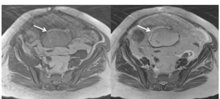
No calcification or cystic lesions were found. According to these findings, our first impression as diagnosis was peritoneal car-cinomatosis. Laboratory work-up revealed high serum CA125, carcinoembryonic antigen CEA and CA19-9 levels (109 U/mL, 12,1 ng/mL and 23.576 U/mL, respectively). She had leukocy-tosis (14,20 K/mm3) with a serum CRP level of 107,30 mg/L. MRI was performed for differential diagnosis: an amorphous, thin-walled, cyst-like right adnexal lesion of 9.8x7.0x5.5 cm was detected. The margins of lesion were discontinuous. Addition-ally restricted diffusion of omentum on diffusion weighted im-ages (DWI), some fat – fluid levels on T2WI and foamy appear-ance of fat signals were found on MRI (Figure 2,3).
High lymphocyte count was detected in the peritoneal fluid on peritoneal irrigation. Surgery revealed a ruptured right ovarian dermoid cyst, peritonitis and bowel adhesions. Omentectomy, total abdominal hysterectomy and salpingo-oophorectomy were performed. The frozen biopsy sections of peritoneum and right ovary were reported to be benign. Early postoperative course and 18-month follow-up of the patient was eventless.
Discussion
The typical radiologic finding of dermoid cyst is intratumoral fat. The most common ultrasound finding is a cystic mass with an echogenic tubercule due to hair, teeth, and fat [3]. At CT, intratumoral fat shows negative attenuation [4]. At MRI, intra-tumoral fat shows hyperintense on T1-weighted images, and
drop on fat-saturated T1-weighted images. In addition to in-tratumoral fat, calcification is also commonly seen in ovarian dermoid cysts, but it does not always indicate ovarian dermoid cyst. Therefore, the presence of intratumoral fat is important in the diagnosis of ovarian dermoid cysts [3,5]
The ovarian dermoid cysts have keratinoid content, therefore, they have been shown to exhibit lower ADC values than any other benign or malignant adnexal lesion, a characteristic that may help diagnose lesions with a paucity of fat [6]. We detected restricted diffusion on ADC maps in the both lesion and omental areas, therefore, we thought malignancy.
Spontaneous rupture is an extremely rare complication of der-moid cyst (also known as mature cystic teratoma). Because, it usually has a thick capsule. The reported causes of rupture are torsion with infarction of the tumor, infection, malignancy and rapid growth of the cyst, direct trauma, or prolonged pressure during pregnancy [7]. At ultrasound, CT and MRI, the integrity of the tumor wall should be carefully evaluated for accurate di-agnosis of a ruptured ovarian dermoid cyst [8].Similar to these findings , the integrity of the tumor wall in our case was dis-trupted.
There has been two possible clinic presentations for ruptured dermoid cysts reported in the literature. First presentation is spillage of tumor contents into the abdominal cavity due to rupture, it can lead severe acute chemical peritonitis. Second, chronic granulomatous peritonitis due to chronic cyst content leak is characterized by multiple small white peritoneal lesions, dense adhesions, and ascites mimicking carcinomatosis or tu-berculous peritonitis [9].
Chronic granulomatous peritonitis usually has unusual radio-logic findings: ascites and omental infiltration. These findings simulate carcinomatous or tuberculous peritonitis. Ovarian and gastrointestinal system malignancies usually manifest with as-cites and peritoneal carcinomatosis.
Serum tumor markers (CA19-9, CEA, CA 125) tend to be high in gastrointestinal system lesions. In our case, high tumor
mark-Figure 1. Precontrast and postcontrast axial computed tomography images show intraperitoneal free fluid and ‘omental cake’ appearance.
Figure 2. Axial T1 and T2 weighted images demonstrate amorphous, thin-walled, cyst-like right adnexal lesion. In addition, there are fat - fluid levels and foamy appearance.
Figure 3. On diffusion weighted magnetic resonance images and apparent diffu-sion coefficient maps, there is the restricted diffudiffu-sion as a pitfall.
| Journal of Clinical and Analytical Medicine
702
| Journal of Clinical and Analytical Medicine
Peritoneal Karsinomatozis, Rüptüre Ovaryan Dermoid Kist / Peritoneal Carcinomatosis, Ruptured Ovarian Dermoid Cyst
3
ers, ascites, “omental cake” appearance were suggestive of a malignant tumor.
As a very rare entity, chronic granulomatous peritonitis due to ruptured ovarian dermoid cyst can be easily confused with peri-toneal carcinomatosis. Mild chronic abdominal pain, a very thin cyst wall remnant, fat-fluid levels, and foamy fat signals on MRI are important clues for dermoid cyst rupture and chronic spill-age. Hence, MRI is highly important to make the differential diagnosis.
Competing interests
The authors declare that they have no competing interests. References
1. Tandon A, Gulleria K, Gupta S, Goel S, Bhargava SK, Vaid NB. Mature ovar-ian dermoid cyst invading the urinary bladder. Ultrasound Obstet Gynecol 2010;35(6):751-3.
2. Godara R, Karwasra RK, Garg P, Sharma N. Pilimiction. A diagnostic symptom of ovarian dermoid cyst. Internet J Gynecol Obstet 2006;6(1):62-9.
3. Outwater EK, Siegelman ES, Hunt JL. Ovarian teratomas: tumor types and im-aging characteristics. Radiographics 2001;21(2):475-90.
4. Buy JN, Ghossain MA, Moss AA, Bazot M, Doucet M, Hugol D, et al. Cystic tera-toma of the ovary: CT detection. Radiology 1989;171(3):697-701.
5. Guinet C, Ghossain MA, Buy JN, Malbec L, Hugol D, Truc JB, et al. Mature cystic teratomas of the ovary: CT and MR findings. Eur J Radiol 1995;20(2):137-43. 6. Nakayama T, Yoshimitsu K, Irie H, Aibe H, Tajima T, Nishie A, et al. Diffusion weighted echo-planar MR imaging and ADC mapping in the differential diagnosis of ovarian cystic masses: usefulness of detecting keratinoid substances in mature cystic teratomas. J Magn Reson Imaging 2005;22(2):271–8.
7. Nader R, Thubert T, Deffieux X, Laveaucoupet J, Ssi-Yan-Kai G. Delivery induced intraperitoneal rupture of a cystic ovarian teratoma and associated chron-ic chemchron-ical peritonitis. Case Reports in Radiology 2014;2014:189409. doi:10.1155/2014/189409.
8. Fibus TF. Intraperitoneal rupture of a benign cystic ovarian teratoma: findings at CT and MR imaging. AJR Am J Roentgenol 2000;174(1):261-2.
9. Pantoja E, Noy MA, Axtmayer RW, Colon FE, Pelegrina I. Ovarian dermoids and their complications: comprehensive historical review. Obstet Gynecol Surg 1975;30:1-20.
How to cite this article:
Erbay G, Ozen M, Karadeli E. Ruptured Ovarian Dermoid Cyst Mimicking Perito-neal Carcinomatosis: CT and MRI. J Clin Anal Med 2015;6(suppl 5): 701-3.
Journal of Clinical and Analytical Medicine | 703