Vitamin D deficiency is related to thyroid
antibodies in autoimmune thyroiditis
Aslı Dogruk unAl
1, ozlem TArCın
1, HulyA PArılDAr
2, ozlem Cıgerlı
2,
HACer eroglu
3, nılgun guvener DemırAg
11Department of Endocrinology and Metabolism, Baskent University Istanbul Hospital, Istanbul, Turkey 2Department of Family Medicine, Baskent University Istanbul Hospital, Istanbul, Turkey
3Department of Biochemistry, Baskent University Istanbul Hospital, Istanbul, Turkey
Abstract
Introduction: ıt has been known that vitamin D has some immunomodulatory effects and in au-toimmune thyroid diseases, vitamin D deficiency was more prevalent. ın this study, our aim was to investigate the relationship between thyroid autoantibodies and vitamin D.
Material and methods: Group 1 and 2 consisted of 254 and 27 newly diagnosed Hashimoto’s thyroiditis (HT) and Graves’ disease (GD) cases, respectively; age-matched 124 healthy subjects were enrolled as controls (group 3). All subjects (n = 405) were evaluated for 25OHD and thyroid autoanti-body [anti-thyroid peroxidase (anti-TPo) and anti-thyroglobulin (anti-tg)] levels.
Results: Group 2 and group 1 patients had lower 25OHD levels than group 3 subjects 14.9 ±8.6 ng/ml, 19.4 ±10.1 ng/ml and 22.5 ±15.4 ng/ml, respectively (p < 0.001). Serum 25OHD levels inversely correlated with anti-Tg (r = –0.136, p = 0.025), anti-TPO (r = –0.176, p = 0.003) and parathormone (PTH) (r = –0.240, p < 0.001). Group 2 patients had higher anti-Tg and anti-TPo levels than group 1 and 3 (p < 0.001).
Conclusions: ın this study, we found that patients with autoimmune thyroid disease (AıTD) present with lower vitamin D levels and gD patients have higher prevalence. since we found an inverse cor-relation between vitamin D levels and thyroid antibody levels, we may suggest that vitamin D deficiency is one of the potential factors in pathogenesis of autoimmune thyroid disorders.
Key words: vitamin D, thyroid, autoimmunity.
(Centr Eur J Immunol 2014; 39 (4): 493-497)
Introduction
Vitamin D is a lipid soluble vitamin which affects via vitamin D receptor (VDR). Vitamin D receptor is an in-tracellular receptor which belongs to the steroid/thyroid nuclear receptor family. This receptor is located in many immune cells, such as neutrophils, macrophages, dendritic cells, T and B cells. In recent years, apart from its primary role in bone and mineral homeostasis, it has been shown that vitamin D has potent immunomodulatory effects both on the innate and adaptive immune system [1-4]. Vitamin D inhibits pro-inflammatory processes by suppressing the over-activity of CD4+, Th1, Th2 and Th17 cells and the production of their related cytokines by the activation of VDR [1, 5]. Epidemiological studies have shown a relation between vitamin D deficiency and autoimmune diseases, such as rheumatoid arthritis, systemic sclerosis, systemic lupus erythematosus and autoimmune thyroiditis [6-8].
Autoimmune thyroid diseases (AITDs), including Graves’ disease (GD), Hashimoto’s thyroiditis (HT) and
postpartum thyroiditis, are the most frequently seen auto-immune diseases affecting more than 5% of population. In recent years, there have been a few studies demonstrating an increase in vitamin D deficiency in HT [9, 10].
In this study, our aim was to compare vitamin D lev-els of newly diagnosed AITDs (GD and HT) and healthy controls and investigate the relation between thyroid auto-antibodies and vitamin D deficiency.
Material and methods
This study was approved by the Baskent University Institutional Review Board and Ethics Committee (Project no. KA13/176) and supported by the Baskent University Research Fund. Informed consent was obtained from all patients and healthy controls.
Study population
This study population consisted of newly diagnosed AITD adult patients and controls. According to
diagno-sis, the study population was separated into three different groups. Hashimoto’s thyroiditis patients (diagnosed by elevated antithyroid peroxidase and antithyroglobulin an-tibodies (TPOAb, TgAb) and basal thyrotrophic hormone (TSH) as well as typical hypoechogenicity of the thyroid in high resolution sonography) were included in group 1, GD patients [diagnosed by elevated free thyroxine (fT4) and suppressed TSH levels and the presence of diffused goiter and thyroid receptor antibody (TRAb) positivity] in group 2 and controls in group 3. Patients with primary liver and renal failure, diabetes mellitus, metabolic bone disorders, hyperparathyroidism, malignancy, previously known thy-roid disorders and on oral contraceptive, anticonvulsant, anti-osteoporotic therapy and other medications that might alter 25(OH)D or 1,25(OH)2D metabolism and thyroid func-tions had been excluded from the study.
Laboratory investigation
For measuring 25(OH)D, TSH, anti-TPO and anti-tg, a blood sample was collected by venipuncture at the fast-ing state, the serum was separated by centrifugation and then stored at –70oC for a week until analysed.
Vitamin D: Vitamin D status was evaluated by mea-surement of serum 25(OH)D levels with a chemilumines-cent immunoassay method (Architect i1000 system); nor-mal range was 8.8-46.3 ng/ml (winter); and the intra-assay CV ranged from 2.6 to 4.0%. Serum 25(OH)D levels be-low 20 ng/ml were considered as deficiency.
Parathormone: Serum PTH levels were measured with an electrochemiluminescent immunoassay method (Architect i2000 system); normal range 15-68 pg/ml; in-tra-assay CV 3.0-6.5%. Serum calcium (Ca) levels were measured with an enzymatic colorimetric assay (C8000 system); intra-assay and inter-assay CV were 0.5-0.6% and 0.3-0.5%, respectively.
Thyrotrophic hormone, anti-Tg, and anti-TPO: TSH, anti-TG, and anti-TPO were also measured with a chemiluminescent immunoassay method (CMIA) (Ar-chitect i2000 system, Abbott, USA). The assays have in-tra-assay precision of 4.3%, 5.8%, and 3.2%, respective-ly. Positive anti-TPO, and anti-tg were defined as a value greater than 5.61 IU/ml and 4.11 IU/ml, respectively.
Statistical analyses
Statistical analyses were performed with the Statistical Package for Social Sciences (SPSS for Windows) software (version 17.0, SPSS Inc., Chicago, IL, USA). All para-metric variables were given as mean ± SD or median and interquartile range according to distribution of variables. Distribution of patients was assessed by using visual (his-tograms, probability plots) and analytical methods (Kolm-ogorov-Smirnov/Shapiro-Wilk’s test). The difference between categorical variables was analyzed with Chi-square test and continuous variables were analyzed with Mann-Whitney u test. As the vitamin D level was not nor-mally distributed even after logarithmic transformation, the data were compared by the non-parametric Mann-Whitney u test. A p-value below 0.05 was considered to be statis-tically significant.
While investigating the associations between non-nor-mally distributed and ordinal variables, the correlation co-efficients and their significance were calculated using the Spearman test.
Results
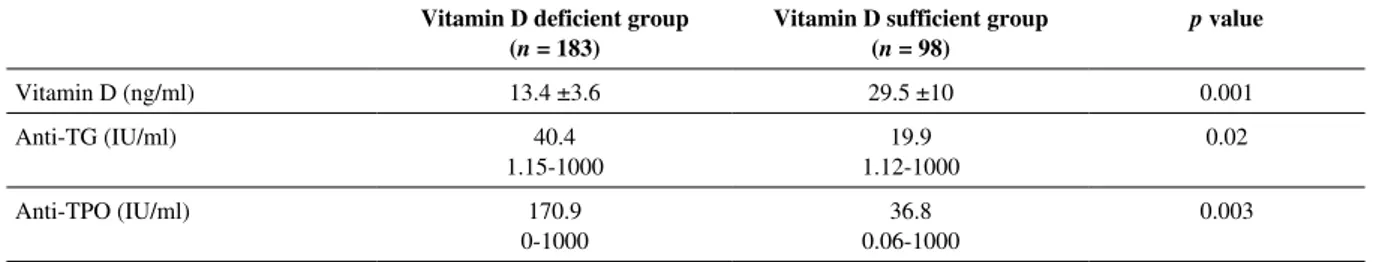
A total of 405 patients were enrolled in our study. The mean age was 44.6 ±13.5 years and 89.4% were women in the study population. There was no age difference between 3 groups (p > 0.05). A mean vitamin D level of all partici-pants was 20.1 ±12 ng/ml. Patients whose serum 25(OH)D levels were below 20 ng/ml were considered as vitamin D deficient. Sixty five percent (183/281) of the AITDs pa-tients were vitamin D deficient. When vitamin D deficient (n = 183) and sufficient (n = 98) AITDs patients were com-pared, anti-TG and anti-TPO levels found significantly high in the vitamin D deficient group (p = 0.02 and p = 0.003, respectively, for anti-tg and anti-TPO) (Table 1).
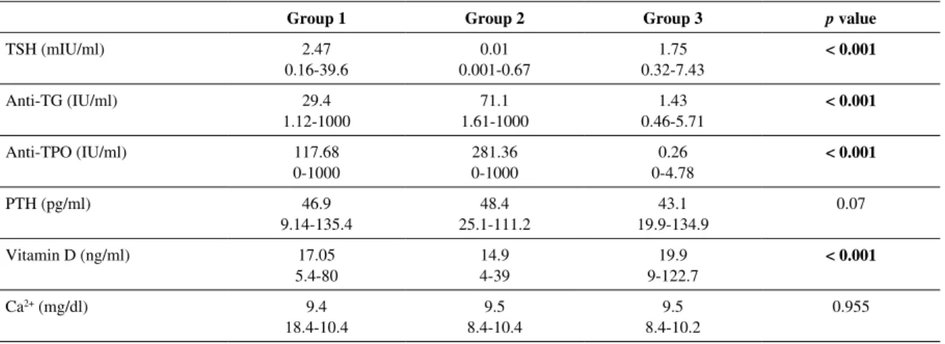
Group 1, group 2 and group 3 consisted of 254, 27 and 124 patients, respectively. The prevalence of vitamin D insufficiency was 63% and 85.2% in group 1 and 2, re-spectively. Although the levels of vitamin D were lowest in group 2 (Fig. 1), calcium and PTH levels were similar between groups. Laboratory results of study population are demonstrated in Table 2.
Table 1. Thyroid autoantibody levels according to the vitamin D status
Vitamin D deficient group (n = 183)
Vitamin D sufficient group (n = 98) p value Vitamin D (ng/ml) 13.4 ±3.6 29.5 ±10 0.001 Anti-TG (IU/ml) 40.4 1.15-1000 19.9 1.12-1000 0.02 Anti-TPO (IU/ml) 170.9 0-1000 36.8 0.06-1000 0.003 Values are expressed as mean ÷ SD or median with interquartile range
Multivariate analysis using logistic regression revealed that independent determinants of vitamin D levels were age and sex (r = 0.17, p = 0.004; r = –0.15, p = 0.01, respectively). As expected, there was a negative correlation between vita-min D and PTH levels (r = –0.24, p < 0.001). When we have investigated the association of thyroid autoantibodies, we found that there was a significant correlation between these autoantibodies and vitamin D and TSH levels (Table 3). Thy-roid autoantibodies tended to be higher with lower vitamin D levels and higher TSH levels. Serum PTH levels, however, were not associated with anti-TG and anti-TPO levels.
Discussion
In this case-control study, patients with autoimmune thyroid diseases (HT and GD) had lower 25(OH)D levels than healthy subjects and vitamin D levels were the lowest in GD patients. Since we have known from our previous and other Turkish studies that vitamin D deficiency has been a prevalent health problem in our country, we adopt-ed a cutoff level of vitamin D level as 20 ng/ml [21-23]. According to this cutoff value, 60.7% of our study popula-tion had vitamin D deficiency.
It has been known that vitamin D deficiency is not correlated with severity of hyperthyroidism [24, 25], and antithyroid drugs have immunosuppressive effects. In ad-dition, thyroid hormones relatively affect renal activity of 1α-hydroxlase and plasma 1,25(OH)2D levels [26, 27]. For these reasons, only newly diagnosed AITDs patients were included and we measured 25(OH)D level instead of 1,25(OH)2D in our study.
Autoimmune thyroid diseases are relatively common organ-specific autoimmune disorders that cause diseases ranging in severity from hypothyroidism (HT) to hyperthy-roidism (GD) [28]. The effects of various environmental
factors and the intrinsic genetic predisposition of an indi-vidual may lead to a loss of self-tolerance and contribute to the initiation of AITDs. In pathological conditions like thyroiditis, infiltrating lymphocytes, cell surface expres-sion of MHCII, Fas-mediated apoptosis and cytokines re-Table 2. Characteristics of the study population
group 1 group 2 group 3 p value
TSH (mIU/ml) 2.47 0.16-39.6 0.01 0.001-0.67 1.75 0.32-7.43 < 0.001 Anti-TG (IU/ml) 29.4 1.12-1000 71.1 1.61-1000 1.43 0.46-5.71 < 0.001 Anti-TPO (IU/ml) 117.68 0-1000 281.36 0-1000 0.26 0-4.78 < 0.001 PTH (pg/ml) 46.9 9.14-135.4 48.4 25.1-111.2 43.1 19.9-134.9 0.07 Vitamin D (ng/ml) 17.05 5.4-80 14.9 4-39 19.9 9-122.7 < 0.001 Ca2+ (mg/dl) 9.4 18.4-10.4 9.5 8.4-10.4 9.5 8.4-10.2 0.955 Values are expressed as median with interquartile range.
TSH – thyroid-stimulating hormone, anti-Tg – thyroglobulin antibody, anti-TPO – anti-thyroid peroxidase antibody, PTH – parathyroid hormone, Ca – calcium
Table 3. Correlation between thyroid autoantibodies and vitamin D and TSH levels
Vitamin D TSH Anti-TG r p –0.13 0.025 0.144 0.018 Anti-TPO r p –0.17 0.003 0.21 0.001 TSH – thyroid-stimulating hormone, Tg – thyroglobulin antibody, anti-TPO – anti-thyroid peroxidase antibody
125 100 75 50 25 0
Hashimoto Graves Control
thyroiditis disease
group
Fig. 1. Vitamin D levels in each group
leased from both immune cells and thyrocytes contribute to amplification and progression of AITD [29-34]. According to this theory, as a result of defective suppressor T cells, Th (CD4) cells are able to activate and cooperate with B lymphocytes. B lymphocytes activated by T lymphocytes produce antibodies that react with thyroid antigens. Stud-ies on HT and GD patients had reported low vitamin D levels [9, 10, 35, 36]. Consistent with the literature, we found a lower vitamin D level in patients with autoimmune thyroiditis (GD group and HT group) than in controls. In addition, this is the first study that found a statistically sig-nificant negative correlation between serum 25(OH)D and anti-TPO, anti-TG levels in both HT and GD patients.
Conclusions
Our findings indicate that patients with AITD present with lower vitamin D levels and GD patients have higher prevalence. Since we found an inverse correlation between vitamin D levels and thyroid antibody levels, we might speculate that there might exist a casual relationship. How-ever, these findings do not clarify whether treatment with vitamin D has any beneficial effect on progression or re-mission of AITD. So, further studies specifically designed to evaluate the beneficial effect of vitamin D supplemen-tation on AITD are needed.
The authors declare no conflict of interest.
References
1. Arnson Y, Amital H, Shoenfeld Y (2007): Vitamin D and au-toimmunity: new aetiological and therapeutic considerations. Ann Rheum Dis 66: 1137-1142.
2. Baeke F, Takiishi T, Korf H, et al. (2010): Vitamin D: mod-ulator of the immune system. Curr Opin Pharmacol 10: 482-496.
3. Deluca HF, Cantorna MT (2001): Vitamin D: its role and uses in immunology. FASEB J 15: 2579-2585.
4. Cantorna MT, Mahon BD (2004): Mounting evidence for vitamin D as an environmental factor affecting autoimmune disease prevalence. Exp Biol Med 229: 1136-1142.
5. Tang J, Zhou R, Luger D, et al. (2009): Calcitriol suppress-es antiretinal autoimmunity through inhibitory effects on the Th17 effector response. J Immunol 182: 4624-4632. 6. Cutolo M, Otsa K, Uprus M, et al. (2007): Vitamin D in
rheu-matoid arthritis. Autoimmun Rev 7: 59-64.
7. Doria A, Arienti S, Rampudda M, et al. (2008): Preventive strategies in systemic lupus erythematosus. Autoimmun Rev 7: 192-197.
8. Kamen DL, Cooper GS, Bouali H, et al. (2006): Vitamin D deficiency in systemic lupus erythematosus. Autoimmun Rev 5: 114-117.
9. Tamer G, Arik S, Tamer I, Coksert D (2011): Relative vitamin D insufficiency in Hashimoto’s thyroiditis. Thyroid 21: 891-896. 10. Bozkurt NC, Karbek B, Ucan B, et al. (2013): The association
between severity of vitamin D deficiency and Hashimoto’s thyroiditis. Endocr Pract 19: 479-484.
11. Cantorna MT, Zhu Y, Froicu M, Wittke A (2004): Vitamin D status, 1,25-dihydroxyvitamin D3, and the immune system. Am J Clin Nutr 80: 1717S-1720S.
12. Li HS, Verginis P, Carayanniotis G (2006): Maturation of dendritic cells by necrotic thyrocytes facilitates induction of experimental autoimmune thyroiditis. Clin Exp Immunol 144: 467-474.
13. Antico A, Tampoia M, Tozzoli R, Bizzaro N (2012): Can sup-plementation with vitamin D reduce the risk or modify the course of autoimmune diseases? A systematic review of the literature. Autoimmun Rev 12: 127-136.
14. Ikeda U, Wakita D, Ohkuri T, et al. (2010): 1alpha,25-Dihy-droxyvitamin D3 and all-trans retinoic acid synergistically in-hibit the differentiation and expansion of Th17 cells. Immunol Lett 134: 7-16.
15. Taher YA, van Esch BC, Hofman GA, et al. (2008): 1al-pha,25-dihydroxyvitamin D3 potentiates the beneficial effects of allergen immunotherapy in a mouse model of allergic asth-ma: role for IL-10 and TGF-beta. J Immunol 180: 5211-5221. 16. Hewison M, Freeman L, Hughes SV, et al. (2003): Differen-tial regulation of vitamin D receptor and its ligand in human monocyte-derived dendritic cells. J Immunol 170: 5382-5390. 17. Piemonti L, Monti P, Sironi M, et al. (2000): Vitamin D3
affects differentiation, maturation, and function of human monocyte-derived dendritic cells. J Immunol 164: 4443-4451. 18. Széles L, Keresztes G, Töröcsik D, et al. (2009): 1,25-dihy-droxyvitamin D3 is an autonomous regulator of the transcrip-tional changes leading to a tolerogenic dendritic cell pheno-type. J Immunol 182: 2074-2083.
19. Correale J, Ysrraelit MC, Gaitán MI (2011): Vitamin D-me-diated immune regulation in multiple sclerosis. J Neurol Sci 311: 23-31.
20. Kivity S, Agmon-Levin N, Zisappl M, et al. (2011): Vitamin D and autoimmune thyroid diseases. Cell Mol Immunol 8: 243-247.
21. Cigerli O, Parildar H, Unal AD, et al. (2013): Vitamin D deficiency is a problem for adult out-patients? A university hospital sample in Istanbul, Turkey. Public Health Nutr 16: 1306-1313.
22. Erkal MZ, Wilde J, Bilgin Y, et al. (2006): High prevalence of vitamin D deficiency, secondary hyperparathyroidism and generalized bone pain in Turkish immigrants in Germany: identification of risk factors. Osteoporos Int 17: 1133-1140. 23. Ralston SH, Binkley N, Boonen S, et al. (2011): Randomized
trial of alendronate plus vitamin D3 versus standard care in osteoporotic postmenopausal women with vitamin D insuffi-ciency. Calcif Tissue Int 88: 485-494.
24. Fournier C, Gepner P, Sadouk M, Charreire J (1990): In vivo beneficial effects of cyclosporin A and 1,25-dihydroxyvitamin D3 on the induction of experimental autoimmune thyroiditis. Clin Immunol Immunopathol 54: 53-63.
25. Misharin A, Hewison M, Chen CR, et al. (2009): Vitamin D deficiency modulates Graves’ hyperthyroidism induced in BALB/c mice by thyrotropin receptor immunization. Endo-crinology 150: 1051-1060.
26. Kozai M, Yamamoto H, Ishiguro M, et al. (2013): Thyroid hormones decrease plasma 1alpha,25-dihydroxyvitamin D levels through transcriptional repression of the renal 25-hy-droxyvitamin D3 1alpha-hydroxylase gene (CYP27B1). En-docrinology 154: 609-622.
27. Pantazi H, Papapetrou PD (2000): Changes in parameters of bone and mineral metabolism during therapy for hyperthy-roidism. J Clin Endocrinol Metab 85: 1099-1106.
28. Hollowell JG, Staehling NW, Flanders WD, et al. (2002): Se-rum TSH, T(4), and thyroid antibodies in the United States population (1988 to 1994): National Health and Nutrition Ex-amination Survey (NHANES III). J Clin Endocrinol Metab 87: 489-499.
29. Ng HP, Banga JP, Kung AW (2004): Development of a mu-rine model of autoimmune thyroiditis induced with homolo-gous mouse thyroid peroxidase. Endocrinology 145: 809-816. 30. McLachlan SM, Nagayama Y, Rapoport B (2005): Insight
into Graves’ hyperthyroidism from animal models. Endocr Rev 26: 800-832.
31. Many MC, Maniratunga S, Varis I, et al. (1995): Two-step development of Hashimoto-like thyroiditis in genetically au-toimmune prone non-obese diabetic mice: effects of iodine-in-duced cell necrosis. J Endocrinol 147: 311-320.
32. Caturegli P, Hejazi M, Suzuki K, et al. (2000): Hypothyroid-ism in transgenic mice expressing IFN-gamma in the thyroid. Proc Natl Acad Sci U S A 97: 1719-1724.
33. Kimura H, Kimura M, Tzou SC, et al. (2005): Expression of class II major histocompatibility complex molecules on thyrocytes does not cause spontaneous thyroiditis but mildly increases its severity after immunization. Endocrinology 146: 1154-1162.
34. Nagayama Y (2005): Animal models of Graves’ hyperthy-roidism. Endocr J 52: 385-394.
35. Yasuda T, Okamoto Y, Hamada N, et al. (2012): Serum vitamin D levels are decreased and associated with thyroid volume in female patients with newly onset Graves’ disease. Endocrine 42: 739-741.
36. Yasuda T, Okamoto Y, Hamada N, et al. (2013): Serum vi-tamin D levels are decreased in patients without remission of Graves’ disease. Endocrine 43: 230-232.