Coronary flow reserve is impaired in
patients with obstructive sleep apnea
Serife Savas Bozbas, Serpil Eroglu1, Berna Akinci Ozyurek2, Fusun Oner Eyuboglu Abstract:
STUDY OBJECTIVES: Obstructive sleep apnea (OSA) is common in adult populations. Accumulating
data indicate that it is independently associated with a variety of cardiovascular diseases and has prognostic importance in affected cases. In this study, we aimed to evaluate coronary flow reserve (CFR) in patients with OSA and controls.
METHODS: Sixty‑one patients undergoing an overnight polysomnography were enrolled in this study.
Patients with an apnea–hypopnea index (AHI) >5 were accepted as OSA group (n = 45) and those
with an AHI <5 were taken as controls (n = 16). Using Doppler echocardiography at baseline and
following dipyridamole infusion, coronary peak flow velocities were obtained. CFR was calculated as the ratio of peak diastolic flow to baseline diastolic flow. A CFR value <2 was accepted as impaired coronary microvascular function.
RESULTS: The mean age was 50.8 ± 10.8 years, of which 16 (26.2%) were female. Both groups
had similar features with regard to demographic and clinical variables. The mean value of CFR was significantly lower in patients with OSA compared to those controls (2.24 ± 0.46 vs. 2.74 ± 0.62, respectively, P = 0.001). An abnormal CFR value was observed in 12 (26.7%) patients with OSA
and in 1 (6.3%) participant in control group.
CONCLUSIONS: The findings of this study indicate that CFR, an indicator of coronary microvascular
function, is significantly impaired in patients with OSA. Coronary microvascular function, an early sign of atherosclerosis, can be evaluated noninvasively in these patients might be used as a predictor of cardiovascular risk.
Keywords:
Cardiac risk, coronary flow reserve, obstructive sleep apnea, polysomnography
O
bstructive sleep apnea (OSA)is a common disorder in adult populations.[1] Accumulating data indicate that it is independently associated with a variety of cardiovascular diseases (CVDs) and has strong prognostic impact in affected cases.[2] Independent of traditional risk factors both cardiovascular and all‑cause mortality has been reported as high in untreated OSA patients.[3] It has been shown that when coronary artery disease (CAD) and OSA coexist, the rate of ischemic events increases during the night.[4] The exact mechanism for such ischemic episodes is not clear. It has been demonstrated that nocturnal ischemic events occur during
the postapneic phase, during which time myocardial oxygen demand increases as a result of elevation in blood pressure and heart rate.[5]
Endothelial dysfunction is accepted as an early manifestation of atherosclerosis and is a predictor of future cardiovascular events. It is accepted as one of the key mechanisms responsible for impairment in endothelial function in OSA patients.[6]
Coronary flow reserve (CFR) determined by transthoracic Doppler echocardiography has been introduced as an indicator of coronary microvascular function in the absence of significant stenosis of epicardial coronary arteries.[7] CFR measured by this Address for
correspondence: Dr. Berna Akinci Ozyurek, Department of Pulmonary Medicine, Ataturk Chest Diseases and Thoracic Surgery Education and Research Hospital, Ankara, Turkey. E‑mail: drberna_1982@ yahoo.com Submission: 01‑06‑2016 Accepted: 10‑06‑2017 Departments of Pulmonary
Medicine and 1Cardiology, Faculty of Medicine, Baskent University, 2Department of Pulmonary Medicine, Ataturk Chest Diseases and Thoracic Surgery Education and Research Hospital, Ankara, Turkey
Access this article online
Quick Response Code:
Website:
www.thoracicmedicine.org
DOI:
10.4103/atm.ATM_195_16
How to cite this article: Bozbas SS, Eroglu S,
Ozyurek BA, Eyuboglu FO. Coronary flow reserve is impaired in patients with obstructive sleep apnea. Ann Thorac Med 2017;12:272‑7.
This is an open access article distributed under the terms of the Creative Commons Attribution‑NonCommercial‑ShareAlike 3.0 License, which allows others to remix, tweak, and build upon the work non‑commercially, as long as the author is credited and the new creations are licensed under the identical terms.
way has been shown to be a reliable and reproducible indicator of coronary microvascular function and has been validated against invasive Doppler wire measurements and positron emission tomography which is the gold standard in the evaluation of CFR.[3‑5] Although the association between OSA and various forms of overt CVD, including CAD, heart failure, hypertension, arrhythmias, and stroke, is well documented, data regarding the effect of OSA on coronary microvascular function are limited. We wondered whether CFR, an early sign of atherosclerosis, is impaired or not in patients with OSA who do not have overt CAD. Therefore, in this study, we aimed to evaluate CFR in patients with OSA and compare with controls.
Methods
Study subjects
Patients with suspicion of OSA who were admitted to the Sleep Disorders Center with symptoms of nocturnal snoring and/or excess daytime sleepiness were included in the study. For this study, 61 consecutive patients who underwent an overnight polysomnography were enrolled in the study. Based on apnea–hypopnea index (AHI) values, participants were divided into two groups. An AHI value >5 is defined as having OSA. Hence, patients with an AHI >5 comprised the study group (n = 45), and participants with AHI <5 (primary snoring) were enrolled as the control group (n = 16).
Exclusion criteria were CAD, aortic stenosis, moderate‑severe aortic or mitral regurgitation, hypertrophic cardiomyopathy, heart failure, uncontrolled hypertension before study, chronic kidney disease, asthma, malignancy, central sleep apnea, and those using any vasoactive drugs.
CAD was defined as the presence of one of the followings: history of myocardial infarction or revascularization, ECG changes for myocardial ischemia, wall motion abnormalities on echocardiography, signs of ischemia on noninvasive stress test, and ≥50% stenosis in any of coronary arteries on coronary angiography.
Polysomnography
S t a n d a r d o v e r n i g h t p o l y s o m n o g r a p h y (Grass‑telefactor, Astro‑Med Inc., West Warwick, RI, USA) was performed in all patients at our sleep center. During polysomnography, electroencephalography electrodes were positioned according to the international 10–20 system. Sleep was recorded and scored according to the standard method.[8] Electrooculography and electromyography were obtained. Oronasal airflow was measured with thermistor and nasal cannula. Electrocardiographic monitorization, thoracoabdominal
movements, body position, and oxygen saturation were also recorded. Blood oxygen saturation was monitored by pulse oximetry. Apnea was defined as a drop ≥90% of baseline in airflow that lasts for longer than 10 s. Hypopnea was defined as a ≥30% reduction in oronasal flow amplitude ≥10 s, accompanied by ≥3% desaturation or arousal. Classification of a hypopnea and apnea as obstructive, central, or mixed performed calibrated respiratory inductance plethysmography. AHI was defined as the total number of apnea and hypopnea episodes per hour of sleep.
Echocardiographic examination and determination of coronary flow reserve
Transthoracic Doppler echocardiographic examinations were performed using an Acuson Sequoia C‑256 (Acuson Corporation, California, USA) machine with a 3.5 MHz transducer. A cardiologist who is blinded to polysomnographic results performed the echocardiographic examinations. CFR was determined with the use of Doppler echocardiography as previously described.[9] Using a modified parasternal long‑axis view, mid to distal left anterior descending blood flow was obtained. Using pulsed wave Doppler, systolic and diastolic coronary blood flows were determined at baseline and during hyperemia. Dipyridamole was used to obtain hyperemic state (0.56 mg/kg over 4 min). The highest three Doppler recordings were averaged for each measurement, and CFR was calculated as the ratio of peak hyperemic diastolic flow to baseline diastolic flow [Figure 1]. A CFR value <2 is accepted as abnormal.[9,10]
All participants provided written informed consent, and the study complied with the Declaration of Helsinki and was approved by the Ethics Committee at Baskent University Hospital.
Statistical analysis
Statistical analyses were performed with Statistical Package for the Social Sciences software, version 15.0 (SSPS Inc., Chicago, IL, USA). Continuous parameters are presented as mean ± standard deviation and the median (interquartile range) as appropriate. Variables with a normal distribution were compared using paired and unpaired t‑tests. Differences between groups for nonhomogenously distributed parameters were tested with Mann–Whitney U‑test. Categorical variables are given as group percentages and the comparison of the two groups were obtained using Chi‑square test. Correlations were established by Pearson’s correlation analysis or Spearman’s rank correlation test as appropriate. Stepwise linear regression analysis was employed to investigate the independent determinants of CFR. All P values are two‑sided, and values <0.05 were considered statistically significant.
Results
Clinical, laboratory, and polysomnographic data
The mean age was 50.8 ± 10.8 years and 16 (26.2%) were female. There were no significant differences between the OSA groups and controls with regard to baseline clinical and demographic characteristics [Table 1]. Polysomnographic data are provided in Table 1. With regard to severity of OSA, 14 patients had mild (AHI 5–15/h), 15 had moderate (AHI 15–30/h), and 16 had severe OSA (AHI >30/h).
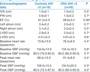
Echocardiographic and coronary flow reserve measurement findings
Comparison of the groups (OSA, controls) with regard to echocardiographic measurements is provided in Table 2. The mean value of left ventricular ejection fraction was similar in both groups. The mean value of CFR was significantly lower in patients with OSA compared to those controls (2.24 ± 0.46 vs. 2.74 ± 0.62, respectively, P = 0.001) [Figure 2]. An abnormal CFR value was observed in 12 (26.7%) patients with OSAS and in 1 (6.3%) participant in control group (P = 0.08). Regarding the severity of OSA subgroups, the mean CFR values were as follows: 2.26 ± 0.42 in mild, 2.35 ± 0.36 in moderate, and 2.07 ± 0.56 in severe OSA. Although it was lowest in severe OSA patients, it did not reach to statistical significance.
CFR was significantly and inversely correlated with body mass index (r = −0.2, P = 0.03), waist circumference
(r = −0.1, P = 0.006), SpO2 < 90% time (r = −0.4, P = 0.01), total AHI (r = −0.3, P = 0.008), and OSA (r = −0.4,
P = 0.001). Age, systolic blood pressure, ejection fraction,
and left ventricular hypertrophy were also inversely correlated with CFR, but these correlations did not reach statistical significance.
Figure 1: Assessment of coronary flow reserve using transthoracic Doppler echocardiography. Mid to distal segment of the left anterior descending coronary artery in
color‑coded and spectral Doppler analysis at rest and during hyperemia induced by dipyridamole infusion
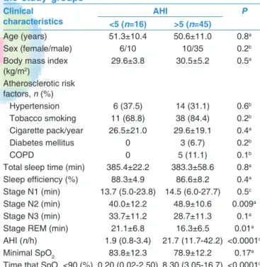
Table 1: Clinical and demographic characteristics of the study groups
Clinical
characteristics <5 (n=16) AHI >5 (n=45) P
Age (years) 51.3±10.4 50.6±11.0 0.8a
Sex (female/male) 6/10 10/35 0.2b
Body mass index (kg/m2) 29.6±3.8 30.5±5.2 0.5a Atherosclerotic risk factors, n (%) Hypertension 6 (37.5) 14 (31.1) 0.6b Tobacco smoking 11 (68.8) 38 (84.4) 0.2b Cigarette pack/year 26.5±21.0 29.6±19.1 0.4a Diabetes mellitus 0 3 (6.7) 0.2b COPD 0 5 (11.1) 0.1b
Total sleep time (min) 385.4±22.2 383.3±58.6 0.8a Sleep efficiency (%) 88.3±4.9 86.6±8.2 0.4a Stage N1 (min) 13.7 (5.0‑23.8) 14.5 (6.0‑27.7) 0.5c Stage N2 (min) 40.0±12.2 48.9±10.6 0.009a
Stage N3 (min) 33.7±11.2 28.7±11.3 0.1a
Stage REM (min) 21.1±6.8 16.3±6.5 0.01a
AHI (n/h) 1.9 (0.8‑3.4) 21.7 (11.7‑42.2) <0.0001c
Minimal SpO2 83.8±12.3 78.9±12.2 0.17a
Time that SpO2 <90 (%) 0.20 (0.02‑2.50) 8.30 (3.05‑16.7) <0.0001c
Data are expressed as the mean±SD, the median (IQR), or frequency counts (%), as appropriate. aStudent’s t‑test, bChi‑square test, cMann‑Whitney U‑test.
AHI=Apnea‑hypopnea index, COPD=Chronic obstructive airway disease, REM=Rapid eye movement, SpO2=Oxygen saturation, IOR=Interquartile range,
Results of multivariate linear regression analyses in which CFR was taken as a dependent variable and potential confounding factors as independent determinants were as follows: OSA (β = −0.38, P = 0.009), age (β = −0.14,
P = 0.35), body mass index (β = −0.09, P = 0.56), diabetes mellitus (β = 0.07, P = 0.62), hypertension (β = 0.14,
P = 0.31), smoking (β = −0.10, P = 0.43), chronic obstructive airway disease (β = −0.08, P = 0.55). OSA was identified as the only independent predictor of CFR.
Discussion
The findings of this study indicate that CFR, an indicator of coronary microvascular dysfunction, is significantly
reduced in patients with OSA than that in controls. This shows that coronary microvascular function is impaired in patients with OSA before significant coronary artery stenosis develops.
Detrimental effects of OSA on the cardiovascular system may be evaluated in two ways. Acute effects can be characterized as an increase in afterload, increased venous return to the right heart, vasoconstriction and elevated blood pressure, decreased vagal tone, and hypoxemia. Chronic effects include hypertension, accelerated atherosclerosis, arrhythmias, coronary ischemia, left ventricular hypertrophy, and diastolic dysfunction. In addition, OSA can decrease nitric oxide production and attenuate vascular conductance leading to the development of endothelial dysfunction. This has been demonstrated in studies where reactive hyperemic blood flow and vascular conductance decreased in patients with OSA on intermittent forearm arterial occlusion.[11]
It is well known that apneic episodes lead to an increase in blood pressure, hypoxemia, and sympathetic overactivation and vasoconstriction. A strong association between sleep‑disordered breathing and CAD has been demonstrated.[2] Furthermore, it has also been identified that the risk of cardiovascular mortality increases as the severity of OSA worsens.[12] Hypoxemia is regarded to be the main mechanism responsible for impairment in endothelial function is OSA patients.[13] Microvascular disease and endothelial dysfunction are accepted as the earliest manifestations of CAD. Nguyen et al.[14] examined therapeutic effect of CPAP on myocardial perfusion reserve and endothelial‑dependent vasodilation in patients with OSA. They enrolled patients with moderate‑to‑severe OSA without documented CAD. It was found that these patients had abnormal myocardial perfusion reserve, an indicator of microvascular disease and abnormal flow‑mediated dilation, an indicator of endothelial dysfunction determined by cardiac magnetic resonance imaging. In line with these findings, we found that echocardiographically determined CFR was impaired in patients with OSA.
The exact mechanism regarding the effects of OSA on coronary vasculature is not fully understood. Vascular endothelial dysfunction, oxidative stress, and increased sympathetic activity are among the mechanisms proposed to be involved in the adverse effects of OSA on coronary microvascular function.[15] Repetitive hypoxia can cause endothelial dysfunction. Regional myocardial perfusion abnormalities may occur during apneic periods. In addition, associated atherosclerotic risk factors such as hypertension, hyperlipidemia, and insulin resistance, which are known to impair vascular function, are common in participants with OSA. It has been shown Table 2: Echocardiographic examination and Doppler
findings of the study groups Echocardiographic
data Controls AHI <5 (n=16) OSA AHI >5 (n=45) P
IVS (cm) 1.0±0.1 1.0±0.2 0.5a PW (cm) 0.9±0.1 1.0±0.2 0.7a EF (%) 61.2±5.0 58.6±3.4 0.06a Left atrium (cm) 3.4±0.2 3.5±0.3 0.7a Right atrium (cm) 3.1±0.2 3.2±0.3 0.5a LVSD (cm) 2.9±0.3 3.0±0.3 0.7a LVDD (cm) 4.51±0.3 4.61±0.4 0.6a
Baseline heart rate
(beats/min) 73.8±13.6 76.2±9.0 0.4
a
Baseline SBP (mmHg) 134.6±14.9 134.4±18.5 0.9a Baseline DBP (mmHg) 80.0 (70.0‑90.0) 80.0 (80.0‑90.0) 0.7b Peak heart rate
(beats/min) 89.2±13.3 91.4±8.9 0.4
a
Peak SBP (mmHg) 129.3±15.5 134.5±20.5 0.3a Peak DBP (mmHg) 80.0 (72.5‑87.5) 80.0 (80.0‑90.0) 0.2b
Data are expressed as the mean±SD, the median (IQR) as appropriate. aStudent’s
t‑test, bMann‑Whitney U‑test. SD=Standard deviation, IQR=Interquartile range,
AHI=Apnea‑hypopnea index, IVS=Interventricular septum, OSA=Obstructive sleep apnea, PW=Posterior wall, LVDD=Left ventricular diastolic diameter, LVSD=Left ventricular systolic diameter, EF=Ejection fraction, SBP=Systolic blood pressure, DBP=Diastolic blood pressure
Figure 2: Comparison of coronary flow values of patients with obstructive sleep
that acetylcholine‑induced endothelial‑dependent vasodilation of resistance vessels was reduced in patients with OSA.[16] In our study, patients with OSA had longer periods of nocturnal hypoxia compared to controls which was thought as the responsible mechanism from impairment in CFR.
We believe that once OSA is diagnosed, early signs of CVD should be evaluated. Depending on the results, appropriate therapy should be initiated as soon as possible. In this way, the development of overt CVD might be delayed so that CV mortality and morbidity attributed to OSA maybe decreased.
Nakashima et al. examined the effects of OSA on myocardial tissue perfusion in patients with ST‑elevation myocardial infarction who underwent successful primary percutaneous coronary intervention (PCI).[17] They determined CFR using Doppler guide wire following PCI. It was found that OSA impairs myocardial tissue perfusion documented by increased incidence of systolic retrograde flow and ST‑segment resolution <50%. They concluded that OSA is associated with microvascular dysfunction in these patients. In a recent study, Wang
et al. reported that compared to controls, invasively
determined CFR is impaired in patients with OSA.[18] They found that higher AHI values were associated with impaired coronary microvascular function. Our findings which were obtained noninvasively are in line with results reported in these papers.
Impaired CFR in patients with OSA shows that coronary microvascular dysfunction – an early finding of atherosclerotic heart disease – is evident in these patients. We believe that our findings are valuable and clinically relevant. These patients, once diagnosed, should be effectively treated so that adverse cardiovascular events may be decreased and overall prognosis improved. In our study, following adjustment for other confounding factors, OSA was identified as the independent predictor of impaired coronary microvascular function. To our knowledge, the present study is the first to compare Doppler echocardiographically obtained CFR in patients with OSA and primary snoring. Detection of endothelial dysfunction is very important because it is accepted as the early treatable/reversible stage of atherosclerosis. Limitations
Small sample size is a limitation of our study. If the number of participants enrolled was higher, we would have reached more conclusive results.
Owing to the invasive nature and lack of indications, coronary angiography was not performed as part of the study protocol.
CFR was determined by transthoracic Doppler echocardiography. If it had been obtained invasively by Doppler wire, the results would be more reliable.[19] However, it has been shown that CFR obtained by Doppler echocardiography is reliable, and reproducible has been validated against invasive Doppler wire measurements and positron emission tomography which is the gold standard in the evaluation of CFR.[20]
Conclusion
Our findings, in line with above‑mentioned literature data on systemic endothelial dysfunction, suggest that coronary microvascular function is impaired in patients with OSA. Studies involving larger numbers of patients and follow‑up data after CPAP therapy would provide more definitive conclusions in this field.
Financial support and sponsorship
All support for this study came from institutional and departmental resources.
Conflicts of interest
There are no conflicts of interest.
References
1. Young T, Palta M, Dempsey J, Skatrud J, Weber S, Badr S. The occurrence of sleep‑disordered breathing among middle‑aged adults. N Engl J Med 1993;328:1230‑5.
2. Shahar E, Whitney CW, Redline S, Lee ET, Newman AB, Nieto FJ, et al. Sleep‑disordered breathing and cardiovascular disease: Cross‑sectional results of the Sleep Heart Health Study. Am J Respir Crit Care Med 2001;163:19‑25.
3. Marshall NS, Wong KK, Liu PY, Cullen SR, Knuiman MW, Grunstein RR. Sleep apnea as an independent risk factor for all‑cause mortality: The Busselton Health Study. Sleep 2008;31:1079‑85.
4. Peled N, Abinader EG, Pillar G, Sharif D, Lavie P. Nocturnal ischemic events in patients with obstructive sleep apnea syndrome and ischemic heart disease: Effects of continuous positive air pressure treatment. J Am Coll Cardiol 1999;34:1744‑9.
5. Hamilton GS, Meredith IT, Walker AM, Solin P. Obstructive sleep apnea leads to transient uncoupling of coronary blood flow and myocardial work in humans. Sleep 2009;32:263‑70.
6. Yeboah J, Crouse JR, Hsu FC, Burke GL, Herrington DM. Brachial flow‑mediated dilation predicts incident cardiovascular events in older adults: The Cardiovascular Health Study. Circulation 2007;115:2390‑7.
7. Schächinger V, Britten MB, Zeiher AM. Prognostic impact of coronary vasodilator dysfunction on adverse long‑term outcome of coronary heart disease. Circulation 2000;101:1899‑906. 8. Berry RB, Budhiraja R, Gottlieb DJ, Gozal D, Iber C, Kapur VK,
et al. Rules for scoring respiratory events in sleep: Update of the 2007 AASM Manual for the Scoring of Sleep and Associated Events. Deliberations of the Sleep Apnea Definitions Task Force of the American Academy of Sleep Medicine. J Clin Sleep Med 2012;8:597‑619.
9. Youn HJ, Foster E. Demonstration of coronary artery flow using transthoracic Doppler echocardiography. J Am Soc Echocardiogr 2004;17:178‑85.
Romeo F. Measurement of coronary flow reserve in the anterior and posterior descending coronary arteries by transthoracic Doppler ultrasound. Am J Cardiol 2002;90:988‑91.
11. Imadojemu VA, Gleeson K, Quraishi SA, Kunselman AR, Sinoway LI, Leuenberger UA. Impaired vasodilator responses in obstructive sleep apnea are improved with continuous positive airway pressure therapy. Am J Respir Crit Care Med 2002;165:950‑3.
12. Marin JM, Carrizo SJ, Vicente E, Agusti AG. Long‑term cardiovascular outcomes in men with obstructive sleep apnoea‑hypopnoea with or without treatment with continuous positive airway pressure: An observational study. Lancet 2005;365:1046‑53.
13. Foster GE, Poulin MJ, Hanly PJ. Intermittent hypoxia and vascular function: Implications for obstructive sleep apnoea. Exp Physiol 2007;92:51‑65.
14. Nguyen PK, Katikireddy CK, McConnell MV, Kushida C, Yang PC. Nasal continuous positive airway pressure improves myocardial perfusion reserve and endothelial‑dependent vasodilation in patients with obstructive sleep apnea. J Cardiovasc Magn Reson 2010;12:50.
15. Bradley TD, Floras JS. Obstructive sleep apnoea and its
cardiovascular consequences. Lancet 2009;373:82‑93.
16. Kato M, Roberts‑Thomson P, Phillips BG, Haynes WG, Winnicki M, Accurso V, et al. Impairment of endothelium‑dependent vasodilation of resistance vessels in patients with obstructive sleep apnea. Circulation 2000;102:2607‑10.
17. Nakashima H, Muto S, Amenomori K, Shiraishi Y, Nunohiro T, Suzuki S. Impact of obstructive sleep apnea on myocardial tissue perfusion in patients with ST‑segment elevation myocardial infarction. Circ J 2011;75:890‑6.
18. Wang N, Li SB, Zhao LS, Li HY, Li ZJ, Shi QW, et al. Relationship between obstructive sleep apnea and coronary microcirculatory function among patients with cardiac syndrome X. Coron Artery Dis 2014;25:35‑9.
19. Caiati C, Montaldo C, Zedda N, Montisci R, Ruscazio M, Lai G, et al. Validation of a new noninvasive method (contrast‑enhanced transthoracic second harmonic echo Doppler) for the evaluation of coronary flow reserve: Comparison with intracoronary Doppler flow wire. J Am Coll Cardiol 1999;34:1193‑200.
20. Saraste M, Koskenvuo J, Knuuti J, Toikka J, Laine H, Niemi P, et al. Coronary flow reserve: Measurement with transthoracic Doppler echocardiography is reproducible and comparable with positron emission tomography. Clin Physiol 2001;21:114‑22.