Endovascular treatment of iliofemoral deep vein thrombosis in
pregnancy using US-guided percutaneous aspiration thrombectomy
Murat Gedikoglu
Levent Oguzkurt
P
regnancy is a risk factor for venous thromboembolism (VTE) and the risk is highest in the postpartum period (1, 2). Anticoagulants are very effective in the prevention of pulmonary embolism (PE) and recurrent thrombosis, but the treatment of deep vein thrombosis (DVT) remains a challenge, because lysis of the thrombus formed in the deep veins is slow and frequently inadequate as a therapy (3, 4). Complete or significant lysis occurs only in 4% of patients treated with heparin alone (3). Persistence of thrombus within deep veins leads to venous hypertension, which is ultimately the cause of post-thrombotic syndrome (PTS) and late disability in 20% to 50% of patients. PTS is a conglomerate of life-style-limiting symptoms that commonly includes chronic leg pain and swelling, heaviness, and/or fatigue, venous claudication, stasis dermatitis, and in advanced cases skin ulcer-ations due to valvular incompetence accompanied by persistent venous outflow obstruc-tion (4–8). Pregnant women are generally younger than other women in the general popu-lation, and they likely suffer a more severe form of PTS for a much longer time.Anticoagulants have been the standard therapy of DVT in pregnant women because of concerns related to administering contrast agent, exposing the fetus to radiation during interventional radiologic procedures, and bleeding complication associated with throm-bolysis (9). Nonetheless, there are a few publications describing thrombolytic therapy of DVT with good success during pregnancy and peri- or postpartum period (10–13). In a previous study (14), a pregnant woman with DVT was treated using percutaneous mechanical thrombectomy under venographic guidance with contrast agent. Percuta-neous aspiration thrombectomy (PAT) with or without fluoroscopy guidance in preg-From the Department of Radiology (M.G.
[email protected]), Başkent University School of Medicine, Adana, Turkey; Department of Radiology (L.O.), Koç University Hospital, Istanbul, Turkey.
Received 20 April 2016; revision requested 26 May 2016; revision received 14 June 2016; accepted 20 June 2016.
Published online 1 November 2016. DOI 10.5152/dir.2016.16199
Diagn Interv Radiol 2017; 23: 71–76
© Turkish Society of Radiology 2017
INTER VENTIONAL R ADIOLOGY
ORIGINAL AR TICLE
PURPOSE
We aimed to describe ultrasonography (US)-guided percutaneous aspiration thrombectomy in pregnant women with iliofemoral deep vein thrombosis.
METHODS
This study included nine pregnant women with acute and subacute iliofemoral deep vein throm-bosis, who were severe symptomatic cases with massive swelling and pain of the leg. Patients were excluded from the study if they had only femoropopliteal deep vein thrombosis or mild symptoms of deep vein thrombosis. US-guided percutaneous aspiration thrombectomy was ap-plied to achieve thrombus removal and uninterrupted venous flow. The treatment was consid-ered successful if there was adequate venous patency and symptomatic relief.
RESULTS
Complete or significant thrombus removal and uninterrupted venous flow from the puncture site up to the iliac veins were achieved in all patients at first intervention. Complete relief of leg pain was achieved immediately in seven patients (77.8%). Two patients (22.2%) had a recurrence of thrombosis in the first week postintervention. One of them underwent a second intervention, where percutaneous aspiration thrombectomy was performed again with successful removal of thrombus and establishment of in line flow. Two patients were lost to follow-up after birth. None of the remaining seven patients had rethrombosis throughout the postpartum period. Symp-tomatic relief was detected clinically in these patients.
CONCLUSION
Endovascular treatment with US-guided percutaneous aspiration thrombectomy can be consid-ered as a safe and effective way to remove thrombus from the deep veins in pregnant women with acute and subacute iliofemoral deep vein thrombosis.
72
• January–February 2017 • Diagnostic and Interventional Radiology Gedikoglu and Oguzkurtnant women with iliofemoral DVT has not been reported to date. We aimed to report the technical feasibility and initial success of ultrasonography (US)-guided PAT as a thrombus removal method in pregnant women with acute and subacute iliofem-oral DVT.
Methods
Patients
Following institutional review board approval, we conducted a retrospective search of pregnant patients with symp-tomatic acute and/or subacute iliofemoral DVT treated with PAT under real-time US guidance. This study was limited to nine pregnant women who had massive leg swelling similar to phlegmasia alba dolens. The exclusion criteria were asymptomatic or mild cases, femoropopliteal DVT only, any previous surgery or intervention con-cerning the leg veins, thrombus in the in-ferior vena cava, symptomatic PE, acute on top of chronic DVT, acute infection at the puncture site, and contraindication to
hep-arin treatment. Mean age of patients was 29.7±5.03 years (range, 24–36 years).
The diagnosis was confirmed by color Doppler US (CDUS). Because ionizing radi-ation is potentially hazardous to the fetus, we did not perform conventional venog-raphy. Iliac, femoral, popliteal, crural veins, as well as great and small saphenous veins (GSV and SSV) were examined with CDUS. Intravenous unfractionated heparin or sub-cutaneous low-molecular weight heparin was started as soon as a diagnosis of DVT was made. Endovascular treatment using PAT under US guidance was proposed to all patients for the best long-term outcome. Procedure
All thrombus removal procedures were carried out using PAT under US guidance. Before initiation of PAT, potential risks and benefits were explained in detail, and a writ-ten consent was obtained from all patients.
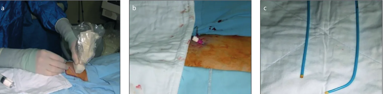
The patients were positioned prone or semi-prone on the operating table and monitored with blood pressure measure-ment and pulse oximetry throughout the procedure. Skin and subcutaneous tissue were infiltrated with 1–2 mL of 1% lido-caine using a 25G needle. After puncture of the ipsilateral popliteal vein under US guidance (Antares, Siemens) with a 21G or 18G needle, a 9 F or 10 F vascular sheath was placed (Fig. 1a, 1b). GSV and SSV were also accessed, when the thrombus extend-ed to this vein. The procextend-edure was carriextend-ed out with local anesthesia in all patients. Propofol (0.5 mg/kg) was administered as intermittent intravenous bolus to achieve procedural sedation, if required. Heparin was administered intravenously as an initial bolus of 5000 U followed by 2500 U every 45 minutes throughout the procedure. PAT was performed cautiously using large bore (7 F to 9 F) guiding catheters (Fig. 1c)
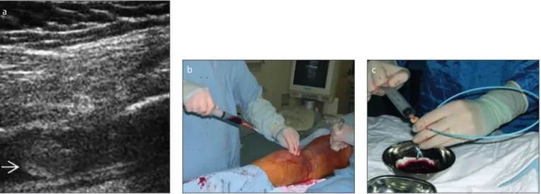
con-nected to a 20 mL syringe under real-time US guidance (Figs. 2 and 3). A straight guid-ing catheter was used to remove thrombus in regular straight veins whereas a guiding catheter with angled tip was preferred in a convoluted vein.
The distance from the puncture site to the estimated proximal common iliac vein was measured and the guiding catheter was intended to aspirate thrombus up to the level of common iliac vein. The guiding catheter was gently advanced without a guidewire. During the advancement of the guiding catheter, negative pressure was ap-plied to aspirate the thrombus. If there was a resistance to the advancement of guiding catheter in a convoluted vein, the catheter was advanced over a hydrophilic guidewire or an angled guiding catheter was rotated for safe advancement to prevent dissec-tion. Aspiration was performed from the caudal to cranial ends of thrombosed veins. The most cranial part was aspirated last to prevent or decrease the risk of PE. The pro-cedure was repeated several times until complete or significant thrombus removal was achieved and uninterrupted venous flow was observed on CDUS. The veins in the thigh could be visualized on CDUS, but it was not possible to visualize the iliac veins from a posterior approach when the patient was lying prone.
The patients were admitted for one day of anticoagulation after the procedure and discharged the next day. Subcutaneous heparin and graduated elastic compression stockings were recommended to all pa-tients until the end of the postpartum pe-riod. Patients were called for follow-up vis-its at three months after delivery. Patency was assessed by physical examination and CDUS. Venography was obtained if there was any clinical doubt regarding the paten-cy of the iliac veins.
Main points
•
Ultrasonography (US)-guided percutaneous aspiration thrombectomy can be used as a rapid and effective method for removal of thrombus in pregnant women with massive leg swelling similar to phlegmasia alba dolens.•
Percutaneous aspiration thrombectomy can be performed safely under US guidance.•
Use of percutaneous aspiration thrombectomymay require experience. Therefore it may not be easily recommended to those who have less experience in using this method.
•
The use of percutaneous aspiration thrombec-tomy under US guidance in pregnant women is justifiable because of the absence of other viable treatment options.Figure 1. a–c. Preprocedure preparation. Panel (a) shows puncture of the popliteal vein under US guidance; (b) shows insertion of a vascular sheath; panel (c) shows straight and angled large bore guiding catheters.
Statistical analysis
Statistical analyses were performed us-ing SPSS v. 17 (SPSS Inc.). Numeric data are expressed as mean±standard deviation or percentage.
Results
Six patients with only femoropopliteal DVT or mild symptoms of DVT were exclud-ed from the study. All nine patients were severe symptomatic cases with massive swelling and pain of the leg. Five patients were in the first trimester of pregnancy, two were in the second trimester, and two in the last trimester. None of the patients had a previous history of DVT or major PE symptoms. The lesion was on the left side in seven patients (77.8%) and on the right side in two patients (22.2%). All patients had anticoagulation therapy with heparin until diagnostic evaluation or potential conse-quent endovascular therapy. DVT was acute (1–14 days) in four patients (44.4%), acute and subacute (15–28 days) in five patients (55.6%). Seven patients had involvement of the iliac, femoral, popliteal, and crural
veins. The popliteal and crural veins were patent in two patients with thrombosis of femoral and iliac veins. Thrombus was also present in the proximal GSV in three pa-tients and in the proximal SSV in a patient because of descending DVT, probably due to May-Thurner syndrome. Initial access to the venous system was through the pop-liteal vein in all patients. Two patients had additional GSV access and one patient had an additional SSV access to remove throm-bus and improve blood flow to the femoral and iliac veins.
Complete or significant thrombus re-moval and uninterrupted venous flow from the puncture site up to the iliac veins were achieved in all patients at first intervention. Complete relief of leg pain was achieved immediately in seven patients (77.8%), but minimal swelling remained around the an-kle. Two patients (22.2%) had a recurrence of thrombosis in the first week postinter-vention. One of the two underwent a sec-ond intervention and PAT was performed again with successful removal of thrombus and establishment of in line flow. The other patient refused a second intervention. None
of the patients required blood transfusion because of blood loss during thrombus ex-traction. The average volume of blood aspi-rated as thrombus ranged 200–400 mL for each patient.
Two patients were lost to follow-up after birth. Two patients were followed with only CDUS and five patients underwent venog-raphy after birth. None of these patients had rethrombosis throughout the postpar-tum period. Symptomatic relief was detect-ed clinically in these seven patients. Venous patency was demonstrated with CDUS and venography (Fig. 4). Venography also re-vealed left common iliac vein compression syndrome (May-Thurner syndrome) in three patients (33.3%).
Discussion
This study showed that PAT can be per-formed safely under US guidance as a thrombus removal method in pregnant women with acute or subacute iliofemoral DVT. We achieved complete or near com-plete thrombus removal and rapid symp-tomatic relief in seven of nine patients
Figure 2. a–c. Aspiration thrombectomy procedure under real-time US guidance. a
b c
Figure 3. a–c. Appearance of thrombus at different stages.
without any complications related to the procedure.
The risk of VTE increases 4- to 5-fold during pregnancy and 20-fold or more in the postpartum period. The main reason for increased risk of VTE is the hypercoagulable state of pregnancy which protects women from bleeding during miscarriage or child-birth. Other underlying factors include ve-nous stasis and vascular damage. DVT and PE are two different manifestations of VTE. PE is the leading cause of maternal deaths (1, 2, 9). Anticoagulation with heparin reduc-es the risk of PE and is currently considered the standard of care for the prevention of PE. However, the management of DVT remains a major clinical problem because this form of therapy does not adequately protect the patient from the development of PTS. Res-olution of the present thrombus formed in the deep veins is slow and frequently in-adequate in this form of therapy (3, 4). In a previous study, 4% of patients treated with heparin had significant or complete lysis, whereas 14% of patients had partial lysis (3). Lysis did not occur in 82% of patients who either failed to improve or worsened. Per-sistence of thrombus in the deep veins leads to venous outflow obstruction and valvular incompetence, which is ultimately the cause of PTS in most patients (4–8). Data from the National Venous Thrombolysis Registry, strongly point to a relationship between the degree of lysis and venous valvular function;
62% of patients with <50% lysis had valvu-lar incompetence, whereas 72% of patients who had complete lysis had normal valve function (15). Patients who progress to PTS could have a poor quality of life and disabili-ty due to clinical features of post-thrombotic sequelae including lifestyle-limiting chronic leg pain and edema, heaviness, and/or fa-tigue, and in severely affected patients ve-nous claudication, varicosities, stasis derma-titis, hyperpigmentation and subsequently venous stasis ulcers (4–8).
A preponderance of the evidence in pa-tients with lower extremity DVT suggests a strategy of early thrombus elimination for fast symptom relief and restoration of venous patency to protect valve function and prevent development of the PTS (4–8, 15–24). Alternative treatment options have been used to remove thrombus from deep veins because of the disappointing results with anticoagulation therapy (16–18). Pub-lished evidence indicates that thrombolytic agents, even when administered systemical-ly, are superior to anticoagulation therapy for achieving early lysis of thrombus. Comerota and Aldridge (3) stated that 45% of patients treated with systemic thrombolysis had sig-nificant or complete lysis compared with 4% of patients treated with heparin. Among those receiving systemic thrombolysis, 18% had partial lysis, while 37% failed to improve or worsened. Despite its potential benefits, thrombolysis is associated with a greater
risk of minor or major bleeding complica-tion than anticoagulacomplica-tion therapy. Cathe-ter-directed thrombolysis (CDT) was recom-mended in selected patients with extensive acute proximal DVT (e.g., iliofemoral DVT, symptoms lasting <14 days, good functional status, life expectancy of >1 year) who have a low risk of bleeding (25). It may be used to reduce acute symptoms and post-throm-botic morbidity if appropriate expertise and resources are available. CDT offers dis-tinct advantages compared with systemic thrombolysis, and anticoagulation alone. Catheter-directed infusion allows higher concentrations of the thrombolytic agent to be delivered directly into the thrombus with a reduced systemic effect (15–19, 24). A recent comparative study (18) provides support for the potential of adjunctive CDT to prevent PTS. In this study, patients with iliofemoral DVT treated with CDT had a sig-nificantly decreased the frequency of PTS and better health-related quality of life com-pared with patients treated with anticoagu-lation alone. A study of systemic thromboly-sis use versus CDT in treating iliofemoral DVT demonstrated that CDT may be superior to systemic thrombolysis in preserving venous valvular function (19). They also found that in CDT group, the needed dose of thrombo-lytic agents was lower and the duration of treatment was shorter. On the other hand, CDT has some potential disadvantages that include the long infusion times required to lyse extensive DVT, lengthy hospital stays, greater bleeding risk and high medication and hospitalization costs (15,16). Recent-ly, percutaneous thrombectomy methods alone or combined with CDT have been widely used as an effective endovascular method for thrombus removal. Compared with CDT, percutaneous thrombectomy methods are able to remove the thrombus more rapidly without the risk of bleeding complication (17, 20–24, 26–29). Vedantham et al. (20) found that the use of adjunctive percutaneous mechanical thrombectomy to augment CDT provides comparable proce-dural success and may reduce thrombolytic dose requirements and infusion times. PAT has been successfully used alone or in com-bination with local thrombolytic infusion to remove thromboembolic material from limb arteries (27, 28). PAT is a cheap and simple method. It proposes a rapid and effective way to remove thrombus. Moreover, it can be used in almost every patient who is unre-sponsive to anticoagulation therapy or has a contraindication to thrombolysis (22, 27–29).
Figure 4. a, b. Venography performed in the postpartum period. Panel (a) shows irregular but patent
femoral veins. The iliac veins completely thrombosed before the procedure seem patent with iliac vein compression (b).
PAT has also been effectively used alone or in combination with thrombolysis to treat lower extremity DVT (22, 29). The efficacy of PAT as the primary method of thrombus removal has been previously shown in 139 patients with acute and subacute iliofemoral DVT (22). In that study, successful recanaliza-tion without the use of CDT was achieved in the majority of patients. CDT was required in only one-fourth of the patients as an ad-junctive treatment following PAT. In other patients who required CDT, the dose of the thrombolytic agent was quite low.
The management of DVT in pregnancy is still challenging for several reasons. DVT is generally treated with intravenous or sub-cutaneous heparin during pregnancy and the postpartum period, but the subsequent risk of post-thrombotic complication devel-opment in this young patient population is a concern. Many interventional radiologic treatment modalities that are extensive-ly used in nonpregnant patients, have not been appropriately validated in pregnancy. Image-guided interventional thrombus re-moval procedures are potentially hazard-ous to the fetus in terms of adverse effects of ionizing radiation and contrast agent. Pregnancy and the peripartum period are generally considered as a contraindication for thrombolytic therapy because of the risk of uncontrolled bleeding complication (9). However, a few case reports have been published describing thrombolytic therapy of DVT during pregnancy and in the peri- or postpartum period (10–13, 30). Henrich et al. (10) performed systemic streptokinase lysis without complications in a pregnant wom-an at 29 weeks of gestation who had acute iliac DVT. They concluded that thrombolytic therapy during pregnancy is possible and may help to prevent serious long-term se-quelae of DVT in these young patients. They also stated that the risk of bleeding must be considered and thrombolytic therapy should only be administered under close observation. In a study by Krishnamurthy et al. (11), a pregnant woman with PE, se-riously ill, and two pregnant women with iliofemoral DVT who failed to respond to treatment with intravenous heparin were treated with catheter-directed urokinase. All three patients had fast symptom relief and successful pregnancy outcomes. They suggested that CDT offered a reasonably safe alternative therapy in these young women and may prevent PTS in the long-term. Patterson et al. (12) treated a patient with iliac vein thrombosis 48 hours after
delivery with catheter-directed urokinase because there was no clinical improvement despite heparin infusion. Acharya et al. (13) used CDT to treat four women with acute postpartum iliofemoral DVT (within 42 days of childbirth) and achieved a successful re-sult and symptom relief in all four patients. Demirturk et al. (30) reported that endovas-cular treatment of postpartum iliofemoral DVT (within 42 days of childbirth) with PAT alone or combined with CDT was rapid, very safe and resulted in a very high rate of thrombus removal in 18 patients. In a pre-vious study (14), a pregnant woman with DVT underwent percutaneous mechanical thrombectomy with the use of venograms because pregnancy was considered a con-traindication to thrombolysis. Thrombus removal using PAT with or without fluoros-copy guidance has not been used to date in pregnant women with iliofemoral DVT. Our institution prefers a strategy of early thrombus elimination as quickly as possi-ble, particularly in young patients, to relieve symptoms and minimize the development of long-term sequelae. For this purpose, we performed PAT under US guidance to treat our pregnant patients with iliofemoral DVT. Full systemic anticoagulation remains an essential element of our treatment to pre-vent PE and recurrent thrombosis.
Our study is limited by its retrospective nature. Technical success and short-term patency were good, but the mid- and long-term results are not known in our patients. Thus, our results must be considered pre-liminary. Use of PAT may require experience. Our center has been using this method for the last 15 years and is quite experienced in using PAT for thrombus removal. Therefore it may not be easily recommended to those who have less experience in using this meth-od. Nonetheless, the use of PAT in pregnant women is justifiable because of the absence of other viable treatment options.
In conclusion, endovascular treatment with US-guided PAT can be considered as a safe and effective way to remove thrombus from the deep veins in pregnant women with acute and subacute iliofemoral deep vein thrombosis.
Conflict of interest disclosure
The authors declared no conflicts of interest.
References
1. Heit JA, Kobbervig CE, James AH, Petterson TM, Bailey KR, Melton LJ. Trends in the incidence of venous thromboembolism during pregnancy or postpartum: a 30-year population-based study. Ann Intern Med 2005; 143:697–706. [CrossRef]
2. James AH, Jamison MG, Brancazio LR, Myers ER. Venous thromboembolism during pregnancy and the postpartum period: incidence, risk fac-tors, and mortality. Am J Obstet Gynecol 2006; 194:1311–1315. [CrossRef]
3. Comerota AJ, Aldridge SC. Thrombolytic ther-apy for deep venous thrombosis: a clinical re-view. Can J Surg 1993; 36:359–364.
4. Comerota AJ, Paolini D. Treatment of acute il-iofemoral deep venous thrombosis: a strategy of thrombus removal. Eur J Vasc Endovasc Surg 2007; 33:351–356. [CrossRef]
5. Akesson H, Brudin L, Dahlström JA, Eklöf B, Oh-lin P, Plate G. Venous function assessed during a 5 year period after acute ilio-femoral venous thrombosis treated with anticoagulation. Eur J Vasc Surg 1990; 4:43–48. [CrossRef]
6. Meissner MH, Manzo RA, Bergelin RO, Markel A, Strandness DE. Deep venous insufficiency: the relationship between lysis and subsequent re-flux. J Vasc Surg 1993; 18:596–608. [CrossRef]
7. Prandoni P, Lensing AW, Cogo A, et al. The long term clinical course of acute deep venous throm-bosis. Ann Intern Med 1996; 125:1–7. [CrossRef]
8. Hull RD, Marder VJ, Mah AF, Biel RK, Brant RF. Quantitative assessment of thrombus burden predicts the outcome of treatment for venous thrombosis: a systematic review. Am J Med 2005; 118:456–464. [CrossRef]
9. Bates SM, Ginsberg JS. How we manage ve-nous thromboembolism during pregnancy. Blood 2002; 100:3470–3478. [CrossRef]
10. Henrich W, Schmider A, Henrich M, Dudenhausen JW. Acute iliac vein thrombosis in pregnancy treat-ed successfully by streptokinase lysis: a case report. J Perinat Med 2001; 29:155–157. [CrossRef]
11. Krishnamurthy P, Martin CB, Kay HH, et al. Cath-eter-directed thrombolysis for thromboembol-ic disease during pregnancy: A viable option. J Matern Fetal Med 1999; 8:24–27. [CrossRef]
12. Patterson DE, Raviola CA, D’Orazio EA, et al. Thrombolytic and endovascular treatment of peripartum iliac vein thrombosis: A case report. J Vasc Surg 1996; 24:1030–1033. [CrossRef]
13. Acharya G, Singh K, Hansen JB, Kumar S, Maltau JM. Catheter-directed thrombolysis for the man-agement of postpartum deep venous thrombo-sis. Acta Obstet Gynecol Scand 2005; 84:155–158.
[CrossRef]
14. Jackson LS, Wang XJ, Dudrick SJ, Gersten GD. Catheter-directed thrombolysis and/or throm-bectomy with selective endovascular stenting as alternatives to systemic anticoagulation for treatment of acute deep vein thrombosis. Am J Surg 2005; 190:864–868. [CrossRef]
15. Mewissen WM, Seabrook GR, Meissner MH, Cynamon J, Labropoulos N, Haughton SH. Catheter-directed thrombolysis for lower ex-tremity deep venous thrombosis: report of a national multicenter registry. Radiology 1999; 211:39–49. [CrossRef]
16. Vedantham S, Millward SF, Cardella JF, et al. So-ciety of interventional radiology position state-ment: treatment of acute iliofemoral deep vein thrombosis with use of adjunctive catheter-di-rected intrathrombus thrombolysis. J Vasc In-terv Radiol 2009; 20:332–335. [CrossRef]
17. Popuri RK, Vedantham S. The role of throm-bolysis in the clinical management of deep vein thrombosis. Arterioscler Thromb Vasc Biol 2011; 31:479–484. [CrossRef]
18. Comerota AJ, Throm RC, Mathias SD, Haughton S, Mewissen M. Catheter-directed thromboly-sis for iliofemoral deep venous thrombothromboly-sis im-proves health-related quality of life. J Vasc Surg 2000; 32:130–137. [CrossRef]
19. Laiho MK, Oinonen A, Sugano N, et al. Preserva-tion of venous valve funcPreserva-tion after catheter-di-rected and systemic thrombolysis for deep venous thrombosis. Eur J Vasc Endovasc Surg 2004; 28:391–396. [CrossRef]
20. Vedantham S, Vesely TM, Parti N, Darcy M, Hovsepian DM, Picus D. Lower extremity ve-nous thrombolysis with adjunctive mechan-ical thrombectomy. J Vasc Interv Radiol 2002; 13:1001–1008. [CrossRef]
21. Kim HS, Patra A, Paxton BE, Khan J, Streiff MB. Catheter-directed thrombolysis with percuta-neous rheolytic thrombectomy versus throm-bolysis alone in upper and lower extremity deep vein thrombosis. Cardiovasc Intervent Radiol 2006; 29:1003–1007. [CrossRef]
22. Oğuzkurt L, Özkan U, Gülcan Ö, Koca N, Gür S. Endovascular treatment of acute and subacute iliofemoral deep venous thrombosis using manual aspiration thrombectomy: long-term result of 139 patients from a single center. Di-agn Interv Radiol 2012; 18:410–416.
23. Vedantham S, Goldhaber SZ, Kahn SR, et al. Rationale and design of the ATTRACT Study: A multicenter randomized trial to evaluate phar-macomechanical catheter-directed throm-bolysis for the prevention of postthrombotic syndrome in patients with proximal deep vein thrombosis. Am Heart J 2013; 165:523–530.
[CrossRef]
24. Meissner MH, Gloviczki P, Comerota AJ, et al. Early thrombus removal strategies for acute deep venous thrombosis: Clinical Practice Guidelines of the Society for Vascular Surgery and the American Venous Forum. J Vasc Surg 2012; 55:1449–1462. [CrossRef]
25. Kearon C, Kahn SR, Agnelli G, Goldhaber S, Raskob GE, Comerota AJ. Antithrombotic therapy for venous thromboembolic disease: American College of Chest Physicians Evi-dence-Based Clinical Practice Guidelines (8th Edition). Chest 2008; 133:454S–545S.
26. Kwon SH, Oh JH, Seo TS, Ahn HJ, Park HC. Per-cutaneous aspiration thrombectomy for the treatment of acute lower extremity deep vein thrombosis: is thrombolysis needed? Clin Radi-ol 2009; 64:484–490. [CrossRef]
27. Starck EE, Mc Dermott JC, Crummy AB, Turnip-seed WD, Acher CW, Burgess JH. Percutaneous aspiration thrombolectomy. Radiology 1985; 156:61–66. [CrossRef]
28. Oguzkurt L, Ozkan U, Gumus B, Coskun I, Koca N, Gulcan O. Percutaneous aspiration throm-bectomy in the treatment of lower extremity thromboembolic occlusions. Diagn Interv Ra-diol 2010; 16:79–83.
29. Rigatelli G, Cardaioli P, Roncon L, Giordan M, Mi-lan T, Zonzin P. Combined percutaneous aspira-tion thrombectomy and rheolytic thrombecto-my in massive subacute vena cava thrombosis with IVC filter occlusion. J Endovasc Ther 2006; 13:373–376. [CrossRef]
30. Demirtürk OS, Oğuzkurt L, Coşkun İ, Gülcan Ö. Endovascular treatment and the long-term re-sults of postpartum deep vein thrombosis in 18 patients. Diagn Interv Radiol 2012; 18:587–593.