ORIGINAL PAPER
Molecular identification of
Theileria and Babesia in sheep
and goats in the Black Sea Region in Turkey
Mehmet Fatih Aydin&Munir Aktas&Nazir Dumanli
Received: 29 March 2013 / Accepted: 1 May 2013 / Published online: 21 May 2013 # Springer-Verlag Berlin Heidelberg 2013
Abstract This study was carried out to investigate presence and distribution of Theileria and Babesia species via micro-scopic examination and reverse line blotting (RLB) tech-niques in sheep and goats in the Black Sea region of Turkey. For this purpose, 1,128 blood samples (869 sheep and 259 goats) were collected by active surveillance from sheep and goats in different provinces of various cities in the region in the years 2010 and 2011. Smears were prepared from the blood samples, stained with Giemsa, and examined under the light microscope for Theileria and Babesia piroplasms. The genomic DNAs were extracted from blood samples. The length of 360–430-bp fragment in the variable V4 region of 18S SSU rRNA gene of Theileria and Babesia species was amplified using the gDNAs. The polymerase chain reaction products were hybridized to the membrane-connected species-specific probes. A total of 38 animals (3.37 %) including 34 sheep (3.91 %) and 4 goats (1.54 %) were found to be positive for Theileria spp. piro-plasms in microscopic examination of smears while Babesia spp. piroplasm could not detected. Infection rates were 34.64 % in sheep, 10.04 % in goats, and totally 28.99 % for Theileria ovis while 0.58 % in sheep and totally 0.44 %
for Babesia ovis. However, Theileria sp. OT3 was detected in 2.65 % of sheep and 2.04 % of all animals; besides Theileria sp., MK had 0.58 % prevalence in sheep and 0.77 % in goats, with a total 0.62 % with RLB. Although T. ovis and Theileria sp. MK were determined in both sheep and goats, B. ovis and Theileria sp. OT3 were observed only in the sheep. These results provide the first detailed molecular data for sheep and goat theileriosis and babesiosis in the region.
Introduction
Tick-borne hemoprotozoan parasites are widespread in trop-ical and subtroptrop-ical climates. Piroplasmosis caused by Theileria and Babesia species leads to clinical and subclin-ical infections in domestic and wild animals with high
mortality and morbidity (Friedhoff 1997; Jongejan and
Uilenberg2004). Theileria lestoguardi, Theileria uilenbergi,
and Theileria luwenshuni are highly pathogenic for small ruminants and cause lenphoproliferative diseases with high mortality and morbidity while Theileria ovis and Theileria
separata have low pathogenicity (Friedhoff1997;
Hoosmand-Rad and Hawa 1973; Luo and Yin 1997; Schnittger et al.
2004; Yin et al.2007,2008). Theileria sp. OT1, Theileria sp. OT3, and Theileria sp. MK are newly described Theileria species; also there is no information about their vector and
pathogenicity (Ahmed et al.2006; Altay et al.2007b; Nagore
et al.2004; Yin et al.2008).
Ovine babesiosis is the most important hemoparasitic tick-borne disease of small ruminants caused by Babesia
ovis, Babesia motasi, B. crassa (Alani and Herbert 1988;
Friedhoff 1997; Levine 1985; Uilengberg et al. 1980;
Hashemi-Fesharki and Uilenburg 1981) Babesia taylori
(Sarwar 1935), Babesia foliata (Ray and Raghavachari
Nucleotide sequence data reported in this paper are available in GenBank, EMBL 21, and DDBJ databases under accession numbers from JQ867384 to JQ867387.
M. F. Aydin (*)
Research Laboratory, Higher Health School, University of Karamanoğlu Mehmetbey, 70100 Karaman, Turkey
e-mail: [email protected] M. Aktas
:
N. DumanliDepartment of Parasitology, Faculty of Veterinary Medicine, University of Firat, 23119 Elazig, Turkey
1941), and recently described Babesia sp. Xinjiang and
Babesia sp. BQ1 (Lintan) (Guan et al.2009). B. ovis and
Babesia sp. Xinjiang are highly pathogenic especially in sheep and cause severe infections which are characterized by fever, anemia, icterus, and hemoglobinuria (Friedhoff 1997; Guan et al.2009).
Diagnosis of theileriosis and babesiosis in acute cases are based on clinical signs and microscopic examination of
Giemsa-stained blood smears (Guo et al.2002; Yin et al.
2003). Animals with theileriosis and babesiosis become porter after infection, and the value of these animals in the population is important in the epidemiology of disease
(Brown 1990). It has been proposed that microscopic
methods are insufficient; also, positive and false-negative results were observed in serological methods for
diagnosis of porter animals (Burridge et al.1974; Gubbels et
al. 1999). For these reasons, it has been needed using molecular diagnostic methods for the epidemiological stud-ies to determine Theileria and Babesia infections (Aktas et al.2005b; Heidarpour Bami et al.2009).
In the recent years, it has began to be used some molec-ular techniques like polymerase chain reaction (PCR)-based reverse line blotting method (RLB) which allows diagnozing several agents simultaneously and discovery of new species and genotypes used to assess parasitic DNA for diagnosis of subclinical Theileria and Babesia infections. Because more than one species could not be detected simul-taneously, new techniques which allow diagnosis multiply agents in blood at the same time as needed. RLB method was developed in 1995 for differentiate four Borrelia
spe-cies in ticks (Rijpkema et al.1995). This method
success-fully used for diagnosis Theileria and Babesia species and is increasing use in the parasitological researches (Schnittger
et al. 2004; Nagore et al. 2004; Altay et al. 2008a, b;
Gubbels et al.1999).
Piroplasmosis has been reported in some parts of Turkey
(Aktas et al.2005b,2007; Altay et al.2007b,2012; Inci et
al.2010), but there is no detailed molecular survey on ovine theileriosis and babesiosis in Black Sea Region of Turkey. In this study, we investigated presence and frequency of Theileria and Babesia species infecting small ruminants in the region using microscopy and RLB.
Materials and methods
Study area and collection of field samples
The Black Sea region covers 18 % of Turkey's land with an
area of 143,537 km2and the third largest region in terms of
area. The region which has the highest rainfall is the first place between the regions with respect to the existence of the forest with the rate of 27 %. The region is dominated by
two different climates. Inland areas of the region have a terrestrial climate, and the stretch of coast has cool summers and mild winters.
This study was planned to represent the whole of Black Sea region. The number of sheep and goats in the provinces was obtained from the database of Turkey Statistical Institute. Sheep and goat populations were seen 1,063,666 and
139,939, respectively, in the region (Anonymous 2011).
Bolu, Kastamonu, Corum, Samsun, Tokat, Giresun, and Bayburt provinces were selected as study areas according to the evaluation criteria of geographical distribution of the provinces, animal population, climatic conditions, and vege-tation. Therefore, at least 121 sheep and 32 goats were
includ-ed from each province in the study (Fig.1and Table1).
Blood samples were collected into tubes containing EDTA from 1128 clinically healthy small ruminants (869 sheep and 259 goat) randomly selected from 43 towns in Bolu (Center, Gerede, Kıbrısçık, Mengen, Seben, and Mudurnu), Kastamonu (Center, Araç, Bozkurt, Çatalzeytin,
Daday, İnebolu, and Taşköprü), Çorum (Center, Alaca,
Dodurga, Kargı, Osmancık, Sungurlu, and Uğurludağ), Samsun (Center, Bafra, Havza, Ladik, Tekkeköy, and Terme), Tokat (Center, Almus, Erbaa, Niksar, Reşadiye, Turhal, and Zile), Gireun (Center, Alucra, Çamoluk,
Dereli, Keşap, Piraziz, and Şebinkarahisar), and Bayburt
(Center, Aydıntepe, and Demirözü) provinces in the Black Sea Region of Turkey in 2010 and 2011, spring and
sum-mer, when ticks were active (Fig.1and Table1).
DNA extraction
Blood samples were defrosted and homogenized at room temperature for 10–15 s. DNA extractions were performed by a commercial DNA isolation kit or the manual method.
In manual method, 125μl of blood was added to 250 μl of
lysis solution (0.32 M sucrose, 0.01 M Tris, 0.005 M
MgCl2, 1 % Triton X-100, pH 7.5). The mixture was
centrifuged at 11,600×g for 1 min. The pellet was washed
three times by centrifugation with 250 μl lysis buffer. The
supernatants were discarded, and the final pellets were
resuspended in 100 μl of PCR buffer (50 mM KCl,
10 mM Tris–HCl (pH 8), 0.1% TritonX-100, pH 8.3).
Proteinase K (50μg/ml) was added to the pellet suspension,
and the mixture was incubated at 56 °C for 1 h. Finally, the samples were heated at 100 °C for 10 min (Aktas et al. 2005a). Genomic DNAs were maintained at −20 °C until use.
Polymerase chain reaction and agarose gel electrophoresis
For the amplification of Theileria and Babesia species, one set of primers was used to amplify an approximately 360– 430 bp fragment of the hypervariable V4 region of the 18S
rRNA gene. The forward [RLB-F2 (5’-GACACAGGGAG GTAGTGACAAG-3’)] and the reverse [RLB-R2 (biotin-5’-CTAAGAATTTCACCTCTGACAGT-3’)] primers were de-scribed by Georges et al. (2001). Touchdown PCR is a modification of conventional PCR. It involves the use of an annealing temperature that is higher than the target opti-mum in early PCR cycles. The annealing temperature was
decreased every second cycle with 2 °C to a“touchdown”
temperature of 57 °C. This allows for the enrichment of the correct product over any non-specific product.
The PCR was performed in a touchdown thermocycler in
a total reaction volume of 25 μl containing PCR buffer
[750 mM Tris–HCl (pH 8.8), 200 mM (NH4)2SO4, 0.1%
Tween 20], 5 mM MgCl2, 125 μM deoxynucleotide
tri-phosphates, 1.25 U Taq DNA polymerase, primers (20 pmol/μl), and template DNA. Five microliters of PCR product were visualized by UV transillumination in a 1.5 % agarose gel after electrophoresis and staining with ethidium bromide.
Reverse line blotting (RLB)
Binding of species-specific oligonucleotides (probes) on the biodyne C membrane
Probes of Theileria/Babesia catchall, Theileria spp., T. ovis, Theileria lestoquardi, T. uilenbergi, T. luwenshuni, Theileria sp. OT1, Theileria sp. OT3, Theileria sp. MK, Babesia spp., B. ovis, B. motasi, and B. crassa were used with a range of 200–400 pmol/150 μl concentration and contain N-terminal N-(trifluoracetamidohexyl-cyanoethyl,N,N-diisopropyl phosphoramidite [TFA])-C6 amino linker in the study
(Gubbels et al. 1999; Nagore et al. 2004; Schnittger et al.
2004; Altay et al.2007b).
Biodyne C membrane was activated in 10 ml of 16 % EDAC 1-ethyl-3-(3-dimethylamino-propyl)carbodiimide for 10 min at room temperature and placed in a miniblotter after washed demineralized water. Residual liquid on the mem-brane was aspirated. One hundred fifty microliters of each probe which diluted to a 50 to 1,200 pmol/150 ml
concen-tration in 500 mM NaHCO3 (pH 8.4) was filled the
miniblotter except for the first and last channel slots. India ink diluted with 2×SSPE and 0.5 % sodium dodecyl sulfate (SDS) with a rate of 1% filled the first and last slots. Then, they were incubated for 10 min at room temperature. Liquids in slots were aspirated after incubation. The mem-brane was inactivated in 100 mM NaOH for 10 min after removing from miniblotter at room temperature. Then it was washed in 2×SSPE/0.1 % SDS for 5 min at 60 °C.
Reverse line blotting
The membrane was washed for 5 min at room temperature with 2× SSPE/0.15 SDS %, and it was placed in the miniblotter with the slots vertical on the previously connected probes. Residual liquid on the membrane was aspirated. Twenty microliters of PCR product was diluted
in 2X SSPE/0.1 % SDS with a total 150 μl volume, and it
was denatured for 10 min at 99 °C. Denaturated PCR products were cooled on ice immediately for uncombined DNA strands again. Denatured PCR samples were filled into the slots and hybridized on a flat surface for 1 h at 42 °C. PCR products on the membrane were aspirated. The mem-brane was washed twice in 2× SSPE / 0.5 % SDS for 10 min at 52 °C. The membrane was incubated in 10 ml of 1:4,000-diluted peroxidase-labeled streptavidin in 2× SSPE/0.5 % SDS for 30 min at 42 °C. Then membrane was washed twice in 2× SSPE/0.5 % SDS for 10 min at 42°C and twice in 2×
T able 1 Distribution of Theileria and Babesia species via RLB and comparison with microscopy and PCR results according to the provinces of the Black Sea Region in sheep and goats Information of samples Microscopy result PCR result RLB result Total infection Single infection Mix infection Province Host Number of samples Theileria spp. Babesia spp. Positive T . ovis Theileria sp. OT3 Theileria sp. MK B. ovis T . ovis Theileria sp. OT3 Theileria sp. MK B. ovis T . ovis + B. ovis T . ovis + Theileria sp. OT3 T . ovis + Theileria sp. MK Bolu Sheep 123 9 – 46 66 6 1 – 63 4 –– – 21 Goat 40 – – – – – – –– – – –– – – ∑ 163 9 – 46 66 6 1 – 63 4 –– – 21 Kastamonu Sheep 125 3 – 22 35 3 – 23 3 2 – 11 1 – Goat 38 –– 11 –– – 1 –– – – – – ∑ 163 3 – 23 36 3 – 23 4 2 – 11 1 – Çorum Sheep 125 10 – 54 101 3 –– 99 1 –– – 2 – Goat 40 – – – – – – –– – – –– – – ∑ 165 10 – 54 101 3 –– 99 1 –– – – – Samsun Sheep 123 7 – 33 51 1 3 1 48 – 2 – 11 1 Goat 35 2 – 36 – 1 – 5 –– – – – 1 ∑ 158 9 – 36 57 1 4 1 53 – 2 – 11 2 Tokat Sheep 123 2 – 20 30 7 1 2 27 6 – 11 1 1 Goat 39 1 – 68 – 1 – 7 –– – – – 1 ∑ 162 3 – 26 38 7 2 2 34 6 – 11 1 2 Giresun Sheep 129 2 – 61 4 –– – 14 –– – – – – Goat 32 1 – 51 0 –– – 10 –– – – – – ∑ 161 3 – 11 24 –– – 24 –– – – – – Bayburt Sheep 121 1 – 44 3 –– 43 –– – – – Goat 35 –– 11 –– – 1 –– – – – – ∑ 156 1 – 55 3 –– 53 –– – – – Total Sheep 869 34 – 185 301 23 5 5 288 16 2 2 3 7 3 Goat 259 4 – 16 26 – 2 – 24 –– – – – 2 ∑ 1,128 38 – 201 327 23 7 5 312 16 2 2 3 7 5
SSPE for 5 min at room temperature. The membrane was incubated in 10 ml of ECL detection fluid for 1 min, and then it was taken on a hard surface and covered with acetate. The membrane was incubated under an ECL hyperfilm for 30 s to 30 min depending on the strength of signals in a dark room. Finally, ECL hyperfilm was processed with developer and fixer solutions. Black spots occurring in rows where PCR products and probes were crossed was evaluated as positive to the related species.
Stripping of PCR-products from membrane
The membrane was washed twice in 1 % SDS for 30 min at 85 °C to strip PCR products from the membrane. Finally, the membrane was rinsed in 20 mM EDTA for 15 min at room temperature and stored in 20 mM EDTA at 4 °C for reuse.
Statistical analysis
A Pearson chi-square (χ2) test was used to evaluate the com-parison of microscopy, PCR, and RLB results, assessment of positive rates in the provinces, and evaluation on the basis of the prevalence of Theileria and Babesia species in the host and cities. Values of P<0.05 were accepted statistically significant. The 15.00 SPSS package program was used for the tests.
Results
Distribution and frequency of Theileria and Babesia species
detected by PCR and RLB are presented in Tables1and2and
results demonstrated in Figs. 2 and 3. Of the 1,128 blood
samples examined, PCR revealed 201 (17.82 %) positive for piroplasms, whereas 362 (32.09 %) of DNA-amplified prod-ucts hybridized with the probes for catchall, genera, and species-specific probes. These results demonstrated that there was a significantly higher rate of detection of Theileria and
Babesia infections (P<0.01) between microscopy PCR and RLB. Single and mixed infections were detected in animals. Dominant species is T. ovis. Theileria sp. OT3 was found as the second dominant species. Theileria sp. MK and B. ovis were also detected in animals.
Microscopic examination results
Theileria spp. piroplasms were detected in 34 sheep, 4 goats (1.54 %), and a total of 38 small ruminants (3.37 %); however, no Babesia spp. piroplasm was detected in blood smears by microscopic examination. Maximum positive rate was deter-mined in Çorum (6.06 %) and the minimum in Bayburt (0.64 %). According to the results of microscopy, the
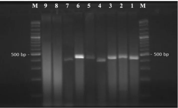
Fig. 2 Ethidium bromide stained agarose gel electrophoresis of am-plification products from Babesia and Theileria species using Babesia-and Theileria-specific primers RLB-F2 Babesia-and RLB-R2. M: marker (100 bp), lane 1 T. ovis, lane 2 Theileria sp. OT3, lane 3 Theileria sp. MK, lane 4 B. ovis, lane 5 T. ovis+B. ovis (mix sample), lane 6 T. ovis positive sample, lane 7 B. ovis positive sample, lane 8 PCR negative control (distilled water), lane 9 extraction negative control (genomic DNA of uninfected sheep)
Fig. 3 Reverse line blot assay of the PCR products generated by amplification of genomic DNA from sheep and goat samples infected with Theileria and Babesia species, and from negative and positive samples as template. Oligonucleotide probes are shown in rows, and samples are applied in columns. Samples bearing identified single and mixed infections and negative and positive controls are showed as follows: lane 1: T. ovis (positive control); 2: T. lestoquardi (positive control); 3: Theileria sp. OT3 (positive control); 4: Theileria sp. MK (positive control); 5: B. ovis (positive control); 6: negative control (genomic DNA of uninfected sheep); 7: negative PCR control (distilled water); 8: T. ovis (field sample); 9: Theileria sp. OT3 (field sample); 10: Theileria sp. MK (field sample); 11: B. ovis (field sample); 12: T. ovis+B. ovis (mix field sample); 13: T. ovis+Theileria sp. OT3 (mix field sample); 14: T. ovis+Theileria sp. MK (mix field sample) Table 2 Provincial distribution of microscopy, PCR, and RLB results
Province Test Microscopy PCR RLB n + % + % + % Bolu 163 9 5.52 46 28.22 73 44.79 Kastamonu 163 3 1.84 23 14.11 41 25.15 Çorum 165 10 6.06 54 32.73 104 63.03 Samsun 158 9 5.70 36 22.78 63 39.87 Tokat 162 3 1.85 26 16.05 49 30.25 Giresun 161 3 1.86 11 6.83 24 14.91 Bayburt 156 1 0.64 5 3.21 8 5.13 Total 1,128 38 3.37 201 17.82 362 32.09
incidence of Theileria spp. in sheep and goats does not have a statistically significant (Pvalue 0.064), whereas there was statistically significant (Pvalue 0.016) between provinces.
PCR results
The results of PCR with RLB-R2-biotin, RLB-F2 revealed that 201 of 1,128 samples were positive for Theileria spp. or Babesia spp. One hundred eighty-five of 869 sheep and 16 of 259 goats were found positive by PCR. The highest rate of PCR positivity was observed in animals from Çorum (32.73 %), and the lowest positivity rate was observed in the animals from Bayburt (3.21 %). Positivity rates in sheep and goats and the differences between provinces were sta-tistically significant.
RLB results
T. ovis, B. ovis, Theileria sp. OT3, and Theileria sp. MK species/genotypes were detected in the region via RLB. Maximum infection rate was detected in Çorum (63.03 %) while minimum rate was in Bayburt (5.13 %).
As a result, molecular prevalence of T. ovis in sheep was 34.64 % (301/869); in goats, 10.04 % (26/259) with a total 28.99 % (327/1,128). Molecular prevalence of B. ovis in sheep was 0.58 % (5/869) and in goats was 0 % (0/259), with a total 0.44 % (5/1128), molecular prevalence of Theileria sp. OT3 in sheep was 2.65 % (23/869) and in goats was 0 % (0/259), with a total 2.04 % (23/1,128). Molecular prevalence of Theileria sp. MK in sheep was 0.58 % (5/869) and in goats was 0.77 %
(2/259) with a total 0.62 % (7/1,128) (Table1). According to
the results of RLB, provincial distribution and frequency of Theileria and Babesia infections in sheep and goats were statistically significant (P<0.01).
Discussion
Diagnosis of piroplasm infections in vertebrate hosts has been mainly carried out by microscopic examination of blood smear. However, piroplasms have similar morpholog-ical features. PCR-based molecular techniques allow detec-tion and discriminadetec-tion of the parasites at low parasitemia rates and mixed infected animals. Additionally, RLB is able to determine more than one parasite in the same samples.
RLB was successfully used for diagnosis of Theileria and Babesia species, with increasing use in the parasitological
researches (Geoges et al.2001; Nagore et al.2004; Altay et
al.2007b,2012; Inci et al.2010).
New Theileria and Babesia species or genotypes were discovered in sheep and goats with molecular techniques in recent years. Theileria sp. OT1 and Theileria sp. OT3 ge-notypes were determined in small ruminants with RLB
technique in Spain (Nagore et al. 2004). Theileria sp. MK
was discovered in sheep and goats in East Anatolia of
Turkey (Altay et al. 2007b). In China, new Babesia
geno-types which are morphologically and genetically different were detected in sheep and goats and named as Babesia sp.
Xinjiang and Babesia sp. Lintan (Liu et al.2007; Guan et al.
2009,2010; Niu et al.2009).
In this study, microscopic prevalence of theileriosis and babesiosis in sheep and goats was determined as 3.37 % and 0 %, respectively. It was shown that microscopic prevalence of these agents was low when compared with other regions
(Inci et al.1998,2002; Sevinc and Dik1996).
Ovine and caprine piroplasmosis has been investigated
by RLB in some parts of Turkey (Altay et al.2007b,2012;
Inci et al.2010). This method was used for the detection of
sheep and goat Theileria and Babesia species in Kayseri province for the first time in Turkey, and B. ovis and T. ovis were detected with a rate of 2.7 % and 34.2 % respectively
(Iça et al.2005).
B. ovis (5.43 %), T. ovis (34.56 %), Theileria sp. MK (1.30 %), and Theileria sp. OT3 (0.43 %) were determined via RLB in sheep and goats in Eastern Anatolia, and Theileria
sp. MK genotype was first defined (Altay et al.2007b). The
results demonstrated that T. ovis (50.55 %) is the most com-mon species, and there is no T. lestoquardi in sheep and goats
in this region (Aktas et al.2005a; Altay et al.2007a).
In the Yeşilhisar province of Kayseri, B. ovis (3.7 %) and T. ovis (37.6 %) were detected with RLB in sheep and goats
(Sarayli et al.2006).
Randomly selected 421 sheep and 152 goats from Kayseri, Sivas, and Yozgat provinces of central Anatolia region were examined with RLB, and B. ovis and T. ovis were determined with a rate of 2.6 % and 33.9 %,
respec-tively (Inci et al.2010). A total of 201 apparently healthy
animals from Artvin, Giresun, Gumushane, and Tokat of East Black Sea Region of Turkey were investigated for the blood protozoans. Theileria piroplasms were identified in nine (4.47 %) samples by microscopic examination, and T. ovis (18.90%), Theileria sp., MK (0.99%), and Theileria sp. OT3 (0.43 %) were detected by RLB.
Theileria sp. MK genotype was determined with a rate of 0.62 % similarly, and Theileria sp. OT3 (2.04 %) was detected higher than to East Anatolia. T. ovis was found the most prevalent species (28.99 %) in Black Sea Region, like the other regions (37.6–33.3 %) of Turkey. In addition, B. ovis was found with very low levels when compared with the other regions.
In this study, B. ovis, T. ovis, Theileria sp. MK, and Theileria sp. OT3 were detected in small ruminants in the region. The other Theileria (T. lestoquardi, Theileria sp. OT1, T. luwenshuni, and T. uilenbergi) B. motasi, and B. crassa were not detected. The most prevalent parasite was T. ovis with 28.99 %. These results organized the preliminary detailed molecular information for sheep and goat theileriosis
and babesiosis in the region and agree with similar the
previ-ous studies (Altay et al.2007a,b,2012; Inci et al.2010).
Acknowledgments We thank Yasin Baykalır, Sezayi Özübek, Çağrı Özçetin, and all veterinarians and technicians also animal breeders in the region for their kind help during sample collection. This study was financially supported by the Scientific and Technical Research Council of Turkey (TUBITAK) with a grant (TOVAG 109 O 766).
References
Ahmed JS, Luo J, Schnittger L, Seitzer U, Jongejan F, Yin H (2006) Phylogenetic position of small ruminant infecting piroplasms. Ann NY Acad Sci 1081:498–504
Aktas M, Altay K, Dumanli N (2005a) Survey of Theileria parasites of sheep in eastern Turkey using polymerase chain reaction. Small Rum Res 60:289–293
Aktas M, Altay K, Dumanli N (2005b) Development of a polymerase chain reaction method for diagnosis of Babesia ovis infection in sheep and goats. Vet Parasitol 133:277–281
Aktas M, Altay K, Dumanli N (2007) Determination of prevalence and risk factors for infection with Babesia ovis in small ruminants from Turkey by polymerase chain reaction. Parasitol Res 100(4):797–802
Alani AJ, Herbert IV (1988) The morphometrics of Babesia motasi (Wales) and its transmission by Haemaphysalis punctata (Canestrni and Fanzago, 1877) to sheep. Vet Parasitol 30:87–95 Altay K, Aktas M, Dumanli N (2007a) Theileria infections in small
ruminants in the east and southeast Anatolia. Turkiye Parazitol Derg 31(4):268–271
Altay K, Dumanli N, Aktas M (2007b) Molecular identification, ge-netic diversity and distribution of Theileria and Babesia species infecting small ruminants. Vet Parasitol 147:161–165
Altay K, Aktas M, Dumanli N, Aydin MF (2008a) Evaluation of a PCR and comparison with RLB for detection and differentiation of Theileria sp. MK and other Theileria and Babesia species of small ruminants. Parasitol Res 103(2):319–323
Altay K, Aydin MF, Dumanli N, Aktas M (2008b) Molecular detection of Theileria and Babesia infections in cattle. Vet Parasitol 158:295–301
Altay K, Dumanli N, Aktas M (2012) A study on ovine tick-borne hemoprotozoan parasites (Theileria and Babesia) in the East Black Sea Region of Turkey. Parasitol Res 111(1):149–153 Anonymous (2011) Livestock statistics data base. Turkey Statistical
Insti tut e Web. ht tp://www.tuik.gov.t r/h a yva nc il i ka p p/ hayvancilik.zul2008. Accessed 10 December 2011
Brown CG (1990) Control of tropical theileriosis (Theileria annulata infection) of cattle. Parassitologia 32(1):23–31
Burridge MJ, Brown CG, Kimber CD (1974) Theileria annulata: cross-reactions between a cell culture schizont antigen and anti-gens of East African species in the indirect fluorescent antibody test. Exp Parasitol 35(3):374–380
Friedhoff KT (1997) Tick-borne diseases of sheep and goats caused by Babesia, Theileria or Anaplasma spp. Parassitologia 39:99– 109
Georges K, Loria GR, Riili S, Greco A, Caracappa S, Jongejan F, Sparagano O (2001) Detection of haemoparasites in cattle by reverse line blot hybridisation with a note on the distribution of ticks in Sicily. Vet Parasitol 99(4):273–286
Guan GQ, Ma ML, Moreau E, Liu JL, Lu BY, Bai Q (2009) A new ovine Babesia species transmitted by Hyalomma anatolicum anatolicum. Exp Parasitol 122:261–267
Guan G, Moreau E, Liu J, Hao X, Ma M, Luo J, Chauvin A, Yin H (2010) Babesia sp. BQ1 (Lintan): molecular evidence of experimental transmission to sheep by Haemaphysalis qinghaiensis and Haemaphysalis longicornis. Parasitol Int 59(2):265–267
Gubbels JM, de Vos AP, van der Weide M, Viseras J, Schouls LM, de Vries E, Jongejan F (1999) Simultaneous detection of bovine Theileria and Babesia species by reverse line blot hybridization. J Clin Microbiol 37:1782–1789
Guo S, Yuan Z, Wu G, Wang W, Ma D, Du H (2002) Epidemiology of ovine theileriosis in Ganan region, Gansu Province, China. Parasitol Res 88:36–37
Hashemi-Fesharki R, Uilenburg G (1981) Babesia crassa n. sp. (Sporozoa, Babesiidae) of domestic sheep in Iran. Vet Quart 2:3–14
Heidarpour Bami M, Haddadzadeh HR, Kazemi B, Khazraiinia P, Bandehpour M, Aktas M (2009) Molecular identification of ovine Theileria species by a new PCR-RFLP method. Vet Parasitol 161(3–4):171–177
Hoosmand- Rad P, Hawa NY (1973) Malignant theileriosis of sheep and goats. Trop Anim Health Pro 5:97–102
Iça A, Yıldırım A, Inci A (2005) Investigation of blood protozoa in sheep by reverse line blotting method in Kayseri province. XIV National Congress of Parasitology, Dokuz Eylul University,İzmir p: 161
İnci A, Yukarı BA, Sayın F (1998) Study on babesiosis and theileriosis agents detected in some sheep and goat flocks using microscopic examination in Çankırı region. Ankara Üniv Vet Fak Derg 45:105–113
İnci A, Karaer Z, İca A (2002) Babesiosis in sheep and goats around Kayseri. FÜ Sag Bil Derg 16(1):79–83
İnci A, İca A, Yıldırım A, Düzlü Ö (2010) Identification of Babesia and Theileria species in small ruminants in Central Anatolia (Turkey) via reverse line blotting. Turk J Vet Anim Sci 34(2):205–210 Jongejan F, Uilenberg G (2004) The global importance of ticks.
Parasi-tology 129(Suppl):3–14
Levine ND (1985) Veterinary protozoology. Iowa State University Press, Ames, Iowa
Liu AH, Yin H, Guan GQ, Schnittger L, Liu ZJ, Ma ML, Dang ZS, Liu JL, Ren QY, Bai Q, Ahmed JS, Luo JX (2007) At least two genetically distinct large Babesia species infective to sheep and goats in China. Vet Parasitol 147:246–251
Luo J, Yin H (1997) Theileriosis of sheep and goats in China. Trop Anim Health Prod 29(4):8–10
Nagore D, García-Sanmartín J, García-Pérez AL, Juste RA, Hurtado A (2004) Identification, genetic diversity and prevalence of Theileria and Babesia species in a sheep population from North-ern Spain. Int J Parasitol 34:1059–1067
Niu Q, Luo J, Guan G, Ma M, Liu Z, Liu A, Dang Z, Gao J, Ren Q, Li Y, Liu J, Yin H (2009) Detection and differentiation of ovine Theileria and Babesia by reverse line blotting in China. Parasitol Res 104(6):1417–1423
Ray HN, Raghavachari K (1941) Observations on Babesia foliata n. sp. from a sheep. Indian J Vet Sci 11:239–242
Rijpkema SG, Molkenboer MJ, Schouls LM, Jongejan F, Schellekens JF (1995) Simultaneous detection and genotyping of three geno-mic groups of Borrelia burgdorferi sensu lato in Dutch Ixodes ricinus ticks by characterization of the amplified intergenic spacer region between 5S and 23S rRNA genes. J Clin Microbiol 33:3091–3095
Sarayli H, Inci A, Ica A, Yildirim A, Duzlu O (2006) Investigation of the Babesia agents in sheep and goats by the reverse line blotting hybridization method around Yeşilhisar. Erciyes Univ Sag Bil Derg 15(3):181–188
Sarwar SM (1935) A hitherto undescribed piroplasm of goats (Piroplas-ma taylori). Indian Jour Vet Sci and Anim Husb 5:171–176
Schnittger L, Yin H, Qi B, Gubbels MJ, Beyer D, Niemann S, Jongejan F, Ahmed JS (2004) Simultaneously detection and differentiation of Theileria and Babesia parasite infecting small ruminants by reverse line blotting. Parasitol Res 92:189–196
Sevinc F, Dik B (1996) The diagnosis of Babesia ovis in sheep in Konya around by ELISA. Veteriner Bilim Derg 12(2):73–79 Uilengberg G, Rombach MC, Perie NM, Zwart D (1980) Blood
para-sites of sheep in The Netherlands. II. Babesia motasi (Sporozoa, Babesiidae). Vet Quart 2:3–14
Yin H, Liu G, Luo J, Guan G, Ma M, Ahmed J, Bai Q (2003) Observation on the schizont stage of an unidentified Theileria sp. in experimentally infected sheep. Parasitol Res 91(1):34–39 Yin H, Schnittger L, Luo J, Seitzer U, Ahmed JS (2007) Ovine theileriosis
in China: a new look at an old story. Parasitol Res 101(2):191–195 Yin H, Liu Z, Guan G, Liu A, Ma M, Ren Q, Luo J (2008) Detection and differentiation of Theileria luwenshuni and T. uilenbergi infection in small ruminants by PCR. Transbound Emerg Dis 55(5–6):233–237