RESEARCH ARTICLE
1
THE EFFECT OF DNASE I, RNASE A, AND PROTEINASE K ON FACULTATIVE THERMOPHILE BREVIBACILLUS AGRI D505B BIOFILMS
Tugba KILIC1*, Arzu COLERI CIHAN2
*1Gazi University, Vocational School of Health Services, Medical Laboratory Techniques Program, 06830, Ankara, [email protected], ORCID: 0000-0002-5474-0288
2Ankara University, Faculty of Science, Department of Biology, 06100, Ankara, [email protected], ORCID: 0000-0002-7289-6251
Received Date:29.01.2020 Accepted Date: 04.12.2020
ABSTRACT
Thermophilic bacteria have been isolated from man-made thermal habitats and natural thermal habitats. Brevibacillus agri D505b was isolated from the geothermal region in Turkey. Thermophilic bacilli can form biofilm in areas such as dairy manufacturing plants, water systems, paper-machine, and can create serious problem due to their spore-forming. Therefore, determining the biofilm-forming properties of these bacteria is very significant for the areas. The aim of this study was to determine the effect of environmental conditions on planktonic growth and biofilm formation, the concentrations of protein, carbohydrate, and extracellular DNA (eDNA) from extracellular polymeric substances (EPSs), and the effects of DNase I, RNase A and proteinase K on eDNA in the biofilm matrix of the isolate. As a result, optimal values of the isolate for planktonic growth and biofilm formation were determined as pH 7.0, 1% NaCl, 50oC, and pH 9.0, 0% NaCl, 45oC, respectively. Genomic DNA (gDNA) and eDNA were isolated, then were treated with DNase I, RNase A and proteinase K. The gDNA was only all degraded by DNase I. However, eDNA was not affected by DNase I, RNase A and proteinase K. Moreover, eDNA was determined to be resistant to all the enzymes tested in this study. The eDNA might be protected by EPS components and/or extracellular membrane vesicles (EVs) structures. In addition, the molecular weights of the gDNA and eDNA were calculated larger than 20 kb. Thus, the presence of eDNA in the biofilm matrix of B. agri was confirmed with agarose gel imaging and spectrophotometric analysis.
Keywords: Brevibacillus agri D505b, Biofilm formation, Extracellular DNA.
1. INTRODUCTION
The Gram-positive spore-forming genus Bacillus occurs in a wide range of environments, from soil to food and dairy processing surfaces [1]. Bacillus spp. spores are often present in raw milk, playing a significant role in the bacterial impairment of milk and milk products. Several spore-forming species have been isolated on dairy farms so far [2]. Zhao et al. [3] identified thermophilic spore-forming bacteria such as Brevibacillus spp., Anoxybacillus spp., and Geobacillus spp. in the dairy industry. In another study, Lücking et al. [4] identified spore-forming bacteria in dairy products, including
Kılıç, T., and Coleri Cihan, A., Journal of Scientific Reports-A, Number 45, 1-11, December 2020.
2
Brevibacillus agri, Bacillus pumilus, and Anoxybacillus flavithermus. Moreover, Brevibacillus sp. was isolated from soil by Vivas et al. [5].
The contamination of industrial plants and products with spore-forming bacteria is a common problem [6]. Spore-forming bacteria have been shown to sporulate within single- and multiple-species biofilms and to release these immensely stress-resistant spores, thus increasing the risk of cross-contamination of food [7]. Paperboard and food-packaging paper primarily contain spore-forming bacteria belonging to the genera Bacillus, Paenibacillus, and Brevibacillus as contaminants [8]. In addition, Pereira and Sant’Ana [9] investigated spore-forming bacteria in raw materials such as sugar, cocoa, starch, and milk powder. These raw materials are contaminated by spore-forming bacteria and are significant for the quality of final products. Furthermore, spores of Bacillus species prevalently contaminate the food. Dried foods such as cereal and milk powders are frequently contaminated with spores and when water is present, these spores can germinate, leading to spoilage or food poisoning [10]. Moreover, aerobic spore-forming bacteria have serious impacts on food quality and safety, so the bacteria are a potential cause of disease [11]. B. agri has been associated with human infections [12]. Ogarkov et al. [13] showed that in the final late stages of chronic tuberculosis, samples obtained from patients contained strains of various species of Bacillus spp. and Brevibacillus spp. In addition, B. agri was isolated in association with an outbreak of waterborne illness. In a study, Logan et al. [14] identified B. agri from clinical, dairy, and industrial specimens (gelatin processing plant, antibiotic fermenter, sterilized milk), and 3 strains were associated with the outbreak of waterborne illness.
In our previous studies, we carried out the experiments including biofilm formation on six abiotic surfaces stainless steel, glass, polyvinyl chloride, polypropylene, polystyrene, and polycarbonate) used in industry and biofilm control with sanitation agents of B. agri D505b. We determined that the isolate was a strong biofilm producer [15] and [16]. The objective of this work was to determine the effect of environmental conditions on planktonic growth and biofilm formation, the concentrations of protein, carbohydrate, and eDNA from EPSs and the effects of DNase I, RNase A and proteinase K enzymes on B. agri D505b biofilms. eDNA is a major structural component of many bacteria. Enzymatic deterioration of eDNA can weaken the biofilm structure and release microbial cells from the surface. DNases could evidence a potent strategy for biofilm control [17].
2. MATERIAL AND METHODS 2.1. Culture Conditions of the Isolate
The endospore-forming, aerobic and facultative thermophilic B. agri D505b was isolated from sediment sample in the hot spring (Dikili, Izmir, Turkey). We used the isolate, which was previously isolated in our lab, to study its biofilm properties. The 16S rRNA gene of the isolate was registered with GenBank Accession Number FJ430048 [18]. The isolate was first cultured in Tryptic Soy Agar (TSA, Merck, Germany) at 55oC for 18 h and was subsequently incubated in Tryptic Soy Broth (TSB, Merck, Germany) for 18 h at 55oC in a shaking incubator (170 rpm). The culture was again incubated in TSB at 55oC for 6 h under shaking. All biofilm assays were carried out with a culture that was 6 h old in the mid-exponential growth phase.
2.2. The Effects of Environmental Conditions on Planktonic Growth and Biofilm Formation The effects of pH (4.0, 5.0, 6.0, 6.5, 7.0, 7.5, 8.0, 8.5, 9.0), salinity (0.0, 1.0, 1.5, 2.0, 2.5, 3.0, 3.5, 4.0%), and temperature (37, 40, 45, 50oC) on planktonic growth and biofilm formation were determined in 96-well polystyrene microtiter plates (LP, Italy) spectrophotometrically (OD 595 nm) for 0, 6, 18, 24,
Kılıç, T., and Coleri Cihan, A., Journal of Scientific Reports-A, Number 45, 1-11, December 2020.
3
and 48 h in TSB. In addition, the crystal violet (CV) staining method was applied for optimal biofilm growth assay at the end of 48 h of incubation. For the pH adjust, NaOH and HCl solutions were used. In this study, we first-time determined optimal biofilm values, and the values were used in all experiments. 2.3. Quantification of Biofilm Formation with Crystal Violet in Microtiter Plates
This assay was carried out using the CV staining assay described by Woodward et al. [19] and Stepanović et al. [20] with a few changes. To the wells of 96-well polystyrene microtiter plates were added 90 µL of TSB without NaCl and 10 µL of bacterium culture. The plates were washed two times with physiological saline at the end of 48 h of incubation; thus, planktonic cells were removed. The remaining adhered cells were fixed with 95% methanol (Merck, Germany) and were incubated at 22oC temperature for 15 min. Then the wells were stained with 1% crystal violet dye (Merck, Germany) at 22oC for 30 min. Then, the plates were washed under running tap water to remove residues of stain and were air-dried. Afterward, the dye bound to the biofilm cells was dissolved with ethanol:acetone (Merck, Germany), and the amount of biofilm was measured at an optical density (OD) of 595 nm using a microplate reader (BioTek Elisa reader, µQuant, Biotek Inc., USA). The negative controls contained only TSB.
2.4. The Determination of Concentrations of Protein, Carbohydrate, and eDNA
The D505b isolate was grown at its optimal conditions on TSA plates for 18 h and 0.1 g of bacterium biomass was collected with sterile plastic loops. The biomass was entirely disintegrated by vortexing at maximum speed for 2 min in 2 mL of physiological saline, containing glass bead (diameter 3 mm). After vortexing, the suspension was centrifuged at 32.000 x g for 7 min. The pellet was used for gDNA isolation. The supernatant was primarily filtered through a 0.22 µm membrane filter (Sartorius, France) and used for protein, carbohydrate, and eDNA assays. Polysaccharide concentration was quantified by the phenol-sulfuric acid method [21]. Glucose was used as a standard for the determination of the calibration curve. The Lowry method was applied for the determination of the protein concentration. Bovine serum albumin (BSA) was used as a standard [22]. eDNA isolation was conducted partially by the method described by Wilson [23]. The filtered supernatant was treated with chloroform and isoamyl alcohol (volume 24:1) for eDNA isolation. Afterward, the solution was centrifuged and treated with phenol-chloroform-isoamyl alcohol (volume 25:24:1). The solution was again centrifuged. Isopropanol was added to the supernatant and was waited for 20 min at -20oC. The solution was centrifuged and was added cold ethanol (70%) on precipitate. The solution was centrifuged, and the supernatant was poured. Tris-EDTA buffer was added on eDNA and was solved. The gDNA extraction was performed using a genomic DNA purification kit (Fermentas K0512, Thermo Fisher Scientific Inc., USA). Finally, gDNA and eDNA qualities were measured with the absorbance values at 260 nm/280 nm with a NanoDrop Spectrophotometer (Thermo Scientific NanoDrop Lite, USA). The DNA samples were subjected to 1.5% agarose gel electrophoresis at 120 V for 45 min. Then, the products were visualized with a Quantum ST4 Gel Documentation System (Vilber Lourmat, France). The molecular weights of DNA samples were determined via the Quantum-Capp software system (Vilber Lourmat).
2.5. The Treatment of gDNA and eDNA with DNase I, RNase A, and Proteinase K Enzymes In this assay, gDNA and eDNA were first isolated according to section 2.4. Afterward, 10 µL of gDNA (295.5 ng/µL) or eDNA (604.6 ng/µL) samples were treated with DNase I (1.45, 1.7, 2.5, and 3.0 mg/mL) (Sigma-Aldrich, DN25, USA), RNase A (0.90 mg/mL) (Sigma-Aldrich, R6513, USA), and proteinase K (0.85 mg/mL) (Sigma-Aldrich, P2308, USA) enzymes at 37˚C for 1 h in 96-well polystyrene microtiter plates. Finally, agarose gel electrophoresis (1.5%) was applied at 120 V for 45 min. The products were visualized with the Vilber Lourmat Quantum ST4 Gel Documentation System.
Kılıç, T., and Coleri Cihan, A., Journal of Scientific Reports-A, Number 45, 1-11, December 2020.
4
The molecular weights of DNA samples were determined with the Quantum-Capp software system. As negative controls were used samples without enzyme treatment.
2.6. The Effect of DNase I on Mature Biofilm
This assay was conducted as defined previously by Grande et al. [24] with a few modifications. The purpose of the assay was to determine whether the eDNA was sensitive or resistant to DNase I. First, 95 µL of TSB and 5 µL of bacterium culture were added to a 96-well polystyrene microtiter plates and were incubated for 40 h (mature biofilm) at 45°C. The biofilms were then treated with 100 µL of DNase I (100 µg/mL) (Sigma-Aldrich, DN25) for 2, 4, 8, and 12 h at 37o
C. The plate wells were washed with physiological saline. Subsequently, the CV staining assay was applied to the plates. The biofilm samples were treated with physiological saline for positive controls.
2.7. Statistical Data Analyses
All the experiments were conducted in three replicates on three independent days. All statistical analyses were performed using SPSS 17.0 statistical program (SPSS Inc., Chicago, IL, USA). The one-way analysis of variance (ANOVA) was applied to determine whether there are any statistically significant differences between the means of independent groups. Probability levels of p < 0.05 were considered statistically significant.
3. RESULTS
3.1. Optimization Assays
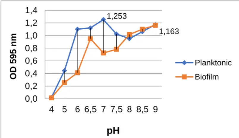
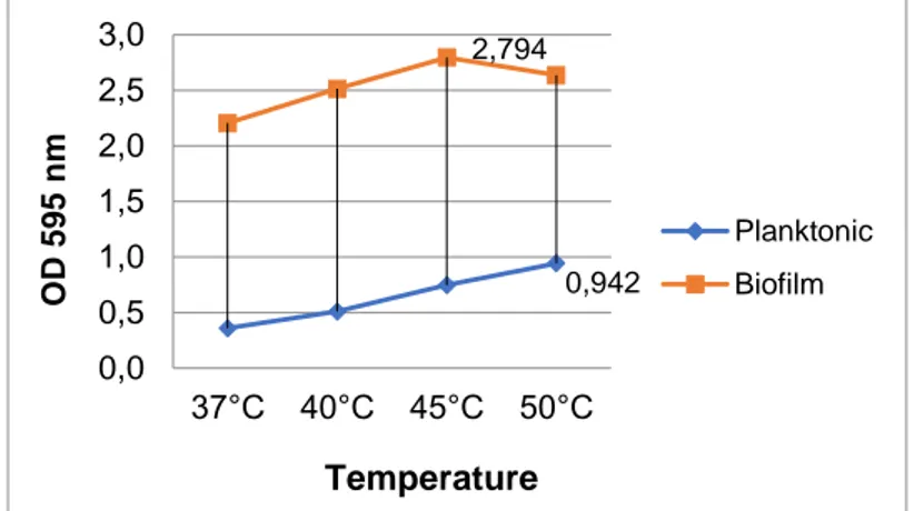
The optimal values of the isolate for planktonic growth were determined as pH 7.0 (OD595nm 1.253 ± 0.003), 1% NaCl (OD595nm 1.174 ± 0.04), and 50oC (OD595nm 0.942 ± 0.17), whereas optimal values for biofilm formation were measured as pH 9.0 (OD595nm 1.163 ± 0.3), 0% NaCl (OD595nm 2.538 ± 0.2), and 45oC (OD595nm 2.794 ± 0.3) in 96-well microtiter plates (Figs. 1, 2, and 3). All parameters were found different for the optimal planktonic growth and the biofilm formation of the isolate. The pH value for biofilm formation was much higher than that for optimal planktonic growth. In addition, the isolate preferred an alkaline and salt-free environment for optimal biofilm production. The biofilm formation decreased with the increased salt amount.
Figure 1. The effects of pH on the planktonic growth and biofilm formation of the isolate.
1,253 1,163 0,0 0,2 0,4 0,6 0,8 1,0 1,2 1,4 4 5 6 6,5 7 7,5 8 8,5 9 O D 59 5 n m
pH
Planktonic BiofilmKılıç, T., and Coleri Cihan, A., Journal of Scientific Reports-A, Number 45, 1-11, December 2020.
5
Figure 2. The effects of salinity (NaCl) on the planktonic growth and biofilm formation of the isolate.
Figure 3. The effects of temperature on the planktonic growth and biofilm formation of the isolate. 3.2. Protein, Carbohydrate, and eDNA Analysis
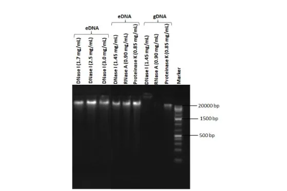
The concentrations of total protein and carbohydrate were quantified as 600 µg/mL and 18.4 µg/mL, respectively. The amount of eDNA was measured as 604.6 ng/µL, whereas the gDNA was quantified as 295.5 ng/µL. Furthermore, the molecular weights of the gDNA and eDNA were calculated larger than
20 kb (Fig. 4). Thus, the presence of eDNA in the biofilm matrix of B. agri was confirmed for the first
time with agarose gel imaging and spectrophotometric analysis.
3.3. The Treatment with DNase I, RNase A, and Proteinase K of gDNA and eDNA
The gDNA was only all degraded by DNase I. On the other hand, eDNA was not affected by any of the enzymes. To confirm the resistance, eDNA was treated with higher concentrations of DNase I (1.7, 2.5, and 3.0 mg/mL). But the results did not change (Fig. 4). Our results showed that the strong biofilm producer B. agri D505b was very resistant to DNase I, RNase A, and proteinase K.
0,942 2,794 0,0 0,5 1,0 1,5 2,0 2,5 3,0 37°C 40°C 45°C 50°C O D 59 5 n m Temperature Planktonic Biofilm 1,174 2,538 0,0 0,5 1,0 1,5 2,0 2,5 3,0 0 1 1,5 2 2,5 3 3,5 4 O D 59 5 n m Salinity Planktonic Biofilm
Kılıç, T., and Coleri Cihan, A., Journal of Scientific Reports-A, Number 45, 1-11, December 2020.
6
Figure 4. Agarose gel electrophoresis image of the eDNA and gDNA of D505b; different DNase I concentrations applied to eDNA (1.7, 2.5, and 3.0 mg/mL); DNase I (1.45 mg/mL), RNase A (0.90 mg/mL), and proteinase K (0.85 mg/mL) treatment of the eDNA and gDNA. Marker (Fermentas Gene
Ruler 1 kb Plus DNA Ladder, 75-20000 bp). 3.4. The Effects of DNase I on Mature Biofilm
DNase I enzyme (100 µg/ mL) was applied to the mature biofilm (40 h) for 2, 4, 8, and 12 h in 96-well polystyrene microtiter plates. DNase I had no significant effect on the biofilm biomass of B. agri D505b. According to the results, the residual biofilm amounts measured after the DNase I treatment were as 16.56% for 2 h and 20.04% for 12 h.
4. DISCUSSION
The optimal values the isolate for planktonic growth were determined as pH 7.0, 1% NaCl, and 50oC, whereas optimal biofilm formation was measured as pH 9.0, 0% NaCl, and 45oC in 96-well polystyrene microtiter plates (Figs 1, 2, and 3). It was seen that all parameters were different for the planktonic growth and biofilm formation of the isolate. The pH value for biofilm formation was much higher than that for optimal growth. Furthermore, the isolate preferred an alkaline and salt-free environment for optimal biofilm production. The biofilm formation decreased as the salt amount increased. High salt concentration in the growth medium resulted in lower environmental water activity (aw), which is destructive to cellular activities.Moreover,osmotic drift influences the response of the tested Bacillus species’ biofilm growth on polystyrene [25].
Extracellular DNA (eDNA) of microbial origin is common in natural terrestrial and aquatic environments [26]. Catlin and Cunningham [27] obtained eDNA from Staphylococcus, Pseudomonas and Alcaligenes faecalis. The presence of eDNA was indicated for the facultative thermophile Brevibacillus agri for the first time in our study. Several studies demonstrated that the eDNA of biofilm
Kılıç, T., and Coleri Cihan, A., Journal of Scientific Reports-A, Number 45, 1-11, December 2020.
7
cells could be sensitive to the DNase I of eDNA. Nijland et al. [28] demonstrated that DNase I (above 15 ng/mL) caused the dispersal of biofilm of Bacillus licheniformis. We determined that the DNase I activity had limited effect at a concentration of 100 µg/mL within 12 h on the mature biofilm of the isolate (20.04%). On the other hand, our results showed that DNase I, RNase A and proteinase K enzymes had no effect on eDNA in the biofilm matrix of B. agri D505b (Fig. 4). Apparently, the eDNA of B. agri D505b could be protected from these enzymes. This could be explained by the rigid pellicle structure and strong biofilm structure of the isolate. We detected in our previous study that B. agri D505b was a strong biofilm producer (OD595 nm: 3.365) [16]. Moreover, the resistance to DNase I may possibly result from the conserved folded structure of eDNA after purification. The purified eDNA which has lost its carbohydrate and protein interactions might become rearranged after chemical treatments, and gain a more compact and condense structure with additional folding [29]. Similarly, DNase I had no effect on Helicobacter pylori eDNA. The eDNA might be protected by other EPS and/or EV-like structures [30]. The eDNA-containing EVs plays a part in biofilm production, bacterial colonization, and subsequent resistance to removal techniques [31]. EVs in biofilms interact with eDNA to strengthen structural integrity [32]. Rivera et al. [33] reported the isolation of EVs from Gram-positive, spore-forming Bacillus anthracis. The first hint of the presence of EVs in Firmicutes came from the studies on Bacillus cereus and Bacillus subtilis [32]. Soler et al. [34] observed that vesicles and eDNA were produced by hyperthermophilic archaea. In addition, DNA in vesicle preparation was found immensely resistant to DNase treatment. Their observations suggest that eDNA could be less or more strongly associated with the vesicles. Vesicles could be a significant factor determining the DNA stability and protecting it from degradation in natural environments. It is unclear how stable eDNA could exist in hydrothermal environments. However, their results demonstrated that eDNA could be preserved in high-temperature environments via its association with vesicles produced by hyperthermophilic archaea. Blesa and Berenguer [35] determined the long-term protection of thermophilic Thermus thermophilus eDNA from DNase associated with EVs generated by cell lysis. Vesicle-protected eDNA can withstand nuclease activity, which allows the movement of eDNA over long distances. EVs are frequently generated by bacteria during the growth phase or upon integration within a biofilm [36].
The characterization of the eDNA of bacterial biofilms revealed that it consists of fragments of high molecular weight (about 30 kb) [37]. We detected that the molecular weight of eDNA was larger than
20 kb with agarose gel electrophoresis (Fig. 4). On the other hand, Ali Mohammed et al. [38] reported
that the size of the eDNA of Fusobacterium nucleatum and Porphyromonas gingivalis was detected to be about 100 bp. The size is generally larger than the one defined for other biofilms. Moreover, the size of eDNA has been reported to range from less than 100 bp to 10 kb [38]. In conclusion, this paper determined that changing environmental conditions could increase the biofilm formation abilities of the isolate. In addition, the presence of eDNA in the biofilm matrix of B. agri was confirmed with electrophoresis and spectrophotometric methods. It was seen in this study that DNase I, RNase A and proteinase K enzymes had no effect on the B. agri D505b biofilms. Therefore, our data suggest that the effect of different enzyme combinations and concentrations should be tested for the biofilm control of B. agri in future studies. The synergistic effect of enzymatic sanitation agents can be determined for biofilm removal. In addition, revealing the association between eDNA and EVs for Brevibacillus agri is significant. Any enzyme which can degrade eDNA holds a potential to be used along with antibiotics as a co-treatment agent.
Kılıç, T., and Coleri Cihan, A., Journal of Scientific Reports-A, Number 45, 1-11, December 2020.
8 ACKNOWLEDGMENT
This research was supported by the Scientific Research Project Office of Ankara University (project number 14B0430003).
REFERENCES
[1] Vilain, S. and Brözel, V. S., (2006), Multivariate approach to comparing whole-cell proteomes of Bacillus cereus indicates a biofilm-specific proteome, Journal of Proteome Research, 5(8), 1924-1930.
[2] Scheldeman, P., Pil, A., Herman, L., De Vos, P. and Heyndrickx, M., (2005), Incidence and diversity of potentially highly heat-resistant spores isolated at dairy farms, Applied and Environmental Microbiology, 71, 1480-1494.
[3] Zhao, Y., Caspers, M. P., Metselaar, K.I., De Boer, P., Roeselers, G., Moezelaar, R., Groot, M.N., Montijn, R.C., Abee, T. and Kort, R., (2013), Abiotic and microbiotic factors controlling biofilm formation by thermophilic sporeformers, Applied and Environmental Microbiology, 79, 5652-5660.
[4] Lücking, G., Stoeckel, M., Atamer, Z., Hinrichs, J. and Ehling-Schulz, M., (2013), Characterization of aerobic spore-forming bacteria associated with industrial dairy processing environments and product spoilage, International Journal of Food Microbiology, 166(2), 270-279. [5] Vivas, A., Vörös, I., Biró, B., Campos, E., Barea, J. M. and Azcón, R., (2003), Symbiotic
efficiency of autochthonous arbuscular mycorrhizal fungus (G. mosseae) and Brevibacillus sp. isolated from cadmium polluted soil under increasing cadmium level, Environmental Pollution, 126(2), 179-189.
[6] De Clerck, E. and De Vos, P., (2002), Study of the bacterial load in a gelatine production process focussed on Bacillus and related endosporeforming genera, Systematic and Applied Microbiology, 25(4), 611-617.
[7] Alvarez-Ordóñez, A., Coughlan, L. M., Briandet, R. and Cotter, P. D., (2019), Biofilms in food processing environments: Challenges and opportunities, Annual Review of Food Science and Technology, 10, 173-195.
[8] Sjöberg, A. M., Sillanpää, J., Sipiläinen-Malm, T., Weber, A. and Raaska, L., (2002), An implementation of the HACCP system in the production of food-packaging material, Journal of Industrial Microbiology and Biotechnology, 28(4), 213-218.
[9] Pereira, A. P. M. and Sant’Ana, A.S., (2018), Diversity and fate of spore forming bacteria in cocoa powder, milk powder, starch and sugar during processing: A review, Trends in Food Science and Technology, 76, 101-118.
[10] Logan, N. A., (2012), Bacillus and relatives in foodborne illness, Journal of Applied Microbiology, 112, 417-429.
Kılıç, T., and Coleri Cihan, A., Journal of Scientific Reports-A, Number 45, 1-11, December 2020.
9
[11] Gopal, N., Hill, C., Ross, P. R., Beresford, T. P., Fenelon, M. A. and Cotter, P. D., (2015), The prevalence and control of Bacillus and related spore-forming bacteria in the dairy industry, Frontiers in Microbiology, 6, 1418.
[12] Sanders, M. E., Morelli, L. and Tompkins, T. A., (2003), Sporeformers as human probiotics: Bacillus, Sporolactobacillus, and Brevibacillus, Comprehensive Reviews in Food Science and Food Safety, 2(3), 101-110.
[13] Ogarkov, O. B., Badleeva, V., Belkova, N. L., Adelshin, R. V., Tsyrenova, T. A., Khromova, P. A., Sinkov, V. V, Kostjunin, K. Y., Dashatsyrenova, S. B., Koshcheyev, M. E, Zarbuev, A. N. and Zhdanove, S. N., (2017), The phenomenon of the formation of biofilms by Brevibacillus spp. and Bacillus spp. in the presence of clinical strains of Mycobacterium tuberculosis, Molecular Genetics, Microbiology and Virology, 32(3), 148-154.
[14] Logan, N. A., Forsyth, G., Lebbe, L., Goris, J., Heyndrickx, M., Balcaen, A., Verhelst, A., Falsen, E., Ljungh, A., Hansson, H. B. and De Vos, P., (2002), Polyphasic identification of Bacillus and Brevibacillus strains from clinical, dairy and industrial specimens and proposal of Brevibacillus invocatus sp. nov., International Journal of Systematic and Evolutionary Microbiology, 52(3), 953-966.
[15] Kilic, T. and Cihan A. C., (2020), Biofilm formation and control of facultative thermophile Brevibacillus agri D505b, Communications Faculty of Sciences University of Ankara Series C Biology, 29(1), 119-130.
[16] Cihan, A. C., Karaca, B., Ozel, P. B. and Kilic, T., (2017), Determination of the biofilm production capacities and characteristics of members belonging to Bacillaceae family, World Journal of Microbiology and Biotechnology, 33, 118.
[17] Jakubovics, N. S., Shields, R. C., Rajarajan, N. and Burgess, J. G., (2013), Life after death: the critical role of extracellular DNA in microbial biofilms, Letters in Applied Microbiology, 57(6), 467-475.
[18] Cihan, A. C., Tekin, N., Ozcan, B. and Cokmus, C., (2012), The genetic diversity of genus Bacillus and the related genera revealed by 16S rRNA gene sequences and ardra analyses isolated from geothermal regions of turkey, Brazilian Journal of Microbiology, 43(1), 309-324.
[19] Woodward, M. J., Sojka, M., Sprigings, K. A. and Humphrey, T.J., (2000), The role of SEF14 and SEF17fimbriae in the adherence of Salmonella enteric serotype Enteritidis to inanimate surfaces, Journal of Medical Microbiology, 49(5), 481-487.
[20] Stepanović, S., Vuković, D., Dakić, I., Savić, B. and Švabić-Vlahović, M., (2000), A modified microtiter-plate test for quantification of staphylococcal biofilm formation, Journal of Microbiological Methods, 40(2), 175-179.
[21] Dubois, M., Gilles, K. A., Hamilton, J. K., Rebers, P. A. and Smith, F., (1951), A colorimetric method for the determination of sugar, Nature, 168(4265), 167.
Kılıç, T., and Coleri Cihan, A., Journal of Scientific Reports-A, Number 45, 1-11, December 2020.
10
[22] Lowry, O. H., Rosebrough, N. J., Farr, A. L. and Randall, R. J., (1951), Protein measurement with the Folin phenol reagent, Journal of Biological Chemistry, 193, 265-275.
[23] Wilson, K., (2001), Preparation of genomic DNA from bacteria, Current Protocols in Molecular Biology, 56, 2-4.
[24] Grande, R., Di Giulio, M., Bessa, L. J., Di Campli, E., Baffoni, M., Guarnieri, S. and Cellini, L., (2010), Extracellular DNA in Helicobacter pylori biofilm: a backstairs rumour, Journal of Applied Microbiology, 110(2), 490-498.
[25] Elhariry, H. M., (2008), Biofilm formation by endospore-forming bacilli on plastic surface under some food-related and environmental stress conditions, Global Journal of Biotechnology and Biochemistry, 3(2), 69-78.
[26] Ibáñez de Aldecoa, A. L., Zafra, O. and González-Pastor, J. E., (2017), Mechanisms and regulation of extracellular DNA release and its biological roles in microbial communities, Frontiers in Microbiology, 8, 1390.
[27] Catlin, B. W. and Cunningham, L. S., (1958), Studies of extracellular and intracellular bacterial deoxyribonucleic acids, Microbiology, 19, 522-539.
[28] Nijland, R., Hall, M. J. and Burgess, J. G., (2010), Dispersal of biofilms by secreted, matrix degrading, bacterial DNase, PloS One, 5, e15668.
[29] Kilic, T., Karaca, B., Ozel, B. P., Ozcan, B., Cokmus, C. and Coleri Cihan, A., (2017), Biofilm characteristics and evaluation of the sanitation procedures of thermophilic Aeribacillus pallidus E334 biofilms, Biofouling, 33(4), 352-367.
[30] Grande, R., Di Marcantonio, M. C., Robuffo, I., Pompilio, A., Celia, C., Di Marzio, L., Paolino, D., Codagnone, M., Muraro, R., Stoodley, P., Hall-Stoodley, L. and Mincione, G., (2015), Helicobacter pylori ATCC 43629/NCTC 11639 outer membrane vesicles (OMVs) from biofilm and planktonic phase associated with extracellular DNA (eDNA), Frontiers in Microbiology, 6, 1369.
[31] Brown, L., Wolf, J. M., Prados-Rosales, R. and Casadevall, A., (2015), Through the wall: extracellular vesicles in Gram-positive bacteria, mycobacteria and fungi, Nature Reviews Microbiology, 13(10),620-630.
[32] Gill, S., Catchpole, R. and Forterre, P., (2018), Extracellular membrane vesicles in the three domains of life and beyond, FEMS Microbiology Reviews, 43, 273-303.
[33] Rivera, J., Cordero, R. J., Nakouzi, A.S., Frases, S., Nicola, A. and Casadevall, A., (2010), Bacillus anthracis produces membrane-derived vesicles containing biologically active toxins, Proceedings of the National Academy of Sciences, 107(44), 19002-19007.
[34] Soler, N., Marguet, E., Verbavatz, J. M. and Forterre, P., (2008), Virus-like vesicles and extracellular DNA produced by hyperthermophilic archaea of the order Thermococcales, Research in Microbiology, 159(5), 390-399.
Kılıç, T., and Coleri Cihan, A., Journal of Scientific Reports-A, Number 45, 1-11, December 2020.
11
[35] Blesa, A. and Berenguer, J., (2015), Contribution of vesicle-protected extracellular DNA to horizontal gene transfer in Thermus spp., International Microbiology, 18, 177-187.
[36] Blesa, A., Averhoff, B. and Berenguer, J., (2018), Horizontal gene transfer in Thermus spp., Current Issues in Molecular Biology, 29, 23-36.
[37] Martins, M., Uppuluri, P., Thomas, D. P., Cleary, I.A., Henriques, M., Lopez-Ribot, J. L. and Oliveira, R., (2010), Presence of extracellular DNA in the Candida albicans biofilm matrix and
its contribution to biofilms, Mycopathologia, 169, 323-331.
[38] Ali Mohammed, M. M., Nerland, A. H., Al-Haroni, M. and Bakken, V., (2013), Characterization of extracellular polymeric matrix, and treatment of Fusobacterium nucleatum and Porphyromonas gingivalis biofilms with DNase I and proteinase K, Journal of Oral Microbiology, 5 (1), 20015.