Summary
EGF and HB-EGF play crucial roles in embryonic development and peri-implantation. In this study, aim was to characterize expression profiles of EGF and HB-EGF in mare endometrium during estrous cycle and early pregnancy. Endometrium biopsies were obtained from mares on day of ovulation (d0), late diestrus (LD) and after luteolysis (AL) in the estrus phase. In pregnant groups, biopsies were taken on days 14 (P14), 18 (P18), 22 (P22) and 60 (P60). Relative expression levels of genes were quantified using real-time RT-PCR. A mixed model was fitted on the normalized data and Least Significant Difference (LSD) test was employed to determine significantly different group(s). EGF mRNA expression was up-regulated at LD compared to d0 while HB-EGF expression was not changed throughout the cycle. EGF expression was also increased during early pregnancy with the highest expression level observed on P60. Similarly, HB-EGF mRNA level was increased on P60. Pregnancy induced EGF expression on P14 and P18 compared to LD and AL whereas expression of HB-EGF was only significantly higher on P18 than that of AL. These results indicate that EGF expression is up-regulated during the cycle at late diestrus when P4 is high and is increased by pregnancy. HB-EGF expression is induced later in the pregnancy. In conclusion, EGF and HB-EGF appear to involve in the events that happen in the mare endometrium during peri-implantation period.
Keywords: EGF, HB-EGF, Mare, Endometrium, Pregnancy
Kısrak Endometriumunda Epidermal Growth Factor (EGF) ve
Heparin-Binding EGF (HB-EGF) mRNA Ekspresyonu
Özet
EGF ve HB-EGF implantasyon sırasında ve embriyonik gelişim esnasında çok önemli roller oynamaktadır. Bu çalışmanın amacı, erken gebelik ve östrus siklusunda at endometriumunda EGF ve HB-EGF ekspresyon profilini karakterize etmektir. Endometrium biyopsileri östrustaki kısraklardan ovulasyon gününde (d0), geç diöstrusta (LD) ve luteolizis sonrası (AL) dönemlerde toplandı. Gebe kısraklardan ise gebeliğin 14. (P14), 18. (P18), 22. (P22) ve 60. (P60) günlerinde biyopsiler toplandı. Nispi mRNA ekspresyon seviyeleri real-time PZR kullanılarak tespit edildi. Normalize edilmiş veriler karışık model kullanılarak analiz edildi ve istatistiki olarak farklı olan gruplar Asgari Önemli Fark (AÖF) testi ile farklı gruplar tespit edildi. EGF mRNA ekspresyonu LD’de d0 göre artmasına rağmen, HB-EGF ekspresyonu siklus boyunca değişmedi. EGF ayrıca erken gebelik boyunca arttı ve en yüksek seviyeye 60. günde ulaştı. Benzer şekilde HB-EGF mRNA seviyesi 60. günde arttı. Gebelik, EGF ekspresyonunu 14. ve 18. günlerde LD ve AL göre arttırırken, HB-EGF sadece gebeliğin 18. gününde AL’e göre önemli oranda arttı. Bu sonuçlar, EGF’nin P4 seviyesinin yüksek olduğu geç diöstrusta ve gebeliğe bağlı olarak arttığını göstermektedir. HB-EGF ise gebeliğin daha ileri dönemlerinde uyarılmaktadır. Sonuç olarak, kısrak endometriumunda EGF ve HB-EGF’nin peri-implantasyon dönemindeki görülen olaylara katıldığı gözlenmiştir.
Anahtar sözcükler: EGF, HB-EGF, Kısrak, Endometrium, Gebelik
Expression of Epidermal Growth Factor (EGF) and
Heparin-Binding EGF (HB-EGF) mRNA in Mare Endometrium
[1]Mehmet Osman ATLI * Aydın GÜZELOĞLU **&*** Ercan KURAR **&***
Seyit Ali KAYIŞ **** Selim ASLAN ***** Ahmet SEMACAN ****** Sefa ÇELİK *******
[1] * ** *** **** ***** ****** *******
This study was supported by TUBITAK-TOVAG (107O035)
Department of OBGYN, Faculty of Veterinary Medicine, Dicle University, TR-21280 Diyarbakir - TÜRKİYE Department of Genetics, Faculty of Veterinary Medicine, Selcuk University, TR-42031 Konya - TÜRKİYE Advanced Technology Research and Application Center (ILTEK), Selcuk University, TR-42075 Konya - TÜRKİYE Department of Animal Science, Faculty of Agriculture, Selcuk University, TR-42075 Konya - TÜRKİYE
Department of OBGYN, Faculty of Veterinary Medicine, Ankara University, TR-06110 Ankara - TÜRKİYE Department of OBGYN, Faculty of Veterinary Medicine, Selcuk University, TR-42031 Konya - TÜRKİYE
Department of Biochemistry, Faculty of Medicine, Afyon Kocatepe University, TR-03200 Afyonkarahisar - TÜRKİYE
Makale Kodu (Article Code): KVFD-2011-5703
İletişim (Correspondence)
+90 332 2233597INTRODUCTION
The several signaling pathways inside uterus regulated by both embryonic and maternal factors are crucial for establishing and maintaining of pregnancy. As material side, endometrium has to proliferate, reorganize and secrete to support successful pregnancy 1. All these processes are thought to be controlled by ovarian steroid hormones and local growth factors 2. Effects of autocrine, paracrine and endocrine factors are coordinated during this dynamic process. When compared to the other farm animals, mare implantation process starts later and molecule(s) which plays roles in maternal recognition of pregnancy are unknown 3,4. It is only known that equine embryo has to move between uterine horns from day 10 to 16 of pregnancy in order to block luteolytic activity 5. During pre-implantation period, nutritional requirements of embryo have to be supplied by the uterus. Therefore, the secretory products of endometrial glands appear to be a primary source of nutrition for embryonic growth during the first 40 days of early pregnancy in mares 4,6.
The epidermal growth factor (EGF) families, which are secreted signaling molecules, have capacity for inducing proliferation and differentiation in all cell types. They are synthesized as transmembrane protein and need a proteo-lytic process to reach their mature forms 7. These factors bind to their specific cell surface receptors that have a tyrosine kinase domain. When ligand binds, intracellular signaling cascade are activated by phosphorylation of some mitogen-activated protein kinase (MAPK) and Sma- and Mad-related family (SMAD) proteins 8,9. Both EGF and heparin-binding EGF (HB-EGF) belong to EGF family which plays important roles in proliferation and differentiation of several organs 10. EGF is accepted as a powerful mitogen and has several important regulatory roles in endometrium-trophoplast interaction, endometrial and placental growth in sheep, mare, human and rat 11-15. EGF interacts with ErbB gene family-I including ErbB1 (EGF-R), ErbB2, ErbB3 and ErbB4 to transmit its message 8. HB-EGF requires heparine sulfate proteoglycan as a cofactor for binding to its receptor. Similar to EGF, HB-EGF involves in endometrial functions 10. Especially, implantation process is regulated by HB-EGF in rat and human 16,17 but there is no data to demonstrate expression profiles of HB-EGF in equine endometrium during peri-implantation period.
Therefore, the present study aimed to evaluate expression profiles of EGF and HB-EGF genes at mRNA level during early pregnancy and estrous cycle in order to make a contribution to understanding establishment and maintaining of early pregnancy in mares.
MATERIAL and METHODS
All experimental procedures were approved by the Ethics Committee of Faculty of Veterinary Medicine
at Selcuk University (#2007/40 on 23.08.2007). Ten reproductively sound mares and a stallion were used as animal materials. Fertility examination, housing and feeding of animals, insemination procedure, and detection of pregnancy, definition of estrous cycle in mares and collection of biopsy samples were described by Atli et al.18. Biopsy samples were collected from mares on day of ovulation (d0, n=4) and on days of 14 (P14, n=4), 18 (P18, n=4), 22 (P22, n=4), 60 (P60, n=2) of pregnancy. Endometrial biopsies were also obtained from mares on late diestrus (LD, high progesterone [P4], on day of 13.5-14, no edema, n=4), and after luteolysis in the estrus phase (AL, low P4, on day of 17.5-18 obvious edema, n=4) of the cycle that were comparable days to pregnancy sampling days (P14 and P18). Isolation and quality control procedures of RNA and preparation of cDNA samples were described by Kurar et al.19.
Primers for EGF and HB-EGF genes were derived from equine sequences by using Primer3 from NCBI (http://www.
ncbi.nlm.nih.gov/) database. The primer pair sequences
and product sizes are shown on Table 1.
Real-time PCR was used to evaluate the expression profiles of HB-EGF and EGF. The reaction was set up as follows: 10 µl SYBR Green Master Mix (2´), 5 pMol of each primer, 1µl cDNA and ddH2O up to 20 µl of final volume. Thermal cyclic conditions were initial denaturation at 95°C for 10 min followed by 40 cycles of denaturation, annealing and amplification (95°C 30 sec, 60°C 1 min, 72°C 30 sec) on a Mx3005P TM 3005 real-time PCR system. From the RNA extraction to the real-time PCR, whole procedure was performed twice as technical replicate. As for house keeping gene, glyceraldehyde-3-phosphate dehydro-genase (GAPDH) was selected according to Kayis et al.20.
For statistical analysis, data normalization process was performed via 2-C’T method, where C’T = CT, target - CT, reference (where CT, target and CT, reference are threshold cycles for target
and reference genes amplifications, respectively). A mixed model was fitted on the normalized data and least significant difference (LSD) test was employed to determine significantly different groups 18. All analyses were carried out by using Genstat 21 (Release 7).
RESULTS
Expression of EGF and HB-EGF were detected in equine endometrium (Fig. 1). The steady-state concentrations of
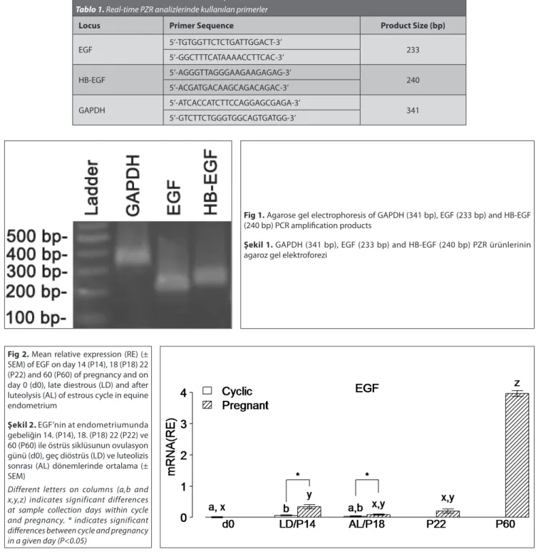
mRNA for EGF are shown in Fig. 2. The concentration of mRNA for EGF was increased significantly at LD during the estrous cycle. Expression of EGF mRNA was also up-regulated both on days P14 and P60. The greatest elevation of EGF expression was observed at P60. When compared to LD, EGF mRNA level was increased significantly on P14.
are shown on Fig. 3. Compared to d0, HB-EGF mRNA did not show significant changes in both LD and AL. There was only a dramatic increase in mRNA concentration for HB-EGF on P60 during the pregnancy. When AL and P18 were compared, expression level of HB-EGF mRNA increased on P18.
DISCUSSION
In the present study, expression of HB-EGF and EGF at mRNA levels were determined in equine endometrium and also their expression profiles during the early pregnancy and estrous cycle were shown. Biopsy day ranges (d0, P14,
P18, P22 and P60) allow us to examine expression of EGF and HB-EGF throughout early pregnancy including pre-implantation and pre-implantation periods. To understand effect of embryo, expression of EGF and HB-EGF on day of P14 and P18 were also compared to LD and AL, respectively. Although expression of HB-EGF mRNA did not show any significant changes during the estrous cycle, EGF mRNA increased at LD. Furthermore, while EGF was up-regulated at both P14 and P60, expression of HB-EGF was only increased at P60. When compared to cyclic days (LD and AL), it can be suggested that EGF expression is regulated by embryonic factors in pre-implantation period.
Table 1. Primers used for real-time PCR analyses Tablo 1. Real-time PZR analizlerinde kullanılan primerler
Locus Primer Sequence Product Size (bp)
EGF 5’-TGTGGTTCTCTGATTGGACT-3’ 233 5’-GGCTTTCATAAAACCTTCAC-3’ HB-EGF 5’-AGGGTTAGGGAAGAAGAGAG-3’ 240 5’-ACGATGACAAGCAGACAGAC-3’ GAPDH 5’-ATCACCATCTTCCAGGAGCGAGA-3’ 341 5’-GTCTTCTGGGTGGCAGTGATGG-3’
Fig 2. Mean relative expression (RE) (± SEM) of EGF on day 14 (P14), 18 (P18) 22 (P22) and 60 (P60) of pregnancy and on day 0 (d0), late diestrous (LD) and after luteolysis (AL) of estrous cycle in equine endometrium
Şekil 2. EGF’nin at endometriumunda gebeliğin 14. (P14), 18. (P18) 22 (P22) ve 60 (P60) ile östrüs siklüsunun ovulasyon günü (d0), geç diöstrüs (LD) ve luteolizis sonrası (AL) dönemlerinde ortalama (± SEM)
Different letters on columns (a,b and x,y,z) indicates significant differences at sample collection days within cycle and pregnancy. * indicates significant differences between cycle and pregnancy in a given day (P<0.05)
Fig 1. Agarose gel electrophoresis of GAPDH (341 bp), EGF (233 bp) and HB-EGF (240 bp) PCR amplification products
Şekil 1. GAPDH (341 bp), EGF (233 bp) and HB-EGF (240 bp) PZR ürünlerinin agaroz gel elektroforezi
Progesterone and embryonic factors (embryonic estrogen, PGF2α, PGE2) play a major role during initial days of equine pregnancy 4. Especially, progesterone regulates endometrial secretions 6. Considering that equine embryo remains un-attached to endometrium until day 40 of pregnancy, critical effects of progesterone regulated embryonic nutrients including uterolipocalin, retinol binding protein and transferrin are more understandable for equine pregnancy 3,22. During the estrous cycle, EGF mRNA increased at LD where progesterone level was high. In contrast to the present study, Gesternberg et al.11 failed to show effect of progesterone on EGF expression in late diestrus mares by in situ hybridization but observed an increase for EGF mRNA at day 36 or more after exogenous progesterone administration. In human, rat and rhesus monkey, regulation of EGF is associated with ovarian steroids 23-25. Similarly, expression of EGF increased in estrus stage compared to proestrus in bitch endometrium 26. Samuel et al.27 proposed that EGF may involve in maintaining endometrial gland integrity and the ongoing histotrophe secretion. In the present study, compared to cyclic days (LD and AL) early increase in EGF mRNA in pregnant endometrium at P14 and P18 may support this idea.
Compared to other large domestic animals, equine embryo has been accepted as the slowest in its achievement of implantation. Non-invasive part of equine trophoblast cells and endometrium start to form the microcotyledonary epitheliochorial placenta around day 40 of pregnancy. Coincident with placenta formation, some part of equine trophoblast cells, invasive components, connect with endo- metrium for the formation of endometrial cups. All those processes are accepted a true implantation 3,4. Considering that a magnitude up-regulation for expression profiles of both EGF and HB-EGF at P60, it can be easily seen that EGF and HB-EGF play critical roles in implantation of equine embryo. Similarly, important roles of HB-EGF during implantation have been detected in uterus of cow 28,29, human 16, baboon 30, hamster 31 and mice 17. HB-EGF regulates stromal cells for accumulating lipid and glycogen vacuoles and induces vascular permeability in rats 32. Moreover, expressions of integrin beta-3 and LIF (leukemia inhibitor factor) mRNA levels are up regulated by HB-EGF 33,34.
Among those, integrin B3 provides embryonic attachment into uterus with osteopontin-1 and LIF induces embryonic developments 35,36. Furthermore, HB-EGF dependent intra-cellular signaling Ca+2 are also important for lysophoshatidic asid (LPA) pathway in blastosist development 37.
EGF expression profiles observed in this study is consistent with former studies 11,38 which also show a dramatic increase for EGF at day 40 of equine pregnancy. HB-EGF was not demonstrated previously in equine endo- metrium and is up-regulated in equine endometrium during the implantation process. However, understanding exact functions of HB-EGF in equine implantation needs more detailed studies. Functions of dramatic increase in EGF during the implantation reveal that EGF has a mitogenic role in the formation and growth of choriallantoic placenta in equine.
There is evidence that HB-EGF mRNA expression in the endometrium of d0, late diestrus and estrus mares is not influenced by both estrogen and progesterone. However, the greatest increase during the implantation process indicated that expression of HB-EGF appears to play important roles for equine implantation as described by former studies for the other mammals 16,17,28,30,31. Similarly, the present study also demonstrated that EGF expression has important roles in equine pregnancy including both earlier (P14) and later (P60) stages. According to these results, it can be suggested that embryonic factors are more effective on regulation of HB-EGF and EGF mRNAs in equine endometrium than steroid hormones.
REFERENCES
1. Hess AP, Nayak NR, Giudice LC: Oviduct and Endometrium: Cyclic Changes in the Primate Oviduct and Endometrium, In, Neill JM (Ed. Chief): Knobil and Neill’s Physiology of Reproduction. 3rd ed., pp; 337-382, Elsevier Academic Press Inc., USA, 2006.
2. Paria BC, Lim H, Das SK, Reese J, Dey SK: Molecular signaling in uterine receptivity for implantation. Cell Dev Biol, 11, 67-76, 2000. 3. Ginther OJ: Equine pregnancy: Physical interactions between the uterus and conceptus. In, 44th Annual Convention of the American
Association of Equine Practitioners, 15-20 July Baltimore, Maryland, USA,
pp. 73-104, 1998.
Fig 3. Mean relative expression (RE) (± SEM) of HB-EGF on day 14 (P14), 18 (P18) 22 (P22) and 60 (P60) of pregnancy and on day 0(d0), late diestrous (LD) and after luteolysis (AL) of estrous cycle in equine endometrium Şekil 3. HB-EGF’nin at endometriumunda gebeliğin 14. (P14), 18. (P18) 22 (P22) ve 60 (P60) ile östrüs siklüsunun ovulasyon günü (d0), geç diöstrüs (LD) ve luteolizis sonrası (AL) dönemlerinde ortalama (± SEM)
Different letters on columns (a, b and x,y,z) indicates significant differences at sample collection days within cycle and pregnancy. * indicates significant differences between cycle and pregnancy in a given day (P<0.05)
4. Allen WR, Wilsher S: A review of implantation and early placentation in the mare. Placenta, 30, 1005-1015, 2009.
5. McDowell KJ, Sharp DC, Grubaugh W, Thatcher WW, Wilcox CJ: Restricted conceptus mobility results in failure of pregnancy maintenance in mares. Biol Reprod, 39, 340-348, 1988.
6. Gerstenberg C, Allen WR, Stewart F: Cell proliferation patterns during development of the equine placenta. J Reprod Fertil, 117, 143-152, 1999. 7. Massagué J, Pandiella A: Membrane-anchored growth factors. Annu
Rev Biochem, 62, 515-541, 1993.
8. Dey SK, Lim H, Das SK, Reese J, Paria BC, Daikoku T, Wang H: Molecular cues to implantation. Endocr Rev, 25, 341-373, 2004.
9. Singh M, Chaudhry P, Asselin E: Bridging endometrial receptivity and implantation: network of hormones, cytokines and growth factors. J
Endocrinology, 210, 5-14, 2011.
10. Lim HJ, Dey SK: HB-EGF: A unique mediator of embryo-uterine interactions during implantation. Exp Cell Res, 315, 619-626, 2009. 11. Gerstenberg C, Allen WR, Stewart F: Factors controlling epidermal growth factor (EGF) gene expression in the endometrium of the mare.
Mol Reprod Dev, 53 (3): 255-265, 1999.
12. Maruo T, Matsuo H, Otani T, Mochizuki M: Role of epidermal growth factor (EGF) and its receptor in the development of the human placenta.
Reprod Fertil Dev, 7, 1465-1470, 1995.
13. Tsutsumi O, Taketani Y, Oka T: The uterine growth-promoting action of epidermal growth factor and its function in the fertility of mice. J
Endocrinol, 138, 437-443, 1993.
14. Lacroix MC, Kann G: Ontogeny and characterization of epidermal growth factor receptors on the fetal area of the sheep placenta. J
Endocrinol, 136, 43-50, 1993.
15. Robertson SA, Seamark RF, Guilbert LJ, Wegmann TG: The role of cytokines in gestation. Crit Rev Immunol, 14, 239-292, 1994.
16. Yoo HJ, Balow DH, Mardon HJ: Temporal and spatial regulation of expression of heparing- binding epidermal-growth factor-like growth factor in the human endometrium: A possible role in blastocyst implantation. Dev Genet, 21, 102-108, 1997.
17. Brown N, Deb K, Paria BC, Das SK, Reese J: Embryo-uterine inter-actions via the neuregulin family of growth factors during implantation in the mouse. Biol of Reprod, 71, 2003-2011, 2004.
18. Atli MO, Kurar E, Kayis SA, Aslan S, Semacan A, Celik S, Guzeloglu A: Evaluation of genes involved in prostaglandin action in equine endo-metrium during estrous cycle and early pregnancy. Anim Reprod Sci, 122, 124-132, 2010.
19. Kurar E, Atli MO, Guzeloglu A, Ozsensoy Y, Semacan A: Comparison of five different RNA isolation methods from equine endometrium for gene transcription analysis. Kafkas Univ Vet Fak Derg, 16 (5): 851-855,
2010.
20. Kayis SA, Atli MO, Kurar E, Bozkaya F, Aslan S, Semacan A, Guzeloglu A: Rating of putative housekeeping genes for quantitative gene expression analysis in cyclic and early pregnant equine endo-metrium. Anim Reprod Sci, 125, 124-132, 2011.
21. Payne RW, Murray DA, Harding SA, Baird DB, Soutar DM: GenStat for Windows. 7th ed., Introduction,VSN International, Hemel Hempstead. 2003.
22. Stewart F, Kennedy MW, Suire S: A novel uterine lipocalin supporting pregnancy in equids. Cell Mol Life Sci, 57 (10): 1373-1378, 2000.
23. Gardner RM, Verner G, Kirkland JL, Stancel GM: Regulation of
uterine epidermal growth factor (EGF) receptors by estrogen in the mature rat and during the estrous cycle. J Steroid Biochem Mol Biol, 32, 339-343, 1989.
24. Imai T, Kurachi H, Adachi K, Adachi H, Yoshimoto Y, Homma H, Tadokoro C, Takeda S, Yamaguchi M, Sakata M, Sakoyama Y, Miyake A: Changes in epidermal growth factor receptor and the levels of its ligands during the menstrual cycle in human endometrium. Biol Reprod, 52, 928-938, 1995.
25. Yue ZP, Yang Zm, Li SJ, Wang HB, Harper MJK: Epidermal growth factor family in Rhesus Monkey uterus during the Menstrual Cycle and early pregnancy. Mol Reprod Dev, 55, 164-174, 2000.
26. Bukowska D, Kempisty B, Jackowska M, Woźna M, Antosik P, Piotrowska H, Jaśkowski JM: Differential expression of epidermal growth factor and transforming growth factor beta isoforms in dog endometrium during different periods of the estrus cycle. Pol J Vet Sci, 14 (2): 259-264, 2011.
27. Samuel CA, Allen WR, Steven DH: Studies on the equine placenta: III. Ultrastructure of the uterine glands and the overlying trophoblast. J
Reprod Fertil, 51, 433-437, 1977.
28. Kleim A, Tetens T, Klonisch M, Grealy M, Fischer B: Epidermal growth factor receptor and lingands in elongating bovine blastocysts.
Mol Reprod Dev, 51, 402-412, 1998.
29. Kizaki K, Ushizawa K, Kimura M, Iga K, Takahashi T, Hashizume K: Active roles of HB-EGF and its receptors in bovine endometrium during implantation. Biol Reprod, 83, 459, 2010.
30. Leach RE, Khalifa R, Armant DR, Brudney A, Das SK, Dey SK, Fazlebas A: Heparin binding-EGF-like growth factor (HB-EGF) modulation by antiprogestin and CG in the baboon (Papio anubis). J Clin Endocrinol
Metab, 86, 4520-4528, 2001.
31. Wang X, Wang H, Matsumoto H, Roy SK, Das SK, Paria DC: Dula source and target of heparin binding EGF-like growth factor during the onset of implantation in the hamster. Development, 129, 4125-4134, 2002. 32. Paria BC, Ma W, Tan J, Raja S, Das SK, Dey SK, Hogan BL: Cellular and molecular responses of the uterus to embryo implantation can be elicited by locally applied growth factors. Proceed Natl Acad Sci USA, 98, 1047-1052, 2001.
33. Lessey BA, Gui Y, Apparao KB, Young SL, Mulholland J: Regulated expression of heparin binding-EGF-like growth factor (HB-EGF) in human endometrium: A potential paracrine role during implantation. Mol Reprod
Dev, 62, 446-455, 2002.
34. Jessmon P, Leach R, Armant DR: Diverse Functions of HBEGF during pregnancy. Mol Reprod Dev, 76, 1116-1127, 2009.
35. Steward CL, Kaspar P, Brunet LJ, Bhartt H, Gadi I, Kontgen F, Abbondanzo SJ: Blastocyst implantation depends on maternal expression of leukemia inhibitory factor. Nature, 359, 76-79, 1992. 36. Apparao KB, Murray MJ, Fritz MA, Meyer WR, Chambers AF, Truong PR, Lessey BA: Osteopontin and its receptor αvβ3 integrin are coexpressed in the human endometrium during the menstrual cycle but regulated differentially. J Clin Endocrinol Metab, 86, 4991-5000, 2001. 37. Liu Z, Armant DR: Lysophosphatidic acid regulates murine blastocyst development by transactivation of receptors for heparin binding-epidermal growth factor. Exp Cell Res, 296, 317-326, 2004.
38. Stewart F, Power CA, Lennard SN, Allen WR, Amet L, Edwards RM: Identification of the horse epidermal growth factor (EGF) coding sequence and its use in monitoring EGF gene expression in the endometrium of the pregnant mare. J Mol Endocrinol, 12 (3): 341-350, 1994.