Purification and Partial Characterisation of Catalase from Chicken
Liver and the Inhibition Effects of Some Chemicals on the
Enzyme Activity
Tülin AYDEMİR1 - Kevser KURU1 - Seda ÇINAR1
Abstract: Catalase plays a major role in the protection of tissues from oxydizing effects of H2O2 and partially reduced oxygen species. It was purified chicken liver by (NH4)2SO4 precipitation, ethanol-chloroform treatment, CM-cellulose and Sephadex G-200 chromatography. The molecular weight of the native chicken liver catalase was estimated by gel filtration to be approximately 235000 Da. SDS-gel electrophoresis results indicated that chicken liver catalase consists of four apparently identical sub-units, with a molecular weight of about 57500 Da. The specific activity was found to be 55765,3 U/mg. The optical spectrum of the purified enzyme shows a Soret band at 407 nm which is the characteristic for heme and there was apparent reduction by dithionite. The Km was found to be 100 mM. The maximal activity of the enzyme was observed between pH 7.0 and 10.0. The enzyme has a optimum reaction temperature at 25 °C. The activity of purified catalase was inhibited by azide, cyanide, β-mercaptoethanol, DTT, GSH and iodoacetamide.
Key words :Chicken liver, catalase, purification, characterisation, inhibitors.
Özet: Katalaz, kısmi indirgenmiş oksijen türleri ve H2O2 nin yükseltgen etkisinden dokuların korunmasında önemli rol oynar. Tavuk karaciğer katalazı, (NH4)2SO4 çöktürmesi, etonol-kloroform muamelesi, CM-Selükoz ve Sephadeks G-200 kromotografisi kullanılarak saflaştırıldı. Jel filitrasyon kromotografisi ile tavuk karaciğer katalazının molekül ağırlığı yaklaşık olarak 235000 Da bulundu. SDS-jel elektroforezi sonucu tavuk karaciğer katalazının molekül ağırlığı 57500 Da olarak bulundu ve 4 alt birimden oluştuğu tespit edildi. Spesifik aktivite 55765.5 U/mg olarak saptandı. Hem içeriğinin kabaca ölçülmesini sağlayan A407/A270 oranı 0.52 olarak bulundu. Enzim için Km değeri 100 mM olarak belirlendi. Enzimin 7.0-10.0 aralığında geniş bir pH optimumuna sahip olduğu görüldü. Tavuk karaciğer katalaz enziminin optimum sıcaklığı 25 °C olarak tespit edildi. Saflaştırılmış enzim aktivitesi KCN, NaN3, β-merkaptoetanol, DTT, iyodoasetamit tarafından inhibe edildi, fakat sodyum dietilditiyokarbamatın enzimi inhibe etmediği görüldü.
Anahtar Kelimeler: Karaciğer, katalaz, purifikasyon
INTRODUCTION
Metabolism in oxygen-enriched environment often results in the generation of reactive oxygen species as superoxide, hydroxyl radical and hydrogen peroxide. Theses reactive oxygen species can oxidize membran fatty acids initiating lipid peroxidation, modify proteins and damage DNA. A great number of chemical and enzymatic scavengers provide antioxidant protection againts these oxygen metabolites [1-4].
Catalase (H2O2: H2O2 oxidoreductase EC1.11.1.6) a ubiquitous heme protein
predominantly in peroxisomal matrix is one of the essential antioxidant enzyme. The iron containing
enzyme catalase catalyzes the breakdown of H2O2, a potencially destructive agent in cells. Most of
the biochemical studies on animal catalase are reported from mammalian liver, lung and blood [5-9].
Catalases have been isolated from a broad range of procaryotic and eucaryotic organisms. Most catalases described so far are tetramers with molecular masses ranging over 220-270 kDa with each subunit containing a protoheme as prosthetic group. These typical catalases are active in the pH range 5-10 and are specifically inactivated by 3-amino-1, 2, 4 triazole [10-12].
During the course of these studies catalase was purified chicken liver and partially characterization. In addition the inhibition effect of some chemicals on the chicken liver catalase activity was examined.
EXPERIMENTAL Chemicals
Hidrogen peroxide, urea, chloroform, ethanol, acrylamide, ammonium sulphate ((NH4)2SO4), sodium azide (NaN3), potassium cyanide (KCN), sodium diethlydithiocarbamate,
sodium chloride (NaCl), sodium dithionite, oxalic acid, lactic acid, CuSO4, MnCl2, FeCl3, ZnSO4,
were purchased from Merck, Germany. Sephadex G-200, bovine serum albumin (BSA), β -mercaptoethanol, dithiotreitol (DTT),sodium dodecyl sulfate (SDS), Triton X-100, iodoacetamide (IA), 3-amino 1,2,4, triazole (AT),Q-Sepharose, 5, 5’dithio-bis (-2-nitrobenzoik acid) (DTNB), glutathione (GSH), molecular weight standarts for gel exclusion and SDS-PAGE were purchased from Sigma Chemical Company, USA. Other reagents are in analytical grade.
Material
Chicken liver tissue was collected on ice from a local slaughter house immediately after killing the animal, to the laboratory in chilled conditions and stored in deep-freezer until used.
Enzyme Assay
Catalase activity was determined spectrophotometrycally, by direct measurement of the decrease in the light absorption at 240 nm caused by the decomposition of hydrogen peroxide by catalase[13]. The reaction mixture contained, in a total volume of 1 mL, 0.01 M H2O2 and 0.05 M
potassium phosphate buffer pH 7.0. The reaction was initiated by the addition of 20 µL enzyme and the rate of a utilization of H2O2 was measured for initial 60 sec. by measuring decrease in
absorbance at 240 nm. Protein Assay
Protein content of the different extracts was determined by the dye-binding method of Bradford using bovine serum albumin as a standard [14].
Enzyme Purification
After washing the liver tissue with 0.9 % NaCl and soaking with Whatman No: 1 filter paper, 10 gr of tissue was homogenized in distilled water using a waring blender for 30 sec. at low speed followed by 60 sec. at high speed. The resulting homogenate (10 % w/v) was filtered the through two layers of muslin and centrifuged at 14000 g for 30 min. Crude extract was brought to 45 % saturation by gradually adding solid (NH4)2SO4 and was stirred for 30 min at 4 °C. The precipitate
was removed by centrifugation at 14000 g for 1h. at 4 °C and resuspended in 20 mM phosphate buffer pH 7.0.
0.5 mL of an ethanol- chloroform mixture (3 part of chloroform to 1 parts of ethanol) was added to the each mL of the supernatant above a mixed rigorously for one minute. The denatured hemoglobin is removed by centrifugation (14000g 15 min ). The ethanol-chloroform mixture is then evaporated in vacuo.
The supernatant of the ethanol-chloroform treatment was applied to a CM-cellulose (1,5x60cm), which was equilibrated with 0.02 M potassium phosphate buffer, pH 7.0, at 4 °C. Elution of the enzyme was achieved with a flow rate of 0.5 ml/min. by establishing a linear gradient of phosphate buffer (0.02-0.05 M, pH 7.0). Fractions containing catalase activity were concentrated by 30000 NMWC ultrafilter. The concentrate samples were applied to the Sephadex G-200 (1.0x90 cm) that had been equilibrated with 0.05 M phosphate buffer pH 7.0 and the enzyme was eluted with a linear gradient of phosphate buffer (0.05-0.1 M pH 7.0).The fractions of 2 mL were collected and assayed catalase activity.
Molecular Weight Determination
The apparent molecular weight of the purified catalase was determined by gel exclusion chromatography on a Sephadex G-200 column [15]. Bovine serum albumin (66000 Da), alcohol dehydrogenase (150000 Da), β-amilase (200000 Da) and apoferritin (443000 Da) were used as molecular weight markers.
Electrophoresis
Sodium dodecyl sulphate polyacrylamide gel electrophoresis (SDS-PAGE) was performed as described by Laemmli by using a vertical slab gel apparatus [16]. Carbonic anhydrase (29000 Da), ovalbumin (45000 Da), albumin bovine plasma (66000 Da), phosphorylase (97000 Da), β -galactosidae (116000 Da), myosin (205000 Da) were used as calibration proteins at the identical subunit moleculer weight proteins were visualized by Commassie Blue.
Effect of pH
Activity profiles of chicken liver catalase were investigated at 25 °C at different pH values in 50 mM acetate (pH 4.0-6.0) phosphate, (pH 6.0-7.5 ) and Tris (pH 7.5-10) buffers for 180 min. at 25
°C. Residual catalase activity was measured under standard assay conditions. Effect of Temperature:
In the thermal stability studies, the purified enzyme was incubated at various temperatures in 0.05 M phosphate buffer for 180 min at pH 7.0. There after retained enzyme activity was measured as indicated in “Enzyme Assay” section.
Enzyme Kinetics
For determination of Michaelis Constant (Km) value of catalase were measured with H2O2 at
varying concentrations 2-25 mM in phosphate buffer pH 7.0 Km value of catalase were calculated
from a the plot of 1/v versus 1/[S] by the method of Lineweaver Burk. Storage Stability
Storage stability of catalase was determined by 16 weeks incubation of enzyme at three different temperatures (-15, 4 and 25 °C). The activity was determined every day for the first weeks and then at weekly intervals. Catalase samples stored –15 °C were kept in separate vials for each observation to avoid repeated freeze-thaw effect on the enzyme.
Inhibition Studies
The effect of inhibitors on the catalase activity was determined by measuring the enzyme activity in various concentrations of inhibitors in 0.05 M phosphate buffer pH 7.0. To determine the
inhibitor concentration that reduced the enzyme activity by 50 % (Ι50) regression analysis graphs
were drawn by using percent inhibition values. Ι50 values were determined from graphs. RESULTS AND DISCUSSION
The purification steps of the catalase from chicken liver is given in Table I. Ammonium sulphate treatment (20-95 % saturation) was investigated to partially remove inactive proteins. Catalase activity of the precipitate obtained with 45 % (NH4)2SO4 gave the highest activity.
Therefore the 45 % for (NH4)2SO4 concentrations was accepted as an optimum conditions and this
concentrations was used in all extraction processes. Ammonium sulphate precipitation steps resulted in 4.16 fold purification of catalase with 51.5 recovery.
TABLE I. Purification steps of catalase from chicken liver
Purification Step Total Unıts Total Protein Specific Activity Yield Purification (mg) (U/mg) (%) (Fold) Crude Extract 119507.2 464 257.55 - - Ammonium Sulfate Fraction 61558.4 56 1099.25 51.50 4.26
Hemoglobine Treatment 35280.6 4.65 7587.22 29.80 29.45 CM-cellulose Chromotography 33332.6 3.12 16794.51 27.89 65.20 Sephadex G-200 Chromotography 25094.0 0.675 55765.30 20.90 216.52
After the precipitation of catalase with (NH4)2SO4, the hemoglobin in the supernatant was
removed by treating with chloroform-ethanol. The precipitation fold was found to 29.45 fold after this step. Like typical catalases, chicken liver catalase is stable to treatment with ethanol chloroform [17, 18]. Chloroform-ethanol treatment of the extract followed by subsequent chromatography steps resulted in 216.52 fold purification of catalase with a yield of 20.90 % (Figure 1 and 2). The purified enzyme gave a single major peak after Sephadex G-200 column chromatography and the specific activity was found to be 55765,3 U/mg.
Figure I: Elution profile of chicken liver catalase from CM –cellulose column chromatography. Absorbance at 280 nm (--ο--); catalase activity (---•--- ).
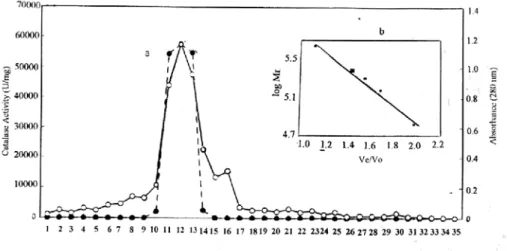
Figure II : Elution pattern of chicken liver catalase from gel filitration chromatography on Sephadex G-200. Absorbance at 280 nm (ο), catalase activity (---•---) Inset : Molecular calibration curve. Protein standard(--•--); purified chicken liver catalase (ο).
The specific activity of goat liver and rat liver catalase have been reported 136000 U/mg and 83132 U/mg respectively [19, 20].
The molecular weight of the purified enzyme was found to be approximately 235000 Da (Figure 3). This value corresponds roughly to those of purified catalase from liver, erythrocyte and other tissues. The molecular weight liver catalase, mouse liver catalase, goat lung catalase and human erythrocyte catalase, and porcine erythrocytes have been reported 240000 Da, 235000 Da, 339000 Da 240000 Da and 250000 Da respectively [19,21-24].
Figure III: SDS/PAGE results for purified chicken liver catalase. Lane A, protein standards, Lane B purified catalase.
The native enzyme was also subjected to SDS-Gel electrophoresis to estimate to molecular weight of the sub-unit the resulting gel showed only a share single bond with a molecular weight of about 57500 Da (Figure 3). This indicates that the chicken liver catalase molecule consist of four identical subunits. Similar results was also observed for the catalases purified from goat liver, human erythrocyte and Aspergillus niger catalase [19,24-25].
Absorption Spectra
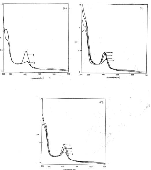
The absorption spectrum of chicken liver catalase exhibits two major peaks, at 270 nm and a soret band at 407 nm.
The peak at 270 nm disappeared at alkaline conditions (pH 11.5). The maxima at 407 nm decreased immediately with the shift of the peak to 415 nm (Figure 4-A).
Figure IV: (A) Absorption spectrum of catalase in alkali. (a) control; (b) alkali denatured (pH: 11.5). (B) The changes in the soret absorption of purified catalase with thiol compounds. (a) control; (b) 10 mM DTT; (c) 5 mM GSH; (d) 10 mM β-mercaptethanol (C) Absorption spectra of catalase with some inhibitors. (a) control; (b) 10 mM KCN; (c) 2 mM sodium dithionite; (d) 8 mM urea.
Treatment of the enzyme with thiol compounds such as β-mercaptoethanol DTT and GSH decreased and shifted the soret band to 414, 412 and 404 nm respectively (Figure 4-B ). Thiol inhibition of catalase has been established by the early workers. Miyahara at al. studied the effect of thiol on goat liver catalase and concluded on the basis of absorption and circular dicroism spectral study that there was interaction between thiol reagents and heme groups in catalase directly and/or with some amino acid residues in the proximity of the heme group causing conformational alteration near the heme group which inhibits the enzyme activity [26].
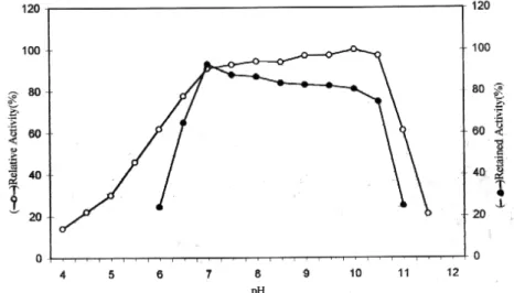
Figure V: The effect of pH and stability on purified chicken liver catalase activity.
Treatment of the enzyme with KCN as shown in Figure 4-C shifted the maximum of the soret band to 416 nm. The native spectrum of the typical catalase of chicken liver and that cyanide complex are similar those found for typical catalases. Reduction of purified enzyme with dithionite caused a decrease of the Soret band and shifted it to 414 nm (Figure 4-C). Treatment of catalase with 8 M urea led to a decrease of Soret band absorbance and shifted it to 412 nm.
The time dependence of changes in the soret absorption spectra of catalase in 1 % SDS is shown. The absorption maximum at 407 nm of purified catalase decreased gradually within one h with an accompanying shift toward 400 nm. The similar results in absorption spectrum obtained in the presence of 1 % SDS have been also reported by Takeda for bovine liver catalase [27,28].
Optimum pH and pH Stability
The maximal activity of the enzyme was observed between 7.0 and 10.0 (Figure 5). pH stability test performed at 25 °C showed that the chicken liver catalase was most stable at pH 7.0 than the other pH values (Figure 5 ) studied. Incubation of the purified enzyme for 180 min at pH 6 and 10.0 caused 10 % and 32 % inhibition, respectively. Several authors have found similar results for bovine liver catalase [17]. Catalase is generally considered as an enzyme without a pH to optimum [18]. In case of other catalases, the enzyme has been shown either exhibit only one sharp optimum pH, a broad pH optima or two pH maxima [29-31].
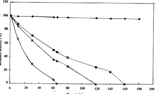
Figure VII: Heat stability of chicken liver catalase at 25°C (--•--), 40 °C (--■--), 50 °C (--▲--), 60 °C (--○--).
Effect of Temperature
The effect of temperatures between 10 - 80 °C on catalase activity showed that the optimum temperature to the purified enzyme was 25 °C. (Figure 6).
Heat stability of the catalase was investigated between 25 °C and 70 °C with a 180 min. duration (Fig 7 ). The enzyme was stable at lower temperature but unstable at higher temperatures. For instance, when the temperatures were increased from 25 to 40 °C, the relative activity of catalase decreased from 100 to 50 % for 60 min. incubation time. However the relative activity decreased from 50 to 5 % when the temperature was raised from 40 to 60 °C for 60 min. It is indicated that the enzyme was rapidly denatured at higher temperatures. The thermal stability of purified enzyme was found similar with bovine liver catalase in the same conditions [17].
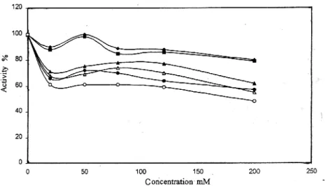
Figure VIII: The effect of ionic strength on chicken liver catalase activity phosphate, pH 6.0 (--▲--), pH 7.0 (--■--), pH 8.0 (--♦--), and chloride, pH 6.0 (--Ο--), pH 7.0 (--∆--), pH 8.0 (--•--), at 25
°C.
The Effect of Ionic Strength
Activity variations depending on the ionic strength phosphate and chloride in the 0.020-0.250 M concentration range were investigated by using the standard activity assay conditions. As seen in Figure 8, similar inhibition values were observed for phosphate at pH 7.0 and 8.0. Monovalent anion chloride was found to be a more effective inhibitor than divalent anion phosphate at pH 7.0.
Storage Stability
The crude enzyme preparation was unstable. At 25 °C, 4 °C and –15 °C, the crude extract lost its activity by 72 h, 24 h and 10 days respectively. The partially purified enzyme ( after (NH4)2SO4 ) precipitation completely lost its activity after 4, 20 and 30 days respectively. Purified
catalase activity was found to be quite stable. At 4 °C, it lost only 10 % enzyme activity on storage for 6 months. At –15 °C, at the and of 6 months storage retained about 95 % of original activity.
Kinetic Properties
The apparent Km value for H2O2 in the catalytic reaction was 0.1 M as determined by
Lineweaver-Burk plot. The apparent Michaelis constant for chicken liver catalase was similar to what has been reported for purified preparation of the goat liver catalase (0.1 M) [15]. The Michaelis constants for beef liver and bovine liver catalase are 0.07 M and 0.28 M respectively [26,29]. Catalases from yeast and Hyalomma dromedarii have Km values of 19.2 and 8.0 mM respectively
[32, 33].
Effect of Some Inhibitor
L-Gly, L-Arg, L-Ala and fenilalanin amino acids concentrations of 6-60 mM did not show any effect on the enzyme activity. L-Asp at 6, 30 and 60 mM concentrations inhibited the enzyme by 5, 30 and 42 % respectively.
3-amino 1,2,4 triazole (AT) showed inhibition effect on the chicken liver catalase. For incubation 60 min, 50 % inhibition was found to be 3 mM. AT has been known for a long time as a specific inhibitor of the catalase activity both in vivo and in vitro. It is assumed that this inhibitor binds irreversible inhibition for 1-2 hours [34]. Similar results concerning the sensitivity to at have
been found for catalase isolated from maize and tobacco. According to Havir the differences in sensitivity to at may be related to modifications in protein structure [35].
The chicken liver catalase inhibited by the heme protein ligands cyanide and azide. 50 % inhibition were observed 1.10-5 M and 5.10-7 M respectively. In the presence of cyanide and azide, Vmax of the enzyme reaction decreased but the Km value remained unchanged showing
non-competitive nature of the inhibition. The Ki values of the enzyme for cyanide and azide, calculated
by Lineweaver Burk plot, were 1,12.10-3 M and 2,25.10-3 M. Goat liver catalase is inhibited by sodium azide non-competitively with Ki value 1.3.10-3 M [19].
The treatment of catalase with β-mercaptoethanol, DTT and GSH caused a gradual reduction in the activity. The 50 % inhibition were observed 2.9 mM, 5.0 mM and 6.0 mM concentrations of respective thiols for 60 min. incubation. The results show that β-mercaptoethanol was the most patent reagent for reduction of enzyme activity. Thiol inhibition of catalases has been established by the early workers [26]. Miyahara at al. studied the effect of thiol on goat liver catalase and concluded on the basis of absorption and circular dichroism spectral study that there was interaction between thiol reagent sand heme groups in catalase directly and/or with some amino acid residues in the proximity of the heme group causing conformational alteration near the heme group which inhibits the enzyme activity [26].
Sulfhydryl blocking reagent DTNB and IA inhibited the enzyme, indicating the presence of free-SH group (s) essential for the enzyme action. 50 % inhibition was found to be 8.10-3 M and 12.10-3 mM concentration of respective sulfhydryl blocking reagent for 30 min. incubation. The inhibition goat liver catalase with DTNB and IA has been reported 50 % inhibition 1,66.10-3 and 20.10-3 M concentration for 30 min. incubation [19].
The purified enzyme was incubated with different concentration of urea for 30 min 50 % inhibition was observed 6.0 M .Samejima and Kita are of interest these outhors reported a complete loss of helical content with time when beef liver catalase was treated with 8 M urea [36].
The enzyme also seemed to be insensitive to 0.03 % and 0.06 % concentrations of Triton X-100 for 1 hours.
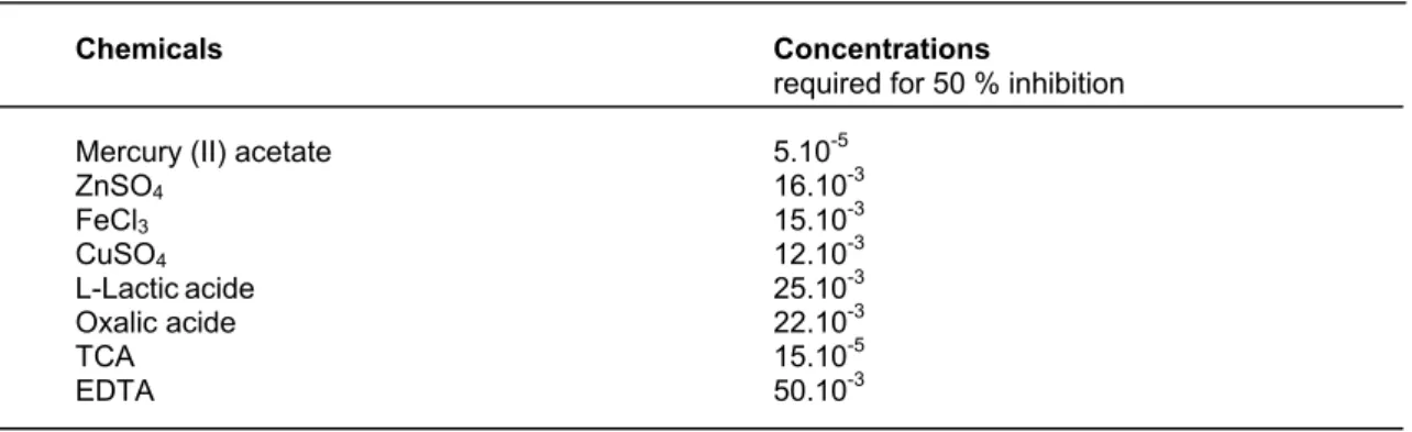
Table II Shows the effect of various chemicals on the activity of purified from chicken liver catalase. The purified enzyme was also sensitive to Cu+2, Zn+2 and Fe+3 ions. 50 % inhibition was observed at 0.07.10-3 M for mercury (II) acetate. This metabolite was more potent reagent for inhibition of enzyme. Certain heavy metal ions, including Hg+2 (mercury II), are examples of non-competitive inhibitors. The toxicity of these common environmental agents is primarily a consequence of their interactions with sulphydryl groups (-SH) and other susceptible functional groups in numerous proteins, including many enzymes [37].
The purified enzyme was incubated with different concentration of SDS for 3 hours. 50 % inhibition was observed 0,006 M. Many proteins combine with SDS to form protein SDS complexes with unfolding of the rigid conformation of proteins [27]. Nelson demonstrated that the rate of binding of SDS with bovine liver catalase was very slow and that the native catalase first combined with a little SDS, than more SDS was easily bound after denaturation of the protein, leading to dissociation of the molecule in to subunits [27]. This SDS is a kind of denaturant for proteins as well as a splitting reagent for protein having subunits.
TABLE II The effect of various chemicals on the activity of purified catalase from chicken liver (for 3 h. incubation).
Chemicals Concentrations
required for 50 % inhibition
Mercury (II) acetate 5.10-5
ZnSO4 16.10-3 FeCl3 15.10-3 CuSO4 12.10-3 L-Lacticacide 25.10-3 Oxalic acide 22.10-3 TCA 15.10-5 EDTA 50.10-3 REFERENCES
1 I. Fridovich, Arch. Biological effects of superoxide radical. Biochem. Biophys.,274,1-5, (1986).
2 A. Hochman and I. Goldberg, Purification and characterization of a Catalase peroxidase and a
typical catalase from the bacterium Klebisealla Pheumioniae. Biochem. et Biophys. Acta., 1077299,
(1991).
3 B. Hallivel and J.M.C. Gutteridge, Free radicals in biology and medicine J. Biochem., 219 (1984). 4 J. Feher, G. Csomos and A. Vereckei, Free Radical Reactions, Springer,9 (1987).
5 A. Pıhl, R. Lange and A. Evang, The ınteraction of cystine and cystein with beef liver catalase. Acta. Chem. Scand., 15 (1961)1271-1279.
6 P., Nicholls, G.R., Schonbaum, Catalases, in Boyer PD, lardy H, Myrback K, (eds), The enzymes (ed) Academic, New York NY (1963), 147.
7 J.V., Eaton, M., Boraas, N.L., Etkin, Catalase activity and red cell metabolism, Adv. Exp. Med. Biol. 28, (1997) 121.
8 T. Miyahara and T. Samejima, Subcellular distribution and characterization of porcine kidney
catalase J. Biochem., 89(1981)919.
9 A. Takeda, T. Miyahara, A. Hachimori and T. Samejima, The interaction of thiol compounds with
porcine erythrocyte catalase. J. Biochem., 87(1980)429-439.
10 R. Bonnichsen, A comperative study of the blood and liver catalases from the horse. Arch. Biochem. Biophys.,12(1947)83-94.
11 M. Nagahisa, Crystallization and alkali-denaturation of porcine blood catalase J. Biochem. (Tokyo).,
51(1962)216-221.
12 H.F.Deutsch, The properties of various crystalline horse erythrocyte catalase preparations. Acta. Chem. Scand., 6(1952)1516-1521.
13 H.E. Aebi, Catalase In:Methods of Enzymatic Analysis. H.U.Bergmeyer., 3(1983)273.
14 M. Bradford, A rapid and a sensitive method for the quantitation of microgram protein utilising the principle of protein-dye binding. Anal. Biochem.,72(1976)248-254.
15 E. Stelwagen, Methods in Enzymology M.O. Deutscher (Ed) Academic Pess, Inc., San Diego.,
182(1990)317.
16 U.K. Laemlli, Nature., Cleavage of structural proteins durind assembly of the head of bacreiophage 227(1970) 680-685.
17 B.P. Wasserman and H.O. Hultin, . Effect of Deglycosylation on the stability of Aspergillus niger
18 B. Change, Effect of pH upon the reaction kinetics of the enzyme-substrate compounds of
catalase., J. Biol. Chem., 194 (1952) 471-478.
19 U. Chatterjee, A.Kumar and G.G. Sanwall, Purification and properties of goat liver catalase: Two pH
optima. Ind. J. Biochem. Biophys., 26(1989)140-147.
20 R.E. Greenfield and U.E. Price, Isolation of catalase from mitocondrial fractions of
polyvinylpyrrolidone sucrose homogenates. J.Biol. Chem., 220(1956)607.
21 H. Aebi, catalase in vitro methods enzymol 105: (1984), 121.
22 G.F., Gaetani, H.N., Kırkman, R., Mangenin, A.M., Ferraris, Importance of catalase in the disposal of hydrogen peroxide with in human erythrocytes, lood, 84, (1994) 325.
23 U. Chatterjee, A.Kumar and G. Sanwall, Purification and characterization of catalase from goat
(capra capra) lung. Molecular and Cellular Biochem., 126(1993)125.
24 H.N., Kirkman, G.F., Gaetani, catalase: A tetrameric enzyme eith fourtighly bound molecules of
NADPH, proc. Natl Acad Sci USA, 81, (1984) 4343
25 H.N., Kirkman, S., Galiano, G.F., Gaetani, The function of catalase bound NADPH, J. Biol. Chem, 262, (1987), 660.
26 T. Miyahara, A. Takeda, A. Hochimori and T. Samejima, On the heterogeneity of catalase from goat
liver. J.Biochem., 84(1978)1267-1276.
27 A. Takeda A. Hochimori, A Muraı, K. Sata and K. Samejima, Effect of sodium dodecyl sulfate on the
dissociation of bovine liver catalase. J.Biochem., 78(1975)911.
28 T.E. Barman, Enzyme Hand Book., 1(1969)232.
29 E.H. Herman and T.N. Burbridge, Initial studies with a brain catalase Life Sci., 9(1970)1389-1396. 30 O. Ito, R. Akuzawa and J. Dairy Purification, crystallization and properties of bovine milk catalase,
Sci., 66(1983)967-973.
31 T.C.M. Seah and J.G. Kaplan, Purification and properties of the catalase Bakers Yeast. J. Biol. Chem., 248(1973)2889.
32 T. Yamada, A. Tanaka, and S. Fukul, Eur. J. Biochem., 125(1982)51521.
33 M.Y. Kamel and R.R. Hamed, Purification and characterization of catalase from developing
embryos of Hyalomma dromedarii (Acarina:Ixodidae), Insect Biochem., 12(1982)481.
34 J.M. Chandlee, A.S. Tsaftaris and J.G. Scandolios, Purification and partial characterizatşon of three
genetically defined catalases of maize. Plant Sci. Lett., 29(1983)117.
35 E.A. Havir, The in vivo and in vitro inhibition of catalase from leaves of nicotiana sylvestris by
3-amino-1,2,4-triazole. Plant Physiol., 99 (1992) 533.
36 T. Samejima and M. Kita, The conformational changes of catalase molecule caused by ligand
molecules, Biochem. Biophys. Acta., 175(1969)24.