M.s. e
hLayeL1,2, a. B
ener3,4Risk Factors of Zinc Deficiency
in Children with Atopic Dermatitis
1Weill Cornell Medical College, Ar-Rayyan, Qatar
2Section of Pediatric Allergy-Immunology, Department of Pediatrics, Hamad Medical Corporation, Doha, Qatar 3Departments of Biostatistics and Medical Informatics, Cerrahpaşa Faculty of Medicine, Istanbul University, Istanbul, Turkey
4Departments of Public Health, Medipol International School of Medicine, Istanbul Medipol University, Istanbul, Turkey
KeyWords
zinc deficiency; risk factor; severe; atopic dermatitis; IgE
Corresponding author
Mohammad S. Ehlayel Section of Allergy-Immunology Department of Pediatrics, Hamad Medical Corporation PO Box 3050, Doha, State of Qatar Phone: +974 4439 2834-22840 Fax: +974 4443 9571 E-mail: [email protected] Doi 10.23822/EurAnnACI.1764-1489.114 Summary
Background and objectives. Zinc deficiency increases risk of infections, allergies
and autoimmunity. We wished to determine risk factors in severe atopic dermatitis (AD) and identify of hypozincemia rate. Materials and methods. Retrospective study done on AD children (≤ 14 years) with serum zinc test. Data included demographic and laboratory tests (serum zinc level, IgE, food-specific IgE), and skin tests. Results. 168 AD children, aged 38.9 months with concomitant allergies in 47 (28%), family history of allergies in 131 (80%), and parental consanguinity in 134 (79.9%). AD was mild in 12 (7.2%, SCORAD 15.8) children, moderate in 41 (24.5%, SCORAD 30.4), and severe in 115 (68.3%, SCORAD 69.4). Hypozincemia was observed in 42 (25%, zinc 8.6 ± 1.1 µmoI/L) children and associated only with severe AD (p = 0.0418) and elevated IgE (p = 0.001). Conclusions. Hypozincemia is rather prev-alent in AD, and severe AD and high IgE increase its risk. An adjunct oral zinc may help reducing severe poorly responsive AD.
is associated with uncommon but significant complications such as infections (4), poor weight gain, marked malnutrition, or trace elements deficiency (5). Published literature on zinc de-ficiency and its association with AD is increasing. The signifi-cance of hypozincemia in AD seems to be poorly understood. There are no studies on how common zinc deficiency is in mod-erate-to-severe AD among children and on the risk factors. The aims of this study were to determine how common zinc defi-ciency is among children with AD, and to determine any risk factors for zinc deficiency in these children.
Materials and methods
In this retrospective study, we reviewed records of all children, 14 years or less, seen at our Pediatric Allergy-Immunology Clin-ics at Hamad General Hospital with severe AD and serum zinc level tested. Serum zinc level was considered low in AD
chil-Introduction
Zinc is a crucial trace element for biological processes of the cells. Zinc plays an important key role in a large number of en-zymes and is involved in cell activities including cell-cell interac-tions, proliferation, and differentiation. It exerts a regulatory role on the immune system, with evidence indicating that zinc deficien-cy propagates inflammation in autoimmune and allergic diseases (1). A recent, large systematic review and meta-analysis on zinc status and autoimmunity indicated that zinc levels were consis-tently lower in autoimmune patients than controls (2). This study included various types of autoimmune diseases, such as alopecia areata, Hashimoto’s thyroiditis, juvenile idiopathic arthritis, mul-tiple sclerosis, pemphigus vulgaris, rheumatoid arthritis, systemic lupus erythematosus, Sjogren’s syndrome, and type 1 diabetes (2). Whereas mild AD represents more than 70-80% of patients, moderate-to-severe AD comprises 20% of cases (3). Severe AD
dren if less than 9.8 umol/L (64 ug/L) (6). We excluded chil-dren with chronic GI disorders (e.g. malabsorptive syndromes, pancreatic disease, cirrhosis, and blind-loop syndrome), dietary problems or restrictions (e.g. total parenteral nutrition, severe-ly restrictive diets, anorexia, and bulimia), trauma (e.g. burns, post-surgery), malignancy, blood transfusions in the preceding 3 months, renal disorders (e.g. tubular disease, nephrotic syn-drome, dialysis), severe chronic infections, certain medications (e.g. anti-metabolites, chelators), diabetes mellitus, hemolytic anemia, collagen vascular disease, acrodermatitis enteropathica, or being on zinc supplements.
Each patient’s record was reviewed and data, collected on a stan-dard form, included patient’s age, sex, clinical presentation, the presence of other allergies, and family history of allergic diseases. SCORing Atopic Dermatitis (SCORAD) was collected. Each pa-tient’s weight and height were collected, from which we calculated body mass index (BMI). We also collected results of CBC, white blood cell count (WBC) with differential counts, total serum IgE. Status of food allergy, whenever available, was reviewed and record-ed as per food allergens tests such as skin prick tests or specific-IgE to a panel of 8 common food allergens, including cow’s milk, egg, wheat, tree nuts, peanut, soy, fish, and seafood. The study was con-ducted in accordance with the Declaration of Helsinki, and the protocol was approved by the Institutional Review Board (IRB) at Hamad Medical Corporation (RMC No. 14193/14).
Results
A total of 168 children with moderate-to-severe AD had zinc level measured. There were 89 (53%) males and 79 (47%) females, with a males-to-female ratio of 1.1:1. Mean age (± SD) was 38.9 ± 38.6 months. Simultaneous other allergic diseases were observed in 47 children (28%), mainly asthma and urticaria. Family history of allergic diseases was positive in 131 (80%) children, with 66 (39.2%) positive for AD, 35 (21%) for asthma, and the rest positive for various combi-nations of asthma, AD, allergic rhinitis, urticaria, and ana-phylaxis. Parental consanguinity was noticed in 134 (79.9%) children. Regarding AD severity, 12 (7.2%) children had mild AD with SCORAD 15.8 ± 3.2 (95% CI 13.3-18.8), 41 (24.5%) moderate, SCORAD 30.4 ± 6.7 (95% CI 28.2-32.5), and 115 (68.3%) severe with SCORAD 69.4 ± 17.0, (95% CI 62-77).
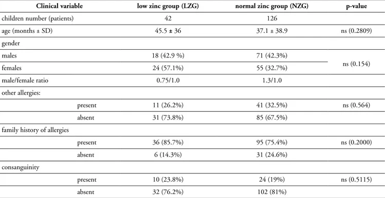
Serum zinc was low in 42 patients (25%), with level of 8.6 ± 1.1 µmoI/L (95% CI 8.1-9.0). Table I shows that there is no significant difference between the low-zinc group compared to the normal-zinc group in term of age, sex, the presence of other allergies or family history of allergic diseases.
AD severity scores, WBC, peripheral eosinophil counts, total IgE levels, number of positive food allergens, and serum zinc levels for both groups are shown in table II.
Table I - Demographic and clinical characteristics of AD children.
Clinical variable low zinc group (LZG) normal zinc group (NZG) p-value
children number (patients) 42 126
age (months ± SD) 45.5 ± 36 37.1 ± 38.9 ns (0.2809) gender males 18 (42.9 %) 71 (42.3%) ns (0.154) females 24 (57.1%) 55 (32.7%) male/female ratio 0.75/1.0 1.3/1.0 other allergies: present 11 (26.2%) 41 (32.5%) ns (0.564) absent 31 (73.8%) 85 (67.5%)
family history of allergies
present 36 (85.7%) 95 (75.4%) ns (0.2000)
absent 6 (14.3%) 31 (24.6%)
consanguinity
present 10 (23.8%) 24 (19%) ns (0.5115)
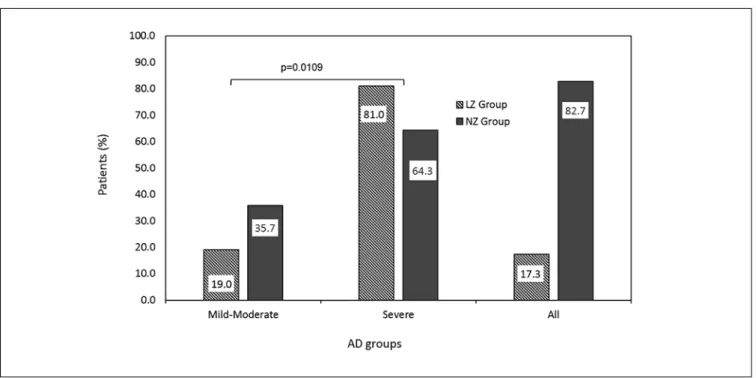
There is a significant difference between the proportion of chil-dren with severe AD in the low-zinc group compared to the normal-zinc group, as depicted in figure 1.
Discussion
The present study demonstrates that zinc deficiency is present in 25% of children with severe AD. They were older than chil-dren with normal serum zinc levels, and family history of aller-gic diseases. We observed that severe AD and high serum IgE are associated with zinc deficiency. The proportion of patients with severe (SCORAD index > 40) AD was significantly higher in
the low-zinc group compared to normal-zinc group. We did not find an association between low zinc in AD and co-existence of other allergic diseases, parental consanguinity, number of food allergens, WBC, and or peripheral blood eosinophilia.
The role of zinc as a micronutrient in AD has been investigated in a limited number of studies, with contradictory results, some investigators reported lower levels (5-7) whereas others found no differences (8-10). However, a recent systematic and me-ta-analysis on and atopic dermatitis conclude that low zinc is as-sociated with AD (11). In 1984, a case-controlled study on 144 children (65 AD, 79 controls) showed that the mean serum zinc of the AD patients was significantly lower (p < 0.0001) than
Figure 1 - Distribution of patients according to AD severity in low-zinc (LZG) compared to normal zinc group (NZG).
Table II - Disease severity index and laboratory variables of children with severe AD.
laboratory variable low zinc group normal zinc group p-value
total SCORAD (number) 63.9 ± 23.3 53.9 ± 25.0 0.0418
no. of positive food allergens 2.1 ± 1.6 1.9 ± 1.7 ns (0.7492) WBC (cells/ul) 11,855.6 ± 4,140.4 11,521.8 ± 3,968.1 ns (0.6476) AEC (cells/ul)1 963.2 ± 860.8 981.9 ± 964.9 ns (0.8256) serum IgE (KU/l) 6,818.7 ± 8,357.2 2,161.7 ± 4,841.1 0.001
vitamin D (ng/ml) 19.2 ± 8.8 8.9 ± 12.9 ns (0.381)
serum zinc (µmol/L) 8.6 ± 1.1 12.4 ± 1.9 < 0.001
that of the controls (12). Endre et al. study on 134 children who were admitted to hospital with AD, found 41 (29.1%) with low serum zinc levels (13). El-Kholy et al. demonstrated that in 18 AD children and 20 controls, serum and hair zinc levels were significantly lower (p < 0.0001) in AD children in compari-son to the control subjects (14). In contrast in 1990, David et al. study on 134 children with atopic eczema and 112 controls failed to prove the hypothesis that atopic eczema is associated with a non-specific decrease in the serum concentration of trace metals, including zinc (8). This study supports previous findings of Endre et al. that serum zinc was low in 29.1% of 134 children who were admitted to hospital with AD (8).
Our results revealed that AD severity is associated with low zinc in AD. These findings are compatible with previous studies (11,15). Karabacak et al. demonstrated in a recent, controlled study on AD patients (n = 67 study patients and 49 controls; mean age 17.9 years) that serum erythrocyte zinc level, but not the serum level, had a significant negative correlation with SCORAD index (15). Although some people take erythrocyte zinc level as the most sensitive clinical marker of zinc level in AD (8), in our study we found that serum zinc was low. This is the first study to assess the association between serum IgE levels and zinc levels in children with AD. Our study shows that study subjects with increased total serum IgE levels had significantly lower zinc levels. Recent data on the participants in the 5th Korean National Health and Nutrition Examination
Survey 2010 (n = 8,958), and on 1,867 adults, confirmed an as-sociation between serum zinc status and allergic sensitization in adults (16). There was a negative correlation between serum zinc levels and total IgE and allergen-specific IgE levels. A con-trolled study on children with food allergy (IgE- and non-IgE mediated), revealed that they had low serum levels of zinc (a cofactor of superoxide dismutase ) and selenium (a cofactor of glutathione peroxidase), and low concentrations of superoxide dismutase and glutathione peroxidase (17). These enzymes in-creased after elimination diet. Agin’s study on a total of 48 sub-jects with allergic (skin prick tested) asthma, of mean age 32.8 ± 9.9 years (range 15-48 years), showed that hypozincemic group (23%) had a markedly higher mean of total IgE level than nor-mozincemic controls (18). Using HR-1 hairless mice, mice fed a diet with low magnesium and zinc developed AD-like (skin dryness, wrinkle-like changes, scratching, reduced skin water content, high transepidermal water loss), and a significantly (p < 0.001) elevated serum IgE compared with control mice fed standard diet (19). Although the exact role of zinc in AD immu-nopathology is not well determined, it seems to work through immune regulation. Zinc deficiency was associated with im-mune dysregulation. Regulatory T (Treg) cells play a key role in immune suppression, promoting tolerance to allergens, and preventing allergic responses including the chronic skin inflam-mation in AD.
They regulate allergen-specific Th2 immune responses and B cell IgE production, block of naïve CD4+ Tconv cells conver-sion into allergen-specific Th2 T cells, control B cells, and block their IgE production (20).
The percentage of Tregs in allergic patients (2.3%) was signifi-cantly lower in AD patients in comparison to healthy controls (4.6%, p = 0.003), even in the asymptomatic AD or food allergy subjects (21). Atopic food-allergic children also had decreased percentages of Treg cells compared with healthy age-matched healthy controls (22). In a recent mouse model study, aller-gen-specific immunotherapy revealed local suppression of Th2 and infiltration of Treg cells into the skin, and induced local and systemic Treg cells and regulatory NK cells (23). In addition to the results that Treg cells percentage and TGF-β level were de-creased in AD lesions, Treg cells percentage negatively correlated with AD severity score (24).
Conversely, zinc supplementation was demonstrated to restore immune regulatory mechanism. In vitro, zinc supplementa-tion significantly diminished the differentiasupplementa-tion of Th9 cells, key players promoting immune-mediated diseases, including allergic inflammation (25), and was capable, by modulating molecular targets Foxp3, KLF-10, and IRF-1, to ameliorate the immune reaction by enhancement of antigen-specific iTreg cells (26). Zinc was also able to induce dendritic cell tolero-genic phenotype and enhanced regulatory T cell-Th17 balance (27). A study on peripheral blood mononuclear cells (PBMCs) from non-atopic and atopic subjects treated with timothy grass allergen pre-incubated with or without zinc, revealed that zinc enhanced regulatory T cell numbers and suppressed their pro-liferation through a significant shift from IL-10 to the Th1 cy-tokine IFN-γ (28).
The strength of this study is that it includes a large sample of AD children, and that all were evaluated by the same physician (ME) thus eliminating any inter-rater difference during AD SCORAD assessment. It may appear that zinc testing in AD children is a possible bias. In fact, it is not. We were prompted to study zinc level as part of workup of poor weight and linear growth in these severe AD children (29). The main limitations of this study include its retrospective design. The rate of zinc de-ficiency might be overrepresented among AD children as these cases are referred to a tertiary care center, but the study con-centrates on those with severe form of AD. It may appear that serum zinc level is a limitation; however, there are numerous studies that used serum samples, not hair samples, as a valid test for determining zinc levels in allergic diseases, including AD.
Conclusions
Zinc deficiency is quite common among AD children. Severe AD and high total IgE are risk factors associated with zinc defi-ciency. In severe AD poorly responsive standard therapy, an
ad-junct oral zinc supplementation might be warranted to reduce disease severity.
Acknowledgements
This study was supported in part by Research Medical Center of Hamad Medical Corporation (RMC No. 14193/14).
Conflict of interest
The authors declare they have no conflict of interest.
References
1. Maywald M, Wessels I, Rink L. Zinc Signals and Immunity. Int J Mol Sci 2017. doi:10.3390/ijms18102222.
2. Sanna A, Firinu D, Zavattari P, Valera P. Zinc status and auto-immunity: a systematic review and meta-analysis. Nutrients 2018; 10(1):68.
3. Arkwright PD, Motala C, Subramanian H et al. Management of difficult-to-treat atopic dermatitis. J Allergy Clin Immunol Pract 2013; 1(2):142-151.
4. Kim KH. Clinical pearls from atopic dermatitis and its infectious complications. Br J Dermatol 2014; 170(Suppl 1):25-30.
5. Toyran M, Kaymak M, Vezir E et al. Trace element levels in chil-dren with atopic dermatitis. J Investig Allergol Clin Immunol 2012; 22(5):341-344.
6. Lin CN, Wilson A, Church BB et al. Pediatric reference intervals for serum copper and zinc. Clin Chim Acta 2012; 413(5-6):612-615.
7. Kim JE, Yoo SR, Jeong MG et al. Hair zinc levels and the efficacy of oral zinc supplementation in patients with atopic dermatitis. Acta Derm Venereol 2014; 94(5):558-562.
8. David TJ, Wells FE, Sharpe TC et al. Serum levels of trace metals in children with atopic eczema. Br J Dermatol 1990; 122(4):485-489.
9. Bruske K, Salfeld K. (Zinc and its status in some dermatologic dis-eases--a statistical assessment). Z Hautkr 1987; 62Suppl1:125-131. 10. Hinks LJ, Young S, Clayton B. Trace element status in eczema and
psoriasis. Clin Exp Dermatol 1987; 12(2):93-97.
11. Gray NA, Dhana A, Stein DJ, Khumalo NP. Zinc and atopic der-matitis: a systematic review and meta-analysis. J Eur Acad Derma-tol Venereol 2019; 33(6):1042-1050.
12. David TJ, Wells FE, Sharpe TC, Gibbs AC. Low serum zinc in children with atopic eczema. Br J Dermatol 1984; 111(5):597-601.
13. Endre L, Gergely A, Osvath P et al. (Incidence of food allergy and zinc deficiency in children treated for atopic dermatitis). Orvosi hetilap 1989; 130(46):2465-2469.
14. el-Kholy MS, Gas Allah MA, el-Shimi S et al. Zinc and copper status in children with bronchial asthma and atopic dermatitis. J Egypt Public Health Assoc 1990; 65(5-6):657-668.
15. Karabacak E, Aydin E, Kutlu A et al. Erythrocyte zinc level in pa-tients with atopic dermatitis and its relation to SCORAD index. Adv Dermatol Allergol /Postȩpy Dermatologii i Alergologii 2016; 33(5):349-352.
16. Seo H-M, Kim YH, Lee JH et al. Serum Zinc Status and Its Associ-ation with Allergic SensitizAssoci-ation: The Fifth Korea NAssoci-ational Health and Nutrition Examination Survey. Sci Rep 2017; 7:12637. 17. Kamer B, Wąsowicz W, Pyziak K et al. Role of selenium and zinc
in the pathogenesis of food allergy in infants and young children. Arch Med Sci 2012; 8(6):1083-1088.
18. Agin Kh. A survey on zinc status among chronic allergic asthma. Int J Forensic Med Toxicol Sci 2015; 5(1):1-7.
19. Makiura M, Akamatsu H, Akita H et al. Atopic dermatitis-like symptoms in HR-1 hairless mice fed a diet low in magnesium and zinc. J Int Med Res 2004; 32(4):392-399.
20. Noval Rivas M, Chatila TA. Regulatory T cells in allergic diseases. J Allergy Clin Immunol 2016; 138(3):639-652.
21. Stelmaszczyk-Emmel A, Zawadzka-Krajewska A, Szypowska A et al. Frequency and activation of CD4+CD25 FoxP3+ regulatory T cells in peripheral blood from children with atopic allergy. Int Arch Allergy Immunol 2013; 162(1):16-24.
22. Prince BT, Devonshire AL, Erickson KA et al. Regulatory T-cell populations in children are affected by age and food allergy di-agnosis. J. Allergy Clin. Immunol. 2017; 140(4):1194-1196.e16. 23. Shin JU, Kim SH, Noh JY et al. Allergen-specific immunotherapy
induces regulatory T cells in an atopic dermatitis mouse model. Allergy 2018; 73(9):1801-1811.
24. Ma L, Xue H-B, Guan X-H et al. The Imbalance of Th17 cells and CD4(+) CD25(high) Foxp3(+) Treg cells in patients with atopic dermatitis. J Eur Acad Dermatol Venereol 2014; 28(8):1079-1086. 25. Maywald M, Wang F, Rink L. Zinc supplementation plays a crucial
role in T helper 9 differentiation in allogeneic immune reactions and non-activated T cells. J Trace Elem Med Biol 2018; 50:482-488.
26. Maywald M, Rink L. Zinc supplementation induces CD4(+) CD25(+)Foxp3(+) antigen-specific regulatory T cells and suppress-es IFN-gamma production by upregulation of Foxp3 and KLF-10 and downregulation of IRF-1. Eur J Nutr 2017; 56(5):1859-1869. 27. George MM, Subramanian Vignesh K, Landero Figueroa JA et al.
Zinc induces dendritic cell tolerogenic phenotype and skews regu-latory T Cell-Th17 balance. J Immunol 2016; 197(5):1864-1876. 28. Rosenkranz E, Hilgers RD, Uciechowski P et al. Zinc enhances
the number of regulatory T cells in allergen-stimulated cells from atopic subjects. Eur J Nutr 2017; 56(2):557-567.
29. Ehlayel M, Ashraf S, De Sanctis V. Linear growth and nutrition-al parameters in adolescents with severe atopic dermatitis. Rivista Italiana di Medicina dell’Adolescenza 2016; 14(1):19-22.