Korean Circulation Journal
Introduction
Diabetes mellitus (DM) has been increasing in both developed and developing communities as a result of epidemic obesity and increas-ingly sedentary lifestyles.1) The relationship between DM and
cardio-vascular diseases2) and the fact that cardiovascular diseases are the http://dx.doi.org/10.4070/kcj.2013.43.2.82
Print ISSN 1738-5520 • On-line ISSN 1738-5555
Evaluation of Cardiac Functions with Tissue
Doppler Imaging in Prediabetic Subjects
Mustafa Kanat, MD
1, Seref Vardi, MD
2, Huseyin Arinc, MD
3, Huseyin Gunduz, MD
3,
Alim Erdem, MD
3, and Yalcin Karagoz, MSc PStat
21Department of Internal Medicine, Istanbul Medipol University, Istanbul,
2Departments of Internal Medicine and 3Cardiology, Izzet Baysal Medical School, University of Abant Izzet Baysal, Bolu, Turkey
Background and Objectives: The aim of the present study was to evaluate left ventricle systolic and diastolic function, using tissue Doppler echocardiography (TDE), in relation to blood glucose status in prediabetic patients who had no evidence of heart disease by conventional echocardiography (CE).
Subjects and Methods: We included 60 patients (30 female, 30 male) and 20 healthy controls (10 male, 10 female). All participants were randomised into four groups according to their oral glucose tolerance test. Group-I consisted of those patients who had only impaired fasting glucose (IFG). group-II consisted of patients who had only impaired glucose tolerance (IGT) and group-III consisted of patients who had both IFG and IGT, that is so-called combined glucose intolerance. Group-IV included the healthy controls. All subjects underwent both CE and TDE.
Results: No significant differences were found among the four groups in terms of CE. There was no significant difference between group-IV and group-I with respect to the early peak diastolic velocity (Ea) of medial mitral annulus (11.65±0.66 vs. 9.72±1.58, p>0.05), whereas a statistically significant difference was found between group-IV and group-II (11.65±0.66 vs. 9.06±1.07, p<0.001) and between group-IV and group-III (11.65±0.66 vs. 9.74±1.09, p<0.05).
Conclusion: Diastolic myocardial dysfunction in prediabetic patients may be identified by quantitative TDE before the appearance of CE in-dices of myocardial dysfunction. (Korean Circ J 2013;43:82-86)
KEY WORDS: Type 2 diabetes mellitus; Diabetic cardiomyopathies; Tissue Doppler imaging; Glucose intolerance.
Received: February 18, 2011 Revision Received: April 11, 2011 Accepted: April 24, 2012
Correspondence: Mustafa Kanat, MD, Department of Internal Medicine,
Medipol Mega Hospital Complex, Bag˘cilar 34214 I˙stanbul, Turkey Tel: 90 542 313 1400, Fax: 90 212 460 7057
E-mail: [email protected]
• The authors have no financial conflicts of interest.
This is an Open Access article distributed under the terms of the Creative Commons Attribution Non-Commercial License (http://creativecommons. org/licenses/by-nc/3.0) which permits unrestricted non-commercial use, distribution, and reproduction in any medium, provided the original work is properly cited.
major reason for morbidity and mortality in diabetics have been kn-own for a long period of time.3) Heart failure is a common and
seri-ous complication of diabetes. The incidence of cardiomyopathy-in-duced congestive heart failure (CHF) has been found to be increased in diabetic patients even in the absence of coronary atherosclero-sis.4-6) Diabetic cardiomyopathy can be described as abnormalities in
myocardial function or structure in the absence of hypertension (HT), alongside coronary artery disease (CAD) and serious valvular heart disease. The development of diabetic cardiomyopathy has been con-sidered to be a multifactorial condition. Increased stiffness due to the accumulation of connective tissue and insoluble collagen on left ventricle (LV) wall,7) autonomic dysfunction,8) endothelial
dysfunc-tion, impairment in various ligand sensitivity9) and anomalies in the
proteins which arrange ion flow, particularly intracellular calcium, are some of the factors that play a role in development of disease.10)11) In
early stages of diabetic cardiomyopathy, the systolic and diastolic functions of LV were found to be preserved on conventional echo-cardiography (CE). Diastolic dysfunction can be sensitively detected
http://dx.doi.org/10.4070/kcj.2013.43.2.82
www.e-kcj.org
at the early stage by tissue Doppler echocardiography (TDE).12)13)
Dia-stolic dysfunction, syDia-stolic dysfunction and myocardial structural changes are three main clinicopathologic features of diabetic car-diomyopathy.
The basis of noninvasive evaluations of diastolic dysfunction is constituted by Doppler studies of transmitral flow, mitral flow velo-cities, deceleration times, isovolumic relaxation time (IVRT) and flow patterns. As the degree of diastolic dysfunction deteriorates, the “early diastolic LV filling (E wave)” decreases and a delayed relaxation pattern appears. However, by increasing left atrial pressure, the E wave can be normalized and turns into a mitral flow wave, in a so-called ‘pseudonormal pattern’ which cannot be distinguished from that of a normal wave. The advantages of this technique are that it achieves perfect temporal resolution and mono-directional correl-ation with progressive cardiac anomalies. In one study, the mitral ear-ly peak diastolic velocities (E) and late peak diastolic velocities (A) of 27 type 1 and 25 type 2 diabetic patients without HT and CAD were examined and the E/A ratio was found to be more markedly de-creased in type 2 diabetics.14) In another study, it was reported that
ventricular filling, particularly early peak filling velocity, was more significantly impaired in type 2 diabetics compared to type 1 diabe-tics.15) Several studies have demonstrated early impaired diastolic
function, despite systolic function parameters remaining normal, in diabetics. This finding may be related to using more sensitive tech-niques in the detection of diastolic dysfunction and the absence of sufficiently adequate techniques to evaluate systolic dysfunction.
Recently, a diabetes-specific cardiomyopathy has been demon-strated in diabetic patients; however the pathophysiology and diag-nostic criteria of this condition have not yet been elucidated. This condition has been described in diabetics without CAD, HT or valvu-lar heart disease, and in the early stage of the disease systolic func-tions are preserved and diastolic dysfunction develops. Although di-abetic cardiomyopathy develops in patients with overt DM, the stage of this condition has not yet been clarified in the prediabetic period. The aim of the present study was to evaluate whether tissue Dop-pler imaging can detect a pre-clinical impairment of diastolic func-tion in prediabetic patients with preserved systolic funcfunc-tions in CE, so as to detect possible early cardiac dysfunction.
Subjects and Methods
Study populationThe patients enrolled in our study were selected between May 2007 and May 2009. Taking excluding criteria into consideration, all par-ticipants were divided into four groups according to oral glucose tolerance test (OGTT). The healthy controls consisted of 20 patients (mean age: 46.40±4.64), who formed the fourth group (group 4).
Ex-clusion criteria were: HT or antihypertensive use, cardiac arrhythmia, presence or history of CAD, presence of findings consistent with CAD in electrocardiography, wall movement defect in CE, serious valvu-lar heart disease or existing artificial valve, smoking, acute disease or psychiatric disease, and poor image quality in echocardiography. Written informed consent was obtained from all the patients and the study was approved by the Abant Izzet Baysal University Ethics Committee.
Method of oral glucose tolerance test and laboratory analysis Oral glucose tolerance test was performed using 75 g of glucose. Before the start of OGTT, a polyethylene catheter was placed into an antecubital vein and blood samples were collected at 0, 30, 60, 90 and 120 minutes for the measurement of plasma glucose.
Samples for serum chemistry were analyzed by a central medical research laboratory. Glucose levels were measured using colorimet-ric enzymatic methods on the Abbott Architect C8200 analyzer (In-tegrated System for ABBOT Diagnostic, Montreal, Canada) and re-agents from the same manufacturer. All blood samples were collected at 08:00 a.m. after a 10 hours fasting period.
Conventional echocardiography
Transthoracic echocardiographic assessment was performed on patients in the left lateral decubitis position during normal respira-tion after five minute resting with a commercially available ultraso-und transducer and equipment (2.5 MHz transducer of Vingmed Vivid System III, Vingmed, General Electric, Horten, Norway). Imag-es were obtained in the standard views of the LV (parasternal long and short axis, apical four- and two-chamber and apical long-axis). Doppler echocardiography was performed in accordance with the recommendations of the American Society of Echocardiography.16)
Measurements were performed at the end of expirium in order to avoid possible artificial effects of respiration. Operators were blind-ed to patients and groups. Transmitral peak early diastolic velocity (E), peak late diastolic (A) velocity, E/A ratio, IVRT and E-wave Edec were measured. The ejection fraction (EF) was calculated as the percent-age change of left ventricular chamber volumes between diastole and systole from apical four- and two-chamber views using modified biplane Simpson’s rule based on three measurements. An EF >55% indicated a normal systolic function. An E-wave DT >140 and <220 ms, an E/A ratio >1 and <2, and IVRT <100 ms indicated normal dia-stolic function.17)
Tissue Doppler echocardiography
Pulsed-wave tissue Doppler measurements were conducted on all patients following CE technique. A total of six measurements were performed from medial and lateral sites of the annular area. Medial
mitral annular systolic velocity (Smed-a), medial mitral annular early (Emed-a) and late (Amed-a) diastolic velocities were measured. Emed- a/Amed-a ratios were detected. Lateral mitral annular systolic velo-city (Slat-a), lateral mitral annular early (Elat-a) and late (Alat-a) dias-tolic velocities were measured.
Elat-a/Alat-a ratios were detected. A total of three measurements were performed from the lateral myocardium. LV Lateral myocardial systolic velocity (Slat-m), lateral myocardial early (Elat-m) and late (Alat-m) diastolic velocities were measured. Elat-m/Alat-m ratios were detected. Velocity at any measurement of annulus was evalu-ated pathologically as <8 cm/sec.
Statistical analysis
Results are given as mean±SD. Kolmogorov-Smirnov test was used. Non-normally distributed data were analyzed by Kruskal-Wal-lis tests and normally distributed data were assessed by analysis of variance. A p<0.05. was considered statistically significant. Statisti-cal analysis was performed with StatistiStatisti-cal Package for the Social Sci-ences (SPSS) for Windows version 11.5 (SPSS Inc., Chicago, IL, USA).
Results
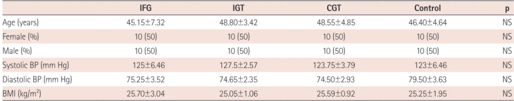
The main features of the subjects studied are summarized in Table Table 1. Demographic and clinical characteristics of groups
IFG IGT CGT Control p
Age (years) 45.15±7.32 48.80±3.42 48.55±4.85 46.40±4.64 NS Female (%) 10 (50) 10 (50) 10 (50) 10 (50) NS Male (%) 10 (50) 10 (50) 10 (50) 10 (50) NS Systolic BP (mm Hg) 125±6.46 127.5±2.57 123.75±3.79 123±6.46 NS Diastolic BP (mm Hg) 75.25±3.52 74.65±2.35 74.50±2.93 79.50±3.63 NS BMI (kg/m2) 25.70±3.04 25.05±1.06 25.59±0.92 25.25±1.95 NS
BP: blood pressure, BMI: Body Mass Index, IFG: impaired fasting glucose, IGT: impaired glucose tolerance, CGT: IFG+IGT, NS: not significant Table 2. CE parameters of groups
CE IFG IGT CGT Control p
LVEDD (mm) 51.35±2.08 50.16±1.96 49.60±2.25 52.20±1.73 NS LVESD (mm) 31.75±1.67 31.23±1.90 30.75±1.77 32.40±1.73 NS LAD (mm) 35.15±2.30 33.87±1.56 34.45±1.84 36.70±1.55 NS IVS (mm) 9.14±0.67 9.53±0.49 9.40±0.54 9.45±0.52 NS PW (mm) 9.12±0.82 8.87±0.65 9.35±0.51 9.50±0.56 NS LVEF (%) 68.15±2.61 66.89±2.73 67.30±3.05 67.45±2.68 NS IVRT (msn) 77.75±3.33 82.30±3.55 79.85±4.49 76.70±4.33 NS Evel (m/sn) 0.77±0.08 0.76±0.15 0.70±0.09 0.87±0.08 NS Avel (m/sn) 0.64±0.09 0.70±0.09 0.63±0.08 0.68±0.05 NS E/A ratio 1.18±0.24 1.07±0.13 1.14±0.22 1.29±0.11 NS Edec (msn) 169.95±21.52 200.05±25.64 179.65±38.06 186.70±14.01 NS
CE: conventionel echocardiographic, IFG: impaired fasting glucose, IGT: impaired glucose tolerance, CGT: IFG+IGT, NS: not significant, LVEDD: left ventricle end-diastolic diameter, LVESD: left ventricle end-systolic diameter, LAD: left atrial diameter, IVS: interventricular septum thickness, PW: posterior wall thick-ness, LVEF: left ventricular ejection fraction, IVRT: isovolumic relaxation time, Evel: transmitral early peak diastolic flow velocity, Avel: transmitral late peak diastolic flow velocity, Edec: deceleration time
Table 3. TDE parameters of groups
TDE IFG IGT CGT Control p
Sa 7.77±0.91 8.39±0.60 7.67±0. 89 7.90±0.57 NS
Ea 9.72±1.58 9.06±1.07* 9.74±1.09* 11.65±0.66* <0.05
Aa 10.34±0.99 10.19±0.95 10.16±1.07 9.06±0.66 NS
Ea/Aa 0.99±0.22 0.93±0.19* 1.02±0.24 1.35±0.11* <0.05
*p<0.05 when compared control group. TDE: tissue Doppler echocardiographic, IFG: impaired fasting glucose, IGT: impaired glucose tolerance, CGT: IFG+IGT, NS: not significant, Sa: mitral medial annulus peak systolic velocity, Ea: mitral medial annulus early peak diastolic velocity, Aa: mitral medial annulus late peak diastolic velocity
http://dx.doi.org/10.4070/kcj.2013.43.2.82
www.e-kcj.org
1. Baseline demographic and clinical characteristic were comparable across the four groups. The CE and TDE parameters of all groups are demonstrated in Table 2 and 3, respectively. The four groups were similar in terms of CE parameters. In TDE evaluation. The early peak diastolic velocity of medial mitral annulus (Ea) of impaired fasting glu-cose (IFG) patients were similar to controls (11.65±0.66 vs. 9.72± 1.58, p=0.084). However, statistically significant difference between controls and impaired glucose tolerance (IGT) patients (11.65±0.66 vs. 9.06±1.07, p≤0.001) and between controls and CGT patients (11.65±0.66 vs. 9.74±1.09, p=0.008) were found. However, there was no difference between groups in terms of measurements from the lateral mitral annulus (SlatA, ElatA, AlatA) and the lateral myo-cardium (SlatM, ElatM, AlatM).
Discussion
The present study has two significant findings. Firstly, it was sh-own that the process of cardiomyopathy initiates in IGT and CGT conditions before overt diabetes develops. In measurements by TDE, we detected that the systolic functions of prediabetics were preserv-ed whereas diastolic functions were impairpreserv-ed. This condition was found to be prominent in IGT and CGT patients. In a study perform-ed with TDE, García-Fernández et al.18) demonstrated that LV
dia-stolic dysfunction might be regional.18) Although in line with this view
we did detect diastolic dysfunction findings in the medial of mitral annulus, diastolic functions were within normal ranges in the lateral annulus and lateral myocardium in the present study. Secondly, we can propose that insulin resistance plays a pivotal role in the effects of diabetes on the myocardium. There is evidence that the cardiac muscle in type 2 diabetics is resistant to insulin, although underlying CAD was not excluded.19) In an animal study by Mizushige et al.20) it
was found that diastolic function in prediabetic rats might be as-sociated with insulin resistance.
The prevalence of CHF and diastolic dysfunction has been increas-ing in diabetic patients.21) LV diastolic dysfunction may present as
the first stage of CHF and is associated with high mortality/morbidi-ty.22) In various studies it has been demonstrated that diabetic
car-diomyopathy contributed to the incidence of increased heart failure in diabetics. Almost all these studies were conducted on overt dia-betics with normal systolic/diastolic function. However, this condi-tion has not been investigated in prediabetics such as those with IFG and IGT. Importantly, CE and normal cardiac functions have not been examined by TDE. Recently, noninvasive techniques that eval-uate diastolic function have improved. However, we do not have en-ough data about early stage of diabetic cardiomyopathy and the pathophysiology of this disease remains unclear.
If physiologic adaptation to metabolic changes and degenerative
changes, which comprise two main aspects of diabetic cardiomyop-athy, are treated in the early stages of diabetes, clinicians may be able to intervene in the progress of the disease. Studies on antihy-perglysemic treatment advocate the view that myocardial function and structural differences are correlated with glysemic control. Po-gátsa et al.23) investigated the effects of antihyperglysemic
treat-ment on diabetic dogs with serious hyperglicemia and it was found that LV passive elastic module (the marker of stiffness) and LV end-diastolic pressure were higher and cardiac output was lower in non-treated dogs. Similarly, in another study it was indicated that diabet-ic cardiomyopathy-induced changes in diabetdiabet-ic rats may be rever-sible with insulin treatment.24) Contrary to these findings, in a study
conducted on diabetic dogs Regan et al.25) suggested that diabetic
cardiomyopathy was not reversible with antihyperglycemic treat-ment.
The current study has some potential limitations that should be noted. First, the study sample size was small. Second, we did not measure the insulin sensitivity of subjects since insulin resistance and hyperinsulinemia are already present in patients with IGT, IFG and CGI. Third, one of the important supplementary methods for assessment of diastolic function is pulmonary venous flow velocity evaluation, which was not reported in this study. Although this stu-dy has several limitations, the results nevertheless provide important information about cardiac functions in prediabetic patients.
In conclusion, there as yet exists no effective treatment for dia-betic cardiomyopathy, which is one of the leading causes of morbi-dity and mortality in patients with overt diabetes. Therefore it is im-portant to prevent the occurrence or progression of the disease. Stu-dies have clearly demonstrated that prediabetes is a reversible condi-tion. In the present study, we obtained data indicating that the effect of prediabetes may be reversible. This finding needs be supported by standardized methods and studies conducted on a wide number of patient populations.
References
1. King H, Rewers M. Global estimates for prevalence of diabetes mellitus and impaired glucose tolerance in adults: WHO Ad Hoc Diabetes Re-porting Group. Diabetes Care 1993;16:157-77.
2. Galderisi M, Anderson KM, Wilson PW, Levy D. Echocardiographic evi-dence for the existence of a distinct diabetic cardiomyopathy (the Fram-ingham Heart Study). Am J Cardiol 1991;68:85-9.
3. Kleinman JC, Donahue RP, Harris MI, Finucane FF, Madans JH, Brock DB. Mortality among diabetics in a national sample. Am J Epidemiol 1988;
128:389-401.
4. Kannel WB, McGee DL. Diabetes and cardiovascular disease: the Fram-ingham Study. JAMA 1979;241:2035-8.
5. Shindler DM, Kostis JB, Yusuf S, et al. Diabetes mellitus, a predictor of morbidity and mortality in the Studies of Left Ventricular Dysfunction
http://dx.doi.org/10.4070/kcj.2013.43.2.82 www.e-kcj.org
(SOLVD) Trials and Registry. Am J Cardiol 1996;77:1017-20.
6. Piccini JP, Klein L, Gheorghiade M, Bonow RO. New insights into dia-stolic heart failure: role of diabetes mellitus. Am J Med 2004;116(Suppl
5A):S64-75.
7. Rodrigues B, Cam MC, McNeill JH. Myocardial substrate metabolism: implications for diabetic cardiomyopathy. J Mol Cell Cardiol 1995;27:
169-79.
8. Fang ZY, Prins JB, Marwick TH. Diabetic cardiomyopathy: evidence, me-chanisms, and therapeutic implications. Endocr Rev 2004;25:543-67.
9. Bell DS. Diabetic cardiomyopathy. A unique entity or a complication of coronary artery disease? Diabetes Care 1995;18:708-14.
10. Golfman LS, Takeda N, Dhalla NS. Cardiac membrane Ca(2+)-transport in alloxan-induced diabetes in rats. Diabetes Res Clin Pract 1996;31
(Suppl):S73-7.
11. Tahiliani AG, McNeill JH. Diabetes-induced abnormalities in the myocar-dium. Life Sci 1986;38:959-74.
12. Sutherland GR, Stewart MJ, Groundstroem KW, et al. Color Doppler myocardial imaging: a new technique for the assessment of myocar-dial function. J Am Soc Echocardiogr 1994;7:441-58.
13. Dokainish H. Tissue Doppler imaging in the evaluation of left ventric-ular diastolic function. Curr Opin Cardiol 2004;19:437-41.
14. Robillon JF, Sadoul JL, Jullien D, Morand P, Freychet P. Abnormalities suggestive of cardiomyopathy in patients with type 2 diabetes of rel-atively short duration. Diabete Metab 1994;20:473-80.
15. Astorri E, Fiorina P, Contini GA, et al. Isolated and preclinical impair-ment of left ventricular filling in insulin-dependent and non-insulin-dependent diabetic patients. Clin Cardiol 1997;20:536-40.
16. Sahn DJ, DeMaria A, Kisslo J, Weyman A. Recommendations regarding quantitation in M-mode echocardiography: results of a survey of echo-cardiographic measurements. Circulation 1978;58:1072-83.
17. How to diagnose diastolic heart failure: European Study Group on Dia-stolic Heart Failure. Eur Heart J 1998;19:990-1003.
18. García-Fernández MA, Azevedo J, Moreno M, et al. Regional diastolic function in ischaemic heart disease using pulsed wave Doppler tissue imaging. Eur Heart J 1999;20:496-505.
19. Iozzo P, Chareonthaitawee P, Dutka D, Betteridge DJ, Ferrannini E, Camici PG. Independent association of type 2 diabetes and coronary artery disease with myocardial insulin resistance. Diabetes 2002;51:
3020-4.
20. Mizushige K, Yao L, Noma T, et al. Alteration in left ventricular diastolic filling and accumulation of myocardial collagen at insulin-resistant prediabetic stage of a type II diabetic rat model. Circulation 2000;101:
899-907.
21. Poirier P, Bogaty P, Garneau C, Marois L, Dumesnil JG. Diastolic dys-function in normotensive men with well-controlled type 2 diabetes: importance of maneuvers in echocardiographic screening for preclini-cal diabetic cardiomyopathy. Diabetes Care 2001;24:5-10.
22. Bhatia RS, Tu JV, Lee DS, et al. Outcome of heart failure with preserved ejection fraction in a population-based study. N Engl J Med 2006;355:
260-9.
23. Pogátsa G, Bihari-Varga M, Szinay G. Effect of diabetes therapy on the myocardium in experimental diabetes. Acta Diabetol Lat 1979;16:
129-38.
24. Litwin SE, Raya TE, Anderson PG, Daugherty S, Goldman S. Abnormal cardiac function in the streptozotocin-diabetic rat. Changes in active and passive properties of the left ventricle. J Clin Invest 1990;86:481-8.
25. Regan TJ, Wu CF, Yeh CK, Oldewurtel HA, Haider B. Myocardial compo-sition and function in diabetes: the effects of chronic insulin use. Circ Res 1981;49:1268-77.