281
Turkish Neurosurgery 2009, Vol: 19, No: 3, 281-284
Önder OKAY1 Ergun DA⁄LIO⁄LU2 Cengiz YAKICIER3 Deniz ÜREN4 Ali DALGIÇ5 Fikret ERGÜNGÖR6
1,2,5,6Ankara Numune Education and Research Hospital, Neurosurgery Department, Ankara, Turkey 3Bilkent University, Department of
Molecular Biology and Genetics, Ankara, Turkey
4Bilkent University, Biotechnology Research and Development Center, Bilkent, Ankara, Turkey
Received : 02.03.2009 Accepted : 27.04.2009
Correspondence address: Ergün DAĞLIOĞLU
E-mail: [email protected]
Choroid Plexus Papillomas in
two Siblings: Case Report
‹ki Kardeflte Görülen Koroid Pleksus
Papillomu: Olgu Sunumu
ABSTRACT
Choroid plexus papilloma (CPP) is a rare, benign epithelial brain tumor of the nervous system seen particularly in infants. Familial cases are extremely uncommon. Some other form of malignant tumors was noted in the relatives of patients with CPPs, and some genetic defects regarding this coincidence were reported in the literature. These neoplasms are occasionally bilateral and hydrocephalus is an associated sign in most of the cases. We report three lateral ventricle CPPs in two siblings, at the age of 7 month and 2 years respectively. All tumors were resected with parietotemporal craniotomy and a superior temporal sulcus approach to the lateral ventricle. To avoid a concomitant need of vent-riculoperitoneal shunt insertion, external ventricular drainage was inserted for a week in the postoperative period relieving symptoms of hydrocephalus. Search for a hereditary defect in the p53 gene of the second infant (7 months old) revealed no mutation. Postoperative courses were uneventful and the patients were followed for three years without any recurrence. Bilateral CPPS are rare and unusual in two siblings. A genetic predisposition such as the p53 mutation should be investigated in bilateral CPPs in particular.
KEY WORDS: Choroid plexus, Papilloma, Familiar, p53-Mutation, Lateral ventricle, Hydrocephalus
ÖZ
Koroid pleksus papillomu (KPP) özellikle bebeklerde görülen sinir sisteminin nadir bir epitelyal beyin tümörüdür. Ailesel olgular çok nadirdir. Bu insidans dikkate alındığında KPP olgularının birinci dereceden akrabalarında bazı diğer malign tümörler ve bazı genetik defektler literatürde bildirilmiştir. Bu tümörler çok nadiren bilateraldir ve olguların büyük bir kısmında hidrosefali eşlik edebilir. Bu yazıda 2 yaşında ve 7 aylık olan iki kardeşte görülen 3 KPP olgusu bildirilmektedir. Tüm tümörler parietotemporal kraniotomi ve süperior temporal sulkus yaklaşımı ile eksize edilmiştir. Herhangi bir ventriküloperi-toneal şant gereksiniminden kaçınmak ve postoperatif dönemde hidrosefali semptomlarının kontrolü için 1 hafta eksternal ventriküler drenaj uygulanmıştır. 7 aylık ikinci olguda p53 geninde olası bir defekt araştırıldı ancak herhangi bir mutasyon saptanamadı. Postoperatif dönemde herhangi bir komplikasyon gelişmedi ve hastalar 3 yıl rekürens açısından yakın izleme alındı. Bilateral KPP nadir olup iki kardeşte görülmesi nadirdir. Özellikle bilateral KPP olguları p53 gen mutasyonu açısından araştırılmalıdır.
ANAHTAR SOZCÜKLER:Koroid pleksus, Papillom, Ailesel, p53 mutasyonu, Lateral ventrikül, Hisdrosefali
INTRODUCTION
Choroid plexus papillomas (CPPs) are congenital neoplasms that are more frequently recorded in the pediatric population. They constitute 3% of all primary pediatric cerebral neoplasms (8, 22). Patients usually present with epileptic seizures and symptoms of hydrocephalus. They make up the most common ventricular tumor of childhood. About two thirds of choroid plexus papillomas are localized in the lateral ventricles (8,13,22). Genetic transition (3,11,17,19,23,24) or bilateral presentation is extremely rare and less than 10 cases have been reported previously (1,4-6,18). Here we report two siblings operated for unilateral and bilateral CPPs.
Case 1
A 2-year-old male infant was admitted with vomiting and epileptic seizures noted within the last 2 weeks. Neurological examination, clinical and family history were completely normal. An intensely enhancing mass in the right lateral ventricle trigone and hydrocephalus were noted on cranial CT. MRI showed a right lateral ventricle mass extending into the third ventricle and the preliminary diagnosis was CPP (Figure 1A). A right parietotemporal craniotomy was performed and the lesion was approached through the posterior portion of the superior temporal sulcus. A grayish-pink lobulated mass was removed grossly as total. An external ventricular drainage system was inserted in case of postoperative hydrocephalus. Drainage was removed a week later and the patient was discharged without any neurological deficit. Histopathological sections revealed typical findings of CPP. The antiepileptic drug was discontinued after 3-year seizure-free follow-up. MRI performed three years after surgery demonstrated total removal without any remnant and hydrocephalus (Figure 1B).
Case 2
A 7-month-old male infant experienced a seizure characterized with loss of consciousness and staring at a fixed point. His parents brought the child to the hospital since their elder son had been operated for the same symptom (Case 1). Further family investigation revealed no history of malignancy or a similar pathology. Neurological examination was unremarkable. CT demonstrated intensely enhancing bilateral lateral ventricle mass. On MRI, the lesions filled the lateral ventricles completely 282
Turkish Neurosurgery 2009, Vol: 19, No: 3, 281-284 Okay: Choroid Plexus Papillomas in two Siblings
and hydrocephalus was prominent (Figure 2A). The infant was operated through superior temporal sulcus approaches bilaterally with an interval of 3 weeks between the operations. The excised lesions were put into liquid nitrogen directly after removal in order to perform genetic studies. Histopathological examination was again compatible with CPP. The patient was discharged 5 weeks after his admission without any neurological deficit. Imaging studies performed 2 years after surgery demonstrate complete removal of both tumors (Figure 2B). Study for a hereditary p53 gene mutation was negative shown by genetic analysis (Figure 3).
DISCUSSION
Choroid plexus papillomas are benign neoplasms which originate from neuroectodermal tissues. They constitute about 0.5 % of whole intracranial tumors.
Figure 1: A. Axial T1-weighted precontrast image demonstrating right lateral ventricular mass and mildly dilated ventricles. B. Postoperative axial T1-weighted postcontrast image shows complete removal of right ventricular mass.
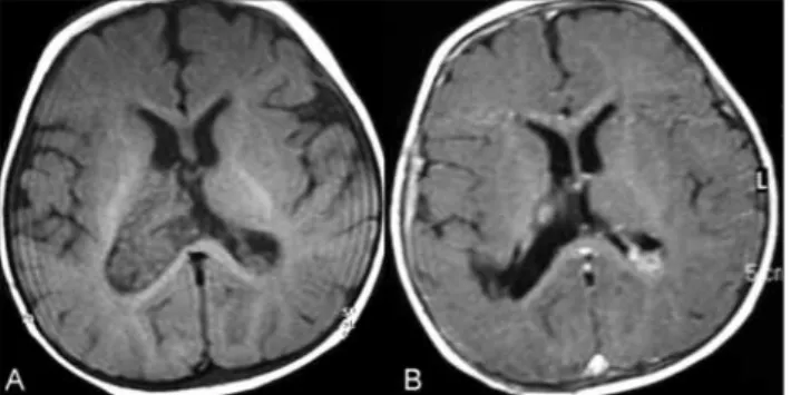
Figure 2: A. Axial T1-weighted postcontrast image demonstrating bilateral trigonal intensely enhanced mass and moderate signs of hydrocephalus. B. Postoperative coronal T1-weighted postcontrast image shows removal of both ventricular masses with a small remnant in the right lateral ventricular mesial wall.
Genetic transition and p53 mutation (location is at locus p53, 191170.0036) have infrequently been reported (17,23). Moreover, hSFN5/IN/1 somatic mutations of chromosome 22 were reported in CPP, atypical CPP and choroid plexus carcinomas were reported in the same family recently (19). CPPs in siblings or identical twins were reported twice previously (3,11). Other malignant tumors were noted in the relatives of patients with CPPs and some genetic defects regarding this coincidence were reported (6,19). We analyzed the p53 gene, which is a well known tumor suppressor gene, for the presence of germline mutations that can be associated with CPP. All coding exons of p53 were sequenced in both patients, which did not reveal any pathogenic mutations. An example of the sequence data where a previously reported mutation at codon 248 (CGG>TGG; Arg>Trp) resides is shown in Figure 3. Other tumor suppressor genes like hSFN5/IN/1 or oncogenes may have played a role in the development of the CPP in these two siblings. Choroid plexus papillomas are usually localized in the lateral ventricles in children and fourth ventricle in adults (3,4,8,22). However, true bilateral lateral ventricle CPPs is rare (4,5,13,18). True bilateral CPPs in two siblings had not been reported until now. At the operation, we did not find any difference with regard to their surgical removal when compared to unilateral CPPs but one should be aware of this finding for operative planning. Another important point with regard to bilateral localization is to differentiate the contralateral papilloma from villous hypertrophy. Histopathological studies may be insufficient to differentiate these two entities and neuroradiological features may be used as an effective tool (7). Association of hydrocephalus, lobulated appearance and homogeneous enhancement are highly suspicious for CPPs.
Hydrocephalus is usually an associated feature of CPPs. In fact, the majority of tumors present with symptoms of hydrocephalus (6,14). Hydrocephalus
283
Turkish Neurosurgery 2009, Vol: 19, No: 3, 281-284 Okay: Choroid Plexus Papillomas in two Siblings
in these patients is thought to be caused by an overproduction of CSF as noted in diffuse villous hyperplasia, and should thus resolve if tumor resection is complete (2). The role of inflammation and obstructive causes remains to be proved. Meticulous microsurgical techniques aid in total removal of tumor as well as prevention of hydrocephalus. The use of temporary external ventricular drainage also assists in prevention of postoperative hydrocephalus as indicated in this report. Drainage of blood and postoperative collections could also contribute to prevention of complications like hydrocephalus and subdural effusions (15). Shunting is an alternative strategy but should be avoided if possible since drop metastases of CPP are a well-known feature (9,10).
Surgical resection is the preferred way of treatment for CPPs. Several surgical approaches were defined previously for lateral ventricle CPPs (12,15,16,20). We prefer a transsulcal approach from the most posterior portion of the superior temporal sulcus. This approach provides a direct access to the posterior one-third of the lateral ventricle, called the trigone. The safety of the approach depends upon the cerebral tissue dissected although this region is anatomically safe, particularly on the non-dominant hemisphere. Tacconi et al. reported a good outcome if the resection was complete with a recurrence rate of less than 10 % (22). With regard to bilateral CPPs, we advocate a time interval of 3 weeks between the two operations if possible. There seems to be a chance of cure and low risk of recurrence for CPPs if the resection is complete. Role of radiotherapy in CPP treatment is still controversial and it is better to reserve this alternative just for recurrent and residual lesions with a risk of high morbidity and mortality (21). Follow-up is therefore the best choice to handle these patients.
REFERENCES
1. Beppu T, Kagawa M, Shimizu T, Matsumori K, Kubo O: Case of congenital bilateral choroid plexus papilloma. (Jpn) (Abstract). No To Shinkei 24: 179–186, 1972
2. Britz GW, Kim DK, Loeser JD: Hydrocephalus secondary to diffuse villous hyperplasia of the choroid plexus. Case report and review of the literature. J Neurosurg 85: 689–691, 1996 3. Coons S, Johnson PC, Dickman CA, Rekate H: Choroid plexus
carcinoma in siblings: A study by light and electron microscopy with Ki-67 immunocytochemistry. J Neuropath Exp Neurol 48: 483–493, 1989
4. Di Rocco C, Iannelli A. Poor outcome of bilateral congenital choroids plexus papillomas with extreme hydrocephalus. Eur Neurol 37: 33–37, 1997
5. Erman T, Göçer AI, Erdoğan S, Tuna M, Ildan F, Zorludemir S: Choroid plexus papilloma of bilateral lateral ventricle. Acta Neurochir (Wien) 145: 139–143, 2003
6. Fujimura M, Onuma T, Kameyama M, Motohashi O, Kon H, Yamamoto K, Ishii K, Tominaga T: Hydrocephalus due to cerebrospinal fluid overproduction by bilateral choroid plexus papillomas. Childs Nerv Syst 20: 485–488, 2004
7. Hirano H, Hirahara K, Asakura T, Shimozuru T, Kadota K, Kasamo S, Kasamo S, Shimohonji M, Kimotsuki K, Goto M: Hydrocephalus due to villous hypertrophy of the choroids plexus in the lateral ventricles. Case report. J Neurosurg 80: 321–323, 1994
8. Horska A, Ulug AM, Melhem ER, Filippi CG, Burger PC, Edgar MA, Souweidane MM, Carson BS, Barker PB: Proton magnetic resonance spectroscopy of choroid plexus tumours in children. J Magn Reson Imaging 14: 78–82, 2001
9. Jinhu Y, Jianping D, Jun M, Hui S, Yepeng F: Metastasis of a histologically benign choroid plexus papilloma: Case report and review of the literature. J Neurooncol 83: 47–52, 2007 10. Kaptanoglu E, Tun K, Celikmez RC, Ozen O, Taskin Y: Spinal
drop metastasis of choroid plexus papilloma. J Clin Neurosci 14: 381–383, 2007
11. Komminoth R, Woringer E, Baumgartner J, Braun JP, LeMaistre D: Papillome intraventriculaire familial: Caracteristiques angiographiques. Neurochirurgie 11: 267–272, 1965
12. Koos WT, Spetzler RF, Lang J: Color atlas of microneurosurgery, New York: Georg Thieme Verlag Theme Medical Publishers Inc. 1993:85
13. Levy ML, Goldfarb A, Hyder DJ, Gonzales-Gomez I, Nelson M, Gilles FH, McComb JG: Choroid plexus tumours in children: significance of stromal invasion. Neurosurgery 48: 303–309, 2001
284
Turkish Neurosurgery 2009, Vol: 19, No: 3, 281-284 Okay: Choroid Plexus Papillomas in two Siblings
14. Miyagi Y, Natori Y, Suzuki SO, Iwaki T, Morioka T, Arimura K, Maeda Y, Shono T, Matsukado K, Sasaki T : Purely cystic form of choroid plexus papilloma with acute hydrocephalus in an infant. Case report. J Neurosurg 105(6 Suppl): 480–484, 2006 15. Nagib MG, O'Fallon MT: Lateral ventricle choroid plexus
papilloma in childhood: Management and complications. Surg Neurol: 54: 366–372, 2000
16. Raimondi AJ: Pediatric neurosurgery, New York: Springer Verlag, 1987:219–223
17. Rutherford J, Chu CE, Duddy PM, Charlton RS, Chumas P, Taylor GR, Lu X, Barnes DM, Camplejohn RS: Investigations on a clinically and functionally unusual and novel germline p53 mutation. Br J Cancer 86: 1592–1596, 2002
18. Sarkar C, Sharma MC, Gaikward S, Saharma C, Singh VP: Choroid plexus papilloma: A clinicopathological study of 23 cases. Surg Neurol 52: 37–39, 1999
19. Sevenet N, Lellouch-Tubiana A, Schofield D, Hoang-Xuan K, Gessler M, Birnbaum D, Jeanpierre C, Jouvet A, Delattre O: Spectrum of hSNF5/INI1 somatic mutations in human cancer and genotype-phenotype correlations. Hum Molec Genet 8: 2359–2368, 1999
20. Shillito J, Matson DD: An atlas of pediatric neurosurgical operations, Philadelphia: W.B. Saunders, 1982:366–367 21. Strojan P, Popoviç M, Surlan K, Jereb B: Choroid plexus
tumors: A review of 28-year experience. Neoplasma 51: 306–312, 2004
22. Tacconi L, Delfini R, Cantore G: Choroid plexus papillomas: Consideration of a surgical series of 33 cases. Acta Neurochir 138: 802–810, 1996
23. Vital A, Bringuier PP, Huang H, San Galli F, Rivel J, Ansoborlo S, Cazauran JM, Taillandier L, Kleihues P, Ohgaki H: Astrocytomas and choroid plexus tumors in two families with identical p53 germline mutations. J Neuropathol Exp Neurol 57: 1061–1069, 1998
24. Zwetsloot CP, Kros JM, Pazy Geuze HD: Familial occurrence of tumours of the choroid plexus. J Med Genet 28: 492–494, 1991