Received: 11 December 2014 Revised: 2 November 2015 Accepted: 3 November 2015 doi: 10.1259/bjr.20140842
Cite this article as:
Baskan O, Silav G, Bolukbasi FH, Canoz O, Geyik S, Elmaci I. Relation of apparent diffusion coefficient with Ki-67 proliferation index in meningiomas. Br J Radiol 2016; 89: 20140842.
FULL PAPER
Relation of apparent diffusion coefficient with Ki-67
proliferation index in meningiomas
1OZDIL BASKAN,MD,2GOKALP SILAV,MD,2FATIH HAN BOLUKBASI,MD,3OZLEM CANOZ,MD,1SERDAR GEYIK,MD and2ILHAN ELMACI,MD
1Department of Radiology, School of Medicine, Istanbul Medipol University, Istanbul, Turkey 2Department of Neurosurgery, School of Medicine, Istanbul Medipol University, Istanbul, Turkey 3Department of Medical Pathology, School of Medicine, Istanbul Medipol University, Istanbul, Turkey
Address correspondence to: Dr Ozdil Baskan E-mail:[email protected]
Objective: The purpose of this study was to investigate the relationship between Ki-67 proliferation indexes and apparent diffusion coefficient (ADC) values of low-grade and atypical/anaplastic (high-grade) meningiomas. Methods: Pre-operative diffusion-weighted imaging and histopathological evaluation of 44 patients with menin-giomas were performed retrospectively. Regions of interest (ROIs) were manually drawn on the ADC images. In total six ROI measurements were taken in three consecutive slices, and the average of the mean ADC value was used. The relationship between the ADC and Ki-67 values was investigated, and the ADC values of the low-grade and high-grade meningiomas were compared.
Results: 31 (70%) patients had low-grade the meningio-mas. 10 (23%) patients had atypical and 3 (7%) had anaplastic meningiomas. ADC values of the low-grade and high-grade meningiomas were 0.816 0.12 3 1023and 0.666 0.08 3 1023mm2s21, respectively. Ki-67 prolifer-ation indexes were 2.19%6 1.14% for low-grade and 11.20%6 9.80% for high-grade meningiomas. A statis-tically significant negative correlation between Ki-67 proliferation index and ADC values of the low-grade and
high-grade meningiomas was detected (r25 0.326, p , 0.001). High-grade meningiomas had lower ADC values than that of low-grade meningiomas. There was statistically significant difference between the ADC values of the low-grade and high-low-grade meningiomas (p , 0.001).
Conclusion: Our data provide an inverse correlation between the ADC and Ki-67 proliferation index values of meningiomas. ADC values can be used for histopath-ological characterization of the meningiomas and pre-surgical planning.
Advances in knowledge: The purpose of this study was to investigate the relationship between Ki-67 proliferation indexes and ADC values of low-grade and atypical/ anaplastic (high-grade) meningiomas. In addition, we compared the ADC and Ki-67 proliferative index values of the low-grade and atypical/anaplastic (high-grade) meningiomas. We concluded that there was an inverse correlation between the ADC and Ki-67 proliferation index values in meningiomas, and we have found statistically significant difference between the ADC values of the low-grade and high-low-grade meningiomas. ADC values can be used for histopathological characterization of the menin-giomas and pre-surgical planning.
INTRODUCTION
Meningiomas are generally slow-growing, intracranial neoplasms, which thought to be arisen from the arachnoid “cap” cells located at the outer layer of the arachnoid mater and the arachnoid villi.1,2 Meningiomas are the most common benign, extra-axial intracranial tumours ac-counting for about 35% of all tumours.3 They are more common in older population and females.3Meningiomas were divided into three grades according to the World Health Organization (WHO) 2007 classification. They were low grade (grade I), atypical (grade II) and anaplastic
1Atypical meningiomas consist of 4.7–7% and
meningiomas.1Low-grade meningiomas are slow growing and consist of 80–90% of all meningiomas.1,2
The recurrence is the main problem and increases patient morbidity and mortality. For that reason, pre-operative evaluation and characterization of meningiomas would be very important before resection, for an appropriate surgical and treatment planning. Atypical and anaplastic meningi-omas are more prone to recurrence.4After surgery, atypical and anaplastic meningiomas recur in about 40% and 50–80% within 5 years, respectively.5
On the other hand,
2007 classification1
mentioned that the presence of brain invasion in histologically benign variants and this group’s prognosis should be considered as grade II.6The recurrence cannot be explained with only the grades of the tumours. The tumours have a tendency to recur, which depends on the grade, brain infiltration, proliferative activity and extent of resection. Immunohistochemistry, using antibodies that are reactive against various proliferating cellular antigens, is one of the ways of determining proliferative activity in tumours.7 The Ki-67 monoclonal antibody (MIB-1) is an important marker of cellular proliferation, which has been widely used in the routine histological evaluation of differ-ent tumours.8Ki-67/MIB-1 monoclonal antibody, which is reactive against the nuclear antigen Ki-67, expressed during cell cycle (G1, S, G2 and M) but absent in G0.7The Ki-67 MIB-1-positive tumour cells are often correlated with the clinical course and prognosis of several types of human neoplasms. An elevated Ki-67 proliferation index is associ-ated with an increased recurrence rate and has prognostic value in meningiomas.2,5,7
On MRI, meningiomas present typically as well-circumscribed mass, with strong enhancement after gadolinium administra-tion, with a peritumoral rim and a dural tail. Atypical imaging features such as heterogeneous enhancement, marked perile-sional oedema, irregular cerebral surface, cystic appearance, intratumoral haemorrhage and parenchymal invasion can be detected but are not specific or reliable diagnostic features for the differentiation of the atypical and malignant meningiomas from benign lesions.9Diffusion-weighted imaging (DWI) is an imaging technique which shows abnormalities of tissue struc-ture by detecting changes in water mobility, which is not possible with other imaging techniques.10Diffusion-weighted (DW) MRI and apparent diffusion coefficient (ADC) meas-urements can be useful in determining tumour cell density and nucleus/cytoplasm ratio, which are related to malign potential of the tumours.5,10,11
The purpose of this study was to investigate the relationship between Ki-67 proliferation indexes and ADC values of low-grade and atypical/anaplastic (high-low-grade) meningiomas. In addition, we compared the ADC and Ki-67 proliferative index values of the low-grade and atypical/anaplastic (high-grade) meningiomas.
METHODS AND MATERIALS Subjects
The study included all patients who had the diagnosis of me-ningioma, had histopathological evaluation with Ki-67 pro-liferation indexes and had undergone pre-operative DWI in Medipol University Hospital between November 2012 and August 2014. None of the patients had undergone any other therapy before the surgery. The local institutional review board approved this retrospective study.
The MRI examinations of 44 patients, who had the inclusion criteria, were retrospectively evaluated. The mean age (6standard deviation) was 53.636 14.45 years with an age range of 28–80 years, and the female/male ratio was 29/15.
Imaging techniques
All MRI examinations were performed on a 3-T clinical scanner (Philips Achieva TX, Philips Healthcare, Best, Netherlands). MRI protocol included: axial and sagittal T1 weighted (T1W)
spin echo (SE) sequence with repetition time (TR)/echo time (TE)5 400/10 ms, slice thickness 5 5 mm, interslice gap5 1 mm and field of view (FOV) 5 24 cm; an axial T2
weighted (T2W) turbo spin echo sequence with TR/TE5 3000/ 80 ms, slice thickness5 5 mm, interslice gap 5 1 mm and FOV5 24 cm; and sagittal three-dimensional fluid-attenuated inversion-recovery sequences with TR/TE5 4500/270 ms and inversion time (TI)5 1650. After the administration of 0.1-mmol kg21gadolinium, contrast-enhanced axial and coronal T1W SE (TR/TE5 400/10 ms) images were obtained.
DWI was performed with a single-shot SE echoplanar imaging sequence with three gradient directions in the axial plan. The following parameters were used: TR/TE5 3190/90 ms, slice thickness5 5 mm, interslice gap 5 1 mm, FOV 5 24 cm and b-values5 0 and 1000 s mm22. ADC maps were automatically generated. We recalled all the results of all the patient exami-nations in order to re-evaluate on the workstation which was provided by the vendor.
Image evaluation
A radiologist who was blinded to the cases and unaware of the histopathological diagnosis randomly reviewed conventional and DW MR images. Regions of interest (ROIs) were manually drawn on the ADC images. The ADC measurements were taken on the workstation which was provided by the vendor. ROIs were drawn in the solid parts of the lesions and cystic–necrotic, and haemorrhagic and calcified parts were avoided. Those areas were identified on the T2W and contrast-enhanced T1W images (Figures 1and2). Totally six ROI measurements were taken in three consecutive slices, and the average of the mean ADC values was used. ROI areas ranged between 0.2 and 1 cm2. According to the size and morphology of the meningioma when three slices were not available, the ADC values of three ROIs in two slices were calculated. The mean ADC value of the normal white matter was calculated by the same method in the centrum semiovale. The mean ADC values of the peritumoral oedemas were calculated by the same method.
Pathology
The pathological diagnoses of the meningiomas were based on the WHO 2007 classification system for brain tumours.1
Tumour proliferation indexes were reported as the percentage of tumour cell nuclei labelled with the Ki-67 (clone MIB-1) monoclonal antibody in formalin-fixed paraffin tissue sections. For each case, areas with the highest number of positive-staining tumour nuclei were selected for counting. The low-grade me-ningiomas were classified into subgroups according to the WHO classification. For further evaluation, we grouped the meningi-omas as low grade (grade I) and high grade (grade II: atypical and grade III: anaplastic).
Statistical analysis
Comparison of the Ki-67 proliferation index values was made between low-grade and high-grade meningiomas, using an
independent unpaired Student’s t-test. Correlation coefficients were also calculated between the mean ADC and Ki-67 pro-liferation index values for all meningiomas using linear re-gression. Correlation analysis was performed using the Pearson’s product–moment correlation with statistical software. A value of p, 0.05 was considered statistically significant.
RESULTS
Totally 44 meningiomas were evaluated retrospectively. 31 (70%) patients had low-grade meningiomas. 10 (23%) patients had atypical meningiomas and 3 (7%) patients had anaplastic me-ningiomas (Table 1). The low-grade meningiomas consisted of 14 (45%) meningothelial, 10 (32%) transitional, 4 (13%) secretory, 2 (7%)fibroblastic and 1 (3%) angiomatous (Table 2). Oedema was detected in 17 meningiomas (39%). The mean ADC value (1.546 0.15 3 1023mm2s21) of the oedema was significantly higher than that of the normal white matter. There was no sta-tistically difference between the ADC values of the oedema of the low-grade and high-grade meningeal tumours (p. 0.05). ADC values of the low-grade and high-grade meningiomas were 0.816 0.12 3 1023 and 0.666 0.08 3 1023mm2s21, re-spectively. Ki-67 proliferation indexes were 2.196 1.14% for low-grade meningiomas and 11.206 9.80% for high-grade meningiomas. A statistically significant negative correlation
between Ki-67 proliferation index and ADC values of the low-grade and high-low-grade meningiomas was detected (r25 0.326, p, 0.001). The mean ADC of the white matter (WM) for low-grade and high-low-grade meningiomas were 0.736 0.03 3 1023 and 0.726 0.027 3 1023mm2s21, respectively. The ratios of the ADC values of the meningiomas to the white matter (ADCm/
ADCwm) were calculated. They were 1.126 0.18 for low-grade
meningiomas and 0.916 0.12 for high-grade meningiomas. A statistically significant negative correlation between Ki-67 proliferation index and ADCm/ADCwmvalues of the low-grade
and high-grade meningiomas was also detected (r25 0.297, p5 0.001) (Table 1).
In low-grade meningiomas, 24 lesions (78%) had an elevated ADC value compared with that of the normal white matter and low Ki-67 proliferation index (Figure 1). On the other hand, the majority (n5 11, 84%) of the high-grade meningiomas showed high Ki-67 proliferation index and a low ADC value compared with that of the normal white matter (Figure 2). There was statistically significant difference between the Ki-67 proliferation indexes of the low-grade and high-grade meningiomas (p5 0.007). There were some exceptions in low-grade and high-grade groups. Two of the meningothelial meningiomas with 4% Ki-67 proliferation index showed lower ADC values than that of the normal white matter (0.683 1023mm2s21). One of these
Figure 2. High-grade meningioma (arrows). (a) Contrast-enhanced coronal T1 weighted image shows enhancing extra-axial
meningioma in the frontal region. (b) Diffusion-weighted MR image. (c). Apparent diffusion coefficient map with region of interest measurement. Max, maximum; Min, minimum; StdDev, standard deviation.
Figure 1. Low-grade meningioma and meningothelial subtype (arrows). (a) Contrast-enhanced coronal T1weighted image shows
enhancing extra-axial meningioma in the right temporo-occipital region. (b) Diffusion-weighted MR image. (c). Apparent diffusion coefficient map with region of interest measurement. Max, maximum; Min, minimum; StdDev, standard deviation.
meningiomas showed progression in 1-year follow-up MRI. One high-grade meningioma had a low Ki-67 proliferation index (1%), with a high ADC value than that of the white matter (0.753 1023mm2s21). Unfortunately, we did not know the clinical course of this case.
High-grade meningiomas had a lower ADC values than that of low-grade meningiomas. There was a statistically significant difference between the ADC values of the low-grade and high grade meningiomas (p, 0.001). A statistically significant difference between the ADCm/ADCwmvalues of the low-grade
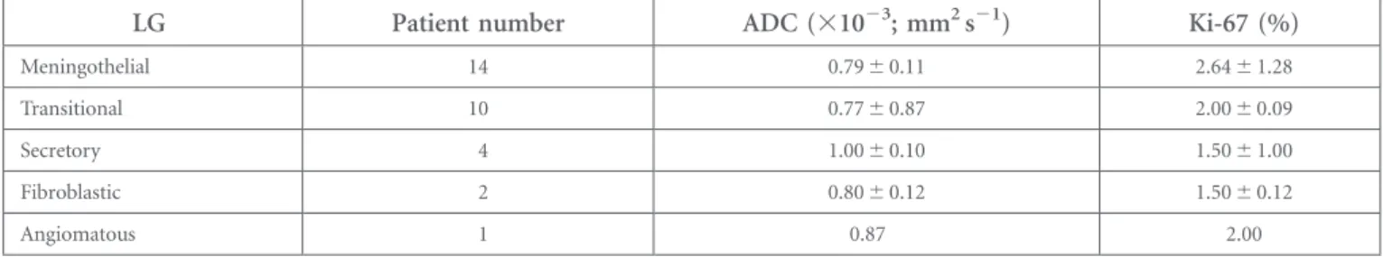
and high-grade meningiomas was also detected (p5 0.002). ADC values of the subtypes of the low-grade meningiomas were 0.796 0.11 3 1023mm2s21 for meningothelial, 0.776 0.8731023mm2s21 for transitional, 1.0060.10 31023mm2s21 for secretory and 0.806 0.12 3 1023mm2s21forfibroblastic. One angiomatous meningioma had an ADC value of 0.873 1023mm2s21 (Table 2). For the subgroups of the low-grade meningiomas, Ki-67 proliferation indexes were 2.646 1.28% for meningothelial, 2.006 0.09% for transitional, 1.50 6 1.00% for secretory, 1.506 0.12% for fibroblastic and 2.00% for an-giomatous meningiomas (Table 2). There was no statistically significant difference between the ADC values of the menin-gothelial (0.796 0.11 3 1023mm2s21) and transitional menin-giomas (0.776 0.87 3 1023mm2s21) (p5 0.664). Statistically significant differences were detected between the mean ADC values of the meningothelial (0.796 0.11 3 1023mm2s21) and secretory meningiomas (1.006 0.10 3 1023mm2s21) (p5 0.016) and between the mean ADC values of the transitional (0.776 0.873 1023mm2s21) and secretory meningiomas (1.006 0.103 1023mm2s21) (p5 0.017).
DISCUSSION
Meningiomas are one of the most common intracranial tumours. Although they are slow growing and most of them are low-grade, the recurrence is the main problem that determines the prognosis. Although tumour grade and subtype are strong prognostic factors in meningiomas, the recurrence is still un-predictable. The Ki-67 proliferation index is an important tool in addition to routine histological evaluation. The Ki-67 pro-liferation index correlates with increased risk of recurrence and appears as an important prognostic factor in meningiomas.5,12,13
The therapeutic approach can change according to the histo-pathological properties of the meningiomas. Determining the histopathological grades, types and proliferative activity of the meningiomas will help in pre-surgical planning, in predicting the prognosis and in the additional use of other therapies such as radiotherapy.14–17 MRI is an important tool for the diagnosis, but conventional MRI techniques are limited in differentiating the low-grade and high-grade (atypical and anaplastic) meningiomas.
DW MRI has been used for the diagnosis and follow-up of the brain tumours. Water motion diffusivity is greater in the ex-tracellular space than in the inex-tracellular space. The cellularity and the nucleus/cytoplasm ratio of the tumour will determine the restriction of the water molecules.10 Malignant lesions are densely cellular neoplasms, which have a greater restriction of water diffusion than less cellular tumours; this results in de-creased ADC values.10The use of ADC values in determining the histopathological grades of the various intracranial tumours was investigated.18,19
Table 1. Characteristics of low-grade (LG) and high-grade (HG) meningiomas
Findings LG HG p-value Patient number 31 13 ADC (31023; mm2s21) 0.816 0.12 0.666 0.08 ,0.001 Ki-67 (%) 2.196 1.14 11.206 9.80 0.007 ADCwm(31023; mm2s21) 0.736 0.03 0.726 0.027 .0.05 ADCm/ADCwm 1.126 0.18 0.916 0.12 0.002
ADC, apparent diffusion coefficient; ADCm/ADCwm, the ratios of the ADC values of the meningiomas to the white matter; ADCwm, the ADC value of the
white matter; Ki-67, Ki-67 proliferation index.
A statistically significant negative correlation between Ki-67 proliferation index and ADC values of the LG and HG meningiomas was detected (r2
5 0.326, p , 0.001).
Table 2. Apparent diffusion coefficient (ADC) and Ki-67 proliferation index (Ki-67) values of the subtypes of the low-grade (LG) meningiomas
LG Patient number ADC (31023; mm2s21) Ki-67 (%)
Meningothelial 14 0.796 0.11 2.646 1.28
Transitional 10 0.776 0.87 2.006 0.09
Secretory 4 1.006 0.10 1.506 1.00
Fibroblastic 2 0.806 0.12 1.506 0.12
We found that there was a statistically significant inverse cor-relation between ADC and Ki-67 proliferation index values in meningiomas (r25 0.326, p , 0.001). Two previous studies16,17 have investigated the relationship of ADC values and Ki-67 pro-liferation index in meningiomas. Tang et al17found statistically significant inverse correlation between ADC and Ki-67 proliferation values, which was consistent with our results. Ginat et al16 detected an inverse relationship between ADC and Ki-67 pro-liferation index values in high-grade meningiomas, which was not statistically significant. They included only the high-grades meningiomas in their study, and the average Ki-67 prolifera-tion index for atypical and anaplastic meningiomas were not significantly different.
Tang et al17had described patterns of relationships between the ADC and Ki-67 proliferation index values. Combination of high ADC values and low Ki-67 proliferation index values of low-grade meningiomas was the common pattern. In our study, 24 (78%) of the low-grade meningiomas showed an elevated ADC value com-pared with that of the normal white matter and low Ki-67 pro-liferation index, as generally expected according to their histopathological properties. On the other hand, two (6%) of the meningothelial meningiomas with 4% Ki-67 proliferation index showed lower ADC values than that of the normal white matter (0.6831023
mm2s21). One of these meningiomas showed pro-gression on 1-year follow-up MRI. The clinical course of the other menangioma was unknown. Thesefindings were consistent with the second described pattern.17 In the high-grade meningiomas, most of them had low ADC and high Ki-67 proliferation index values (84%). This pattern reflects the high cellularity and active proliferation activity2,5,7 and high risk of recurrence.5,12,13In our study group, we had no data that were consistent with the pattern of low ADC and low Ki-67 proliferation index values in aggressive meningiomas. One high-grade meningioma had a low Ki-67 pro-liferation index (1%), with a high ADC value than that of the white matter (0.7531023mm2s21). Unfortunately, the clinical course of this case was unknown. The lack of patients’ follow-up was the limitation of our study. The future prospective studies with larger patient groups could be useful for detecting the correlation of these patterns with the clinical course and long-term imagingfindings.
In the literature, there were studies that have investigated the use of DWI in determining the histopathological grades of the meningiomas.9,11,14,15,18–23The results were controversial. In our study, in addition, we compared the ADC values of the meningiomas. We have found statistically significant differ-ence in ADC and ADCm/ADCwm values between the
low-grade and high-low-grade meningiomas (p, 0.05). Some of these studies14,15,18,22investigating the effectiveness of the DWI in
differentiating the subtypes of benign meningiomas had dif-ferent findings. Hakyemez et al14had found significant dif-ference between the angiomatous and the meningotelial and transitional meningiomas. Thesefindings were not consistent with that of other studies.15,18,22In our study, the low-grade meningiomas consisted of 14 meningothelial, 10 transitional, 4 secretory, 2fibroblastic and 1 angiomatous. Because of the small number of the subtypes, we compared the only ADC values of meningothelial, transitional and secretory menin-giomas. There was no statistically significant difference be-tween the ADC values of the meningothelial and transitional meningiomas (p5 0.664). Statistically significant differences were detected between the mean ADC values of the menin-gothelial and secretory meningiomas (p5 0.016) and between the mean ADC values of the transitional and secretory me-ningiomas (p5 0.017). We found that secretory meningiomas had the highest ADC values than the other subtypes, which was consistent with the previous studies.14,20The ADC values of peritumoral oedema were significantly higher than that of the normal white matter. In this study, no significant differ-ence was found in the ADC values of the peritumoural oe-dema of the low-grade and high-grade meningiomas as that of the previous studies.9,14,22
CONCLUSION
In our study, we concluded that there was an inverse correla-tion between the ADC and Ki-67 proliferacorrela-tion index values in meningiomas, and we have found statistically significant difference in ADC values between the low-grade and high-grade meningiomas. ADC values can be used for histopatho-logical characterization of the meningiomas and pre-surgical planning.
REFERENCES
1. Perry A, Louis DN, Scheithauer BW, Budka H, Von Deimling A. Meningiomas. In: Louis DN, Ohgaki H, Wiestler OD, Cavenee WK, eds. WHO classification of tumours of the central nervous system. Lyon: IARC Press; 2007. pp. 164–72.
2. Mawrin C, Perry A. Pathological classi fica-tion and molecular genetics of meningiomas. J Neurooncol 2010;99: 379–91. doi:10.1007/ s11060-010-0342-2
3. Dolecek TA, Propp JM, Stroup NE, Kruchko C. CBTRUS statistical report: primary brain and central nervous system tumours di-agnosed in the United States in 2005–2009.
Neuro Oncol 2012;14(Suppl. 5): v1–49. doi:
10.1093/neuonc/nos218
4. Durand A, Labrousse F, Jouvet A, Bauchet L, Kalamarid`es M, Menei P, et al. WHO grade II and III meningiomas: a study of prognostic factors. J Neurooncol 2009;95: 367–75. doi:
10.1007/s11060-009-9934-0
5. Riemenschneider MJ, Perry A, Reifenberger G. Histological classification and molecular genetics of meningiomas. Lancet Neurol 2006;5: 1045–54. doi:10.1016/S1474-4422 (06)70625-1
6. Wang DJ, Xie Q, Gong Y, Mao Y, Wang Y, Cheng HX, et al. Histopathological
classification and location of consecutively operated meningiomas at a single institution in China from 2001 to 2010. Chin Med J (Engl) 2013;126: 488–93.
7. Abry E, Thomassen IØ, Salvesen ØO, Torp SH. The significance of Ki-67/MIB-1 labeling index in human meningiomas: a literature study. Pathol Res Pract 2010;206: 810–15. doi:10.1016/j.prp.2010.09.002
8. Chiloiro S, Bianchi A, Doglietto F, de Waure C, Giampietro A, Fusco A, et al. Radically resected pituitary adenomas: prognostic role of Ki 67 labeling index in a monocentric retrospective series and literature review.
Pituitary 2014;17: 267–76. doi:10.1007/ s11102-013-0500-6
9. Nagar VA, Ye JR, Ng WH, Chan YH, Hui F, Lee CK, et al. Diffusion-weighted MR imaging: diagnosing atypical or malignant meningiomas and detecting tumor dediffer-entiation. AJNR Am J Neuroradiol 2008;29: 1147–52. doi:10.3174/ajnr.A0996
10. Schmainda KM. Diffusion-weighted MRI as a biomarker for treatment response in glioma. CNS Oncol 2012;1: 169–80. doi:
10.2217/cns.12.25
11. Watanabe Y, Yamasaki F, Kajiwara Y, Takayasu T, Nosaka R, Akiyama Y, et al. Preoperative histological grading of menin-giomas using apparent diffusion coefficient at 3T MRI. Eur J Radiol 2013;82: 658–63. doi:
10.1016/j.ejrad.2012.11.037
12. Torp SH, Lindboe CF, Grønberg BH, Lydersen S, Sundstrøm S. Prognostic signif-icance of Ki-67/MIB-1 proliferation index in meningiomas. Clin Neuropathol 2005; 24: 170–4.
13. Roser F, Samii M, Ostertag H, Bellinzona M. The Ki-67 proliferation antigen in meningi-omas. Experience in 600 cases. Acta Neuro-chir (Wien) 2004;146: 37–44. doi:10.1007/ s00701-003-0173-4
14. Hakyemez B, Yildirim N, Gokalp G, Erdogan C, Parlak M. The contribution of diffusion-weighted MR imaging to distinguishing typical from atypical meningiomas. Neuro-radiology 2006;48: 513–20. doi:10.1007/ s00234-006-0094-z
15. Santelli L, Ramondo G, Della Puppa A, Ermani M, Scienza R, d’Avella D, et al. Diffusion-weighted imaging does not predict histological grading in meningiomas. Acta Neurochir (Wien) 2010;152: 1315–19. doi:
10.1007/s00701-010-0657-y
16. Ginat DT, Mangla R, Yeaney G, Wang HZ. Correlation of diffusion and perfusion MRI with Ki-67 in highgrade meningiomas. AJR Am J Roentgenol 2010;195: 1391–5. doi:
10.2214/AJR.10.4531
17. Tang Y, Dundamadappa SK, Thangasamy S, Flood T, Moser R, Smith T, et al. Correlation of apparent diffusion coefficient with Ki-67 proliferation index in grading meningioma. AJR Am J Roentgenol 2014;202: 1303–8. doi:
10.2214/AJR.13.11637
18. Kono K, Inoue Y, Nakayama K, Shakudo M, Morino M, Ohata K, et al. The role of diffusion-weighted imaging in patients with brain tumors. AJNR Am J Neuroradiol 2001; 22: 1081–8.
19. Yamasaki F, Kurisu K, Satoh K, Arita K, Sugiyama K, Ohtaki M, et al. Apparent diffusion coefficient of human brain tumors at MR imaging. Radiology 2005;3: 985–91. doi:10.1148/radiol.2353031338
20. Filippi CG, Edgar MA, Ulug AM, Prowda JC, Heier LA, Zimmerman RD. Appearance of meningiomas on diffusion-weighted images: correlating diffusion constants with histo-pathologicfindings. AJNR Am J Neuroradiol 2001;22: 65–72.
21. Pavlisa G, Rados M, Pazanin L, Padovan RS, Ozretic D, Pavlisa G. Characteristics of typical and atypical meningiomas on ADC maps with respect to schwannomas. Clin Imaging 2008; 32: 22–7. doi:10.1016/j.clinimag.2007.07.007
22. Sanverdi SE, Ozgen B, Oguz KK, Mut M, Dolgun A, Soylemezoglu F, et al. Is diffusion-weighted imaging useful in grading and differentiating histopathological subtypes of meningiomas? Eur J Radiol 2012;81: 2389–95. doi:10.1016/j.ejrad.2011.06.031
23. Toh CH, Castillo M, Wong AM, Wei KC, Wong HF, Ng SH, et al. Differentiation between classic and atypical meningiomas with use of diffusion tensor imaging. AJNR Am J Neuroradiol 2008;29: 1630–5. doi: