770
doi: 10.5606/tgkdc.dergisi.2016.12749
Turk Gogus Kalp Dama 2016;24(4):770-772
Case Report / Olgu Sunumu
Rib fracture posing risk for aortic rupture
Aort yaralanması riski oluşturan kaburga kırığı
Nesimi Günal,1 Ekin Zorlu,1 Tevfik Kaplan,2 Koray Dural,1 Berkant Özpolat1ÖZ
Elli yaşında kadın hasta trafik kazası sonrası nefes darlığı ve yelken göğüs bulguları ile acil servise getirildi. Toraks ve batın bilgisayarlı tomografisinde sol hemopnömotoraks, dalakta kanama şüphesi ve çoklu kaburga kırıkları görüldü. Sol altıncı kaburganın kırık ucu inen aortta yönelik laserasyon tehdidi oluşturuyor idi. Tanısal laparatomi sonrası sol torakotomi uygulandı, kırık kaburga ucu herhangi bir aortik yaralanma oluşmadan önce rezeke edildi ve yelken göğüs stabilize edildi. Bu yazıda, aortta yaralanma tehdidi oluşturan arka kaburga kırıklarına erken müdahalenin önemine dikkat çekmek ve olası mekanizmayı açıklamak amaçlandı.
Anah tar söz cük ler: Göğüs yaralanmaları; inen aort; yelken göğüs; kaburga kırıkları.
ABSTRACT
A 50-year-old female patient was referred to emergency department with findings of shortness of breath and flail chest after a traffic accident. Her chest and abdomen computed tomography scan showed left hemopneumothorax, suspicious splenic bleeding, and multiple rib fractures. The fractured edge of the left sixth rib was posing a laceration threat against the descending aorta. After diagnostic laparotomy, left thoracotomy was performed, the fractured rib edge was resected before occurrence of any aortic injury, and the flail chest was stabilized. This article aims to draw attention to the importance of early intervention in posterior rib fractures posing injury threat against aorta and explain the possible mechanism.
Keywords: Chest injuries; descending aorta; flail chest; rib fractures.
Rib fractures are the most frequent complications of blunt chest trauma and have been considered as a risk factor for intrathoracic and abdominal solid organ injuries. Aortic injury due to direct penetration of left posterior rib fractures is uncommon but a life-threatening complication. In the English literature, a few cases regarding aortic injury from rib fractures were reported and unfortunately a delay in diagnosis and treatment was mentioned.[1-3] This
case is important to point out the importance of early surgical management of posterior rib fractures threatening to lacerate the aorta, which could be missed in daily clinical practice.
CASE REPORT
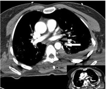
A 50-year-old female patient was referred to emergency service after a traffic accident with signs of severe thoracic trauma including shortness of breath and left flail chest. Thorax computed tomography (CT) demonstrated left sided hemopneumothorax and multiple fractures of left ribs from one to nine posteriorly and from two to eight laterally, fractures of right ribs from eight to 11 laterally and spinous process fractures of thoracic vertebrae from one to eight. The posterior sharp edge of the sixth rib was touching the descending aorta; however, there was no extravasation
Received: November 30, 2015 Accepted: January 16, 2016
Correspondence: Nesimi Günal, MD. Kırıkkale Üniversitesi Tıp Fakültesi, Göğüs Cerrahisi Anabilim Dalı, 71450 Yahşihan, Kırıkkale, Turkey.
Tel: +90 505 - 640 30 02 e-mail: [email protected] Available online at
www.tgkdc.dergisi.org
doi: 10.5606/tgkdc.dergisi.2016.12749 QR (Quick Response) Code
Institution where the research was done:
Medical Faculty of Kırıkkale University, Kırıkkale, Turkey
Author Affiliations:
1Department of Thoracic Surgery, Medical Faculty of Kırıkkale University, Kırıkkale, Turkey 2Department of Thoracic Surgery, Medical Faculty of Ufuk University, Ankara, Turkey
Günal et al. Rib fracture posing risk for aortic rupture
771 of contrast medium (Figure 1). There was a suspicion
of splenic rupture and bleeding on abdominal CT. A chest tube was inserted in the operating room with a 300 mL initial hemorrhagic drainage. An urgent median laparotomy was performed within minutes but there was no obvious bleeding or injury in the abdominal organs. Afterwards, a left posterolateral thoracotomy was performed with a cautious positioning of the patient to avoid an aortic injury. Upon exploration, minimal hemorrhagic fluid in the pleural space was encountered, but marked hemorrhage on descending aortic adventitia suggesting a direct injury of the rib was obvious. No aortic procedure was performed. Resection of broken sharp edge of the sixth rib was performed as well as multiple rib stabilization with titanium clips for flail chest. The patient was extubated on the first postoperative day and discharged on the seventh day uneventfully.
DISCUSSION
Aortic injury due to rib fracture is a rare occurrence, while the majority of the reported cases were delayed ones.[1-4] With the widespread use of CT in trauma
patients, the detection rate of rib fractures have increased while 50% of rib fractures may be missed with plain radiography.[5] Regarding the sudden exsanguinating
instances in previous reports, it seems more reasonable to obtain tomography scans in patients with posterior left rib fractures particularly with flail chest.
The mechanism of aortic injury and whether it occurs at the time of trauma or during patient’s
movements in hospital are not known well. Bruno and Batchelor[1] reported a patient who was discharged
with uncomplicated left eighth rib fracture on X-ray, nevertheless, on the sixth day, the patient underwent an urgent thoracotomy due to aortic laceration by the sharp fragment of the left eighth rib. They considered that the aortic puncture occurred at the time of initial trauma but the clot was dislodged later. Morimoto et al.[6] also believed that even though the aortic injury
occurred in trauma, bone fragments re-penetrated the aorta even with careful changes in the patient’s position. Furthermore, some authors reported sudden bleeding after changing patients’ position.[2,3] Our case
supports the latter hypothesis. Preoperative CT which was obtained in supine position demonstrated the sharp edge of the sixth rib pushing the descending aorta. The intraoperative exploration demonstrated an obvious hemorrhage on the aortic adventitia while the sharp edge of the sixth rib was not in contact to the aorta and was found in its original position. Thus repositioning of the rib by pushing it from the posterior chest wall was required to resect its sharp edge sufficiently. It is obvious that lateral decubitus position widens the anteroposterior diameter of the chest by pushing the anterior and posterior aspects of the ribs outwards, unlike supine position. We suggest that, if possible, patients with such left posterior rib fractures should be positioned accordingly to prevent an injury to the aorta until an exact diagnosis and treatment are achieved.
Boyles et al.[2] reported a case with literature review
and drew attention to the delayed management of aortic injury in most cases with sudden deterioration and bleeding after a stable period of two to 15 days. They reported a patient with left posterior rib fractures exerting direct pressure on the aorta. An open reduction and internal fixation of the flail chest were planned for the following day. However, due to sudden hemorrhagic drainage from chest tube and cardiac arrest, they performed a bedside thoracotomy and discovered the site of the aortic injury by fractured rib. Unfortunately, the patient did not survive.
In conclusion, we suggest an early resection of rib fragment to prevent undesirable aortic injuries when left posterior rib fracture is in close proximity to the descending aorta due to the unpredictable course of this condition.
Declaration of conflicting interests
The authors declared no conflicts of interest with respect to the authorship and/or publication of this article.
Funding
The authors received no financial support for the research and/or authorship of this article.
Figure 1. Thorax computed tomography shows indentation of
descending aorta by edge of broken left sixth rib (arrows) and hemothorax.
Turk Gogus Kalp Dama
772
REFERENCES
1. Bruno VD, Batchelor TJ. Late aortic injury: a rare complication of a posterior rib fracture. Ann Thorac Surg 2009;87:301-3.
2. Boyles AD, Taylor BC, Ferrel JR. Posterior rib fractures as a cause of delayed aortic injury: a case series and literature review. Injury Extra 2013;44:43-5.
3. Yanagawa Y, Kaneko N, Hagiwara A, Kimura T, Isoda S. Delayed sudden cardiac arrest induced by aortic injury with a posterior fracture of the left rib. Gen Thorac Cardiovasc Surg 2008;56:91-2.
4. Kitamura R, Colon M, Dave J. Delayed presentation of aortic laceration from seventh rib fracture: an uncommon complication. J Thorac Cardiovasc Surg 2012;144:121-3. 5. Bhattacharya B, Fieber J, Schuster K, Davis K, Maung A.
“Occult”s rib fractures diagnosed on computed tomography scan only are still a risk factor for solid organ injury. J Emerg Trauma Shock 2015;8:140-3.
6. Morimoto Y, Sugimoto T, Sakahira H, Matsuoka H, Yoshioka Y, Arase H. Successful management of threatened aortic rupture late after rib fracture caused by blunt chest trauma. Ann Vasc Surg 2014;28:1035.