304 Kulak Burun Bogaz Ihtis Derg 2016;26(5):304-306
Case Report / Olgu Sunumu
B
EH
BUT
CEVA
NŞİR KULAK BURUN BOĞ
AZHA STAL IK LA RI VE BAŞ B OYUN CERRAHİSİDERN EĞİ . . doi: 10.5606/kbbihtisas.2016.04810
Treatment of seborrheic keratosis in bilateral external
auditory canal using fiber CO2 laser
Fiber CO2 lazer kullanarak iki taraflı dış kulak kanalında seboreik keratoz tedavisi
Raşit Cevizci, MD.,1 Selin Üstün Bezgin, MD.,2 Gökhan Altın, MD.,1Gülbin Oran, MD.,3 Yıldırım Ahmet Bayazıt, MD.1
ABSTRACT
In this article, we report a 33-year-old female patient with lesions of bilateral external auditory canals who was treated using fiber CO2 laser without canaloplasty. Histopathological examination confirmed the diagnosis of irritated type seborrheic keratosis, which is rare in external auditory canal. Twelve months after the treatment, the patient showed no signs of recurrence.
Keywords: External auditory canal; laser; seborrheic keratosis.
Seborrheic keratosis (SK) is one of the most common, benign skin lesions in older patients that can involve any part of the body except the palms and soles. The most frequently affected sites are the chest, back, head (near the temples), and neck.[1] The presence of SK in the external auditory canal (EAC) is rarely seen.[1,2] To date, only a few reports of SK in the EAC have been published. Generally, in all these cases, SK occurred unilaterally and was treated by complete surgical excision with or without skin
graft.[3-6] Here, we describe bilateral EAC SK that were removed using fiber CO2 laser.
CASE REPORT
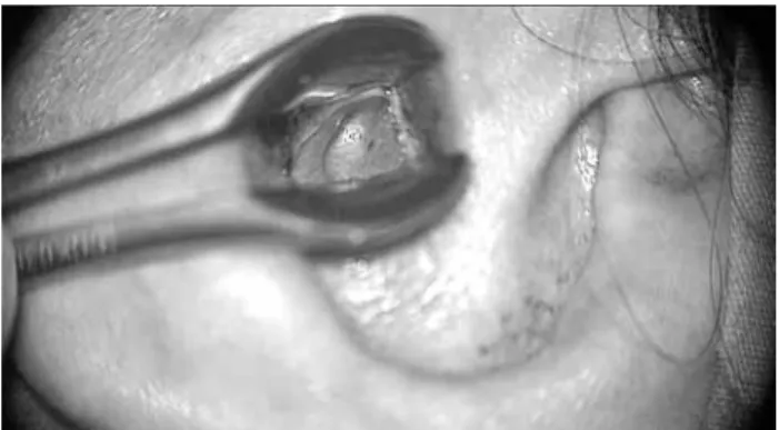
A 33-year-old woman presented with one-month history of lesions in the right and left EAC with bilateral conductive hearing loss. On otoendoscopy, skin-colored, verrucous lesions were found originating from all walls of the EACs and totally occluding them bilaterally (Figure 1, for left EAC). The review of history revealed that 1Department of Otolaryngology, İstanbul Medipol University Medicine Faculty, İstanbul, Turkey
2Department of Otolaryngology, Kanuni Sultan Süleyman Training and Research Hospital, İstanbul, Turkey 3Department of Pathology, İstanbul Medipol University Medicine Faculty, İstanbul, Turkey
Received / Geliş tarihi: February 11, 2016 Accepted / Kabul tarihi: May 04, 2016 Correspondence / İletişim adresi: Selin Üstün Bezgin, MD. Kanuni Sultan Eğitim ve
Araştırma Hastanesi Kulak Burun Boğaz Kliniği, 34303 Küçükçekmece, İstanbul, Turkey.
Tel: +90 212 - 404 15 00 / 1991 e-mail (e-posta): [email protected] Available online at
www.kbbihtisas.org
doi: 10.5606/kbbihtisas.2016.04810 QR (Quick Response) Code ÖZ
Bu makalede, iki taraflı dış kulak kanallarında lezyonlar olan ve kanaloplasti olmadan fiber CO2 lazer kullanılarak tedavi edilen 33 yaşında bir kadın hasta sunuldu. Dış kulak kanalında nadir görülen irrite tip seboreik keratoz tanısı histopatolojik incelemede doğrulandı. Tedaviden 12 ay sonra hastada herhangi bir nüks belirtisi görülmedi.
305 Treatment of seborrheic keratosis in bilateral external auditory canal using fiber CO2 laser
two years before, she presented with lesions in both EACs which were histopathologically diagnosed as SK by incisional biopsy and subsequently removed by electrocoagulation. Here, we chose flexible fiber delivered CO2 laser (Omniguide Inc., Cambridge,. MA, USA) to excise the recurrent lesions without performing incisional biopsy or radiological examination. After excision, both EACs seemed to be smooth and both tympanic membranes were intact. (Figure 2, for left EAC) Histopathological examination confirmed the diagnosis of irritated type SK (Figure 3a, b). There was no recurrence of disease on 12-month follow-up. Written informed consent was obtained from the patient.
DISCUSSION
Seborrheic keratosis is a common, benign epithelial skin tumor that is also known as verruca senilis, senile wart, seborrheic wart,
verruca seborrhoica, basal cell acanthoma, benign acanthokeratoma, and basal cell papilloma. The prevalence of this benign tumor increases particularly in older patients without predilection for either sex.[1] Clinically, lesions appear as sharply demarcated and round or oval shaped, usually elevated and stuck on the skin with a verrucous, uneven surface. Flat forms may exist and often have a smooth surface. The sizes and colors of lesions are variable. They may be skin-colored, yellowish, gray-brown, or black.[1] Although SK is usually asymptomatic, they may cause itching, pain, bleeding and redness.[1,4] They can be generally observed on the chest, back, head and neck.[1] The EAC is a rare affected site of disease that can cause conductive hearing loss.[1,4] In our patient, the lesions were bilateral as distinct from other cases of EAC SK in the literature and there was no symptom except bilateral hearing loss. Figure 1. Skin-colored, verrucous, papillomatous lesion
completely occluding the left external auditory canal. Figure 2. Left external auditory canal after CO2 laser ablation surgery.
Figure 3. (a) Hyperkeratosis, horn cysts, small squamous eddies, proliferation of basaloid cells, and moderate inflammatory infiltrate in the epidermis (H-E x 40). (b) Closed view, horn cysts (arrows) were observed (H-E x 100).
306 Kulak Burun Bogaz Ihtis Derg
The etiology and pathogenesis of SK are not well understood. However, it was believed that SK is more prevalent among Caucasians.[1] In recent years, human papillomavirus infections with risk factors including older age and exposure to ultraviolet light were mentioned in pathogenesis.[1,7] Genetic predisposition as an autosomal dominant pattern has also been suggested although there is a lack of validated data.[1] In addition, FGFR3 gene and PIK3CA oncogene mutations were described in the molecular pathogenesis of SK.[1,8]
Histopathologically, SKs have characterized findings including acanthosis, papillomatosis, hyperkeratosis, horn cysts and horn pseudocysts. There are seven different histological subtypes: acanthotic, hyperkeratotic (also verrucous), adenoid, clonal bowenoid, irritated, and melanoacanthoma.[1] The acanthotic subtype is believed to be the most frequently seen in EAC. Kim et al.[3] reported seven patients with SK in the ear and six of them had the acanthotic subtype. In our case, the irritated subtype of SK was confirmed on histopathological examination, which is rare in the EAC. To best of our knowledge, there is only one report of irritated SK in EAC in the literature.[5] The irritated subtype is characterized by onion-skin like aggregations of eosinophilic squamous epithelial cells (squamous eddies), and inflammatory cell infiltration with partly lichenoid appearances in the dermis. Acantholysis, dyskeratosis and spongiosis may also be present.[1] In our patient, hyperkeratosis, horn cysts, small squamous eddies, proliferation of basaloid cells, and moderate inflammatory infiltrate were observed in the epidermis. In the differential diagnosis of SK, benign lesions such as senile keratosis, papillomas, keratoacanthomas, fibroma and malignant lesions such as malignant melanoma, basal or squamous cell carcinoma should be considered.[3,5,6]
Removal of the SK may be required to exclude the presence of malignancy if clinical appearance is equivocal, or to end the symptoms,
inflammation, and cosmetic problems.[1]
Treatment options include curettage, complete excision and ablative procedures (laser therapy and cryotherapy).[1,3] Treatment using topical and systemic drug therapies have also been reported, but have not yet been proven. Erbium YAG or CO2 laser may be used for removal of SK. In addition, the use of a 532 nm diode laser in
dermatosis papulosa nigra, a clinical variant of SK, has been reported.[1] On literature review, there was only one case of SK in the EAC treated by CO2 laser, which was presented as a poster[9] and here, we describe the second case. Although ablative procedures are known to be inconvenient for obtaining histologic analysis and incisional biopsy can be necessary before surgery, we managed to obtain adequate specimens for diagnosis without performing biopsy before laser treatment. Our knowledge about the previous diagnosis of the lesions and the clinical presentation of the patient consistent with SK led us to choose laser surgery. An advantage of this procedure was having no need for canaloplasty after excision. No recurrence was observed in our patient 12-months post-ablation. However, this tumor always requires clinicians to be alert about potential local recurrence.
Declaration of conflicting interests
The authors declared no conflicts of interest with respect to the authorship and/or publication of this article.
Funding
The authors received no financial support for the research and/or authorship of this article.
REFERENCES
1. Hafner C, Vogt T. Seborrheic keratosis. J Dtsch Dermatol Ges 2008;6:664-77.
2. Elder DE, Elenitsas R, Jhonson BL, Murphy GF. editors. Lever’s Histopathology of the Skin. 9th ed. Philadelphia: Lippincott Williams & Wilkins; 2004. p. 809Y13.
3. Kim KW, Chang J, Lee S, Im GJ, Chae SW, Jung HH, et al. Clinical analysis of seborrheic keratoses in the ear: a retrospective study and literature review. Eur Arch Otorhinolaryngol 2015;272:1113-7.
4. Magliulo G, Ciniglio Appiani M. Seborrheic keratosis, keratotic type, of the external auditory canal. Otolaryngol Head Neck Surg 2011;145:697-8.
5. Konishi E, Nakashima Y, Manabe T, Mazaki T, Wada Y. Irritated seborrheic keratosis of the external ear canal. Pathol Int 2003;53:622-6.
6. Choi JH. Seborrheic keratosis of the external auditory canal. Otol Neurotol 2012;33:27-8.
7. Kennedy C, Bajdik CD, Willemze R, De Gruijl FR, Bouwes Bavinck JN. The influence of painful sunburns and lifetime sun exposure on the risk of actinic keratoses, seborrheic warts, melanocytic nevi, atypical nevi, and skin cancer. J Invest Dermatol 2003;120:1087-93.
8. Yeatman JM, Kilkenny M, Marks R. The prevalence of seborrhoeic keratoses in an Australian population: does exposure to sunlight play a part in their frequency? Br J Dermatol 1997;137:411-4.
9. Ho WL, Chan KC. Seborrheic keratosis of the external auditory canal. Ear Nose Throat J 2011;90:26-7.