Dergi web sayfası:
www.agri.ankara.edu.tr/dergi www.agri.ankara.edu.tr/journalJournal homepage:
TARIM BİLİMLERİ DERGİSİ
—
JOURNAL OF AGRICUL
TURAL SCIENCES
22 (2016) 539-547
Effects of Lead (Pb) and Cadmium (Cd) Elements on Lipid
Peroxidation, Catalase Enzyme Activity and Catalase Gene Expression
Profile in Tomato Plants
Semra SOYDAM AYDINa, İlker BÜYÜKb, Esra GÖKÇE GÜNDÜZERb, Burcu Pelin BÜYÜKb, İrfan
KANDEMİRb, Demet CANSARAN-DUMANc, Sümer ARASb
aMinistry of Health of Turkey, Turkish Medicines and Medical Device Agency, Ankara, TURKEY bAnkara University, Faculty of Science, Department of Biology, Ankara, TURKEY
cAnkara University, Biotechnology Institute, Ankara, TURKEY
ARTICLE INFO Research Article
Corresponding Author: Semra SOYDAM AYDIN, E-mail: [email protected], Tel: +90 (312) 565 53 60 Received: 19 November 2014, Received in Revised Form: 09 August 2015, Accepted: 09 August 2015
ABSTRACT
Heavy metals are significant abiotic stress factor, affecting various response mechanisms in plants. These responses include: changes in membrane composition, production of small molecules and free radicals, and alterations in the activities of antioxidant enzymes and their gene expressions. For this reason, lipid peroxidation levels (MDA), catalase enzyme activity, and gene expression profiles, quantified by real-time PCR, were analyzed in tomato plants exposed to various concentrations (0, 80, 160, 320, 640 and 1280 μM) of Cd2+ and Pb2+. All concentration
of Cd+2 or Pb+2 contamination led to increased lipid peroxidation and catalase enzyme activity, except for 320 and
640 μM Cd+2 contamination levels. As a result, gene expression patterns at the mRNA level and changes in MDA
content under different concentrations of Pb+2 and Cd+2 contamination revealed a positive correlation, although no
correlation was found between gene expression patterns at the mRNA level and catalase enzyme activity. These results might be explained by the regulation of genes at the transcriptional, posttranscriptional, and also translational or posttranslational levels.
Keywords: Tomato (Solanum lycopersicum L.); Lipid peroxidation; CAT enzyme activity; Gene expression; qRT-PCR
Domates Bitkisinde Kurşun (Pb) ve Kadminyumun (Cd) Lipid
Peroksidasyonu, Katalaz (CAT) Enzim Aktivitesi ve Gen Ekpresyon
Profiline Etkisi
ESER BİLGİSİ Araştırma Makalesi
Sorumlu Yazar: Semra SOYDAM AYDIN, E-posta: [email protected], Tel: +90 (312) 565 53 60 Geliş Tarihi: 19 Kasım 2014, Düzeltmelerin Gelişi: 09 Ağustos 2015, Kabul: 09 Ağustos 2015
1. Introduction
Pollution of the environment by heavy metals is a serious problem throughout the world. The most common heavy metals in the environment are cadmium, chromium, copper, mercury, lead and zinc and some of their main sources are emissions from burning fossil fuels, industrial activities, automotive emissions, pesticides usage and domestic wastes (Kabata-Pendias & Pendias 1989).
Heavy metal contamination of economically important plants, such as fruits and vegetables, poses a threat to their quality and leads to alterations in the health statuses of humans and animals. Several researches have indicated that consumption of vegetables loaded with heavy metals such as Cd+2 and
Pb+2 can generate carcinogenic effects (Trichopoulos
1997; Türkdoğan et al 2002). Climatic changes, nature of the soil, time of harvest, and concentrations of heavy metals in the soil are significant impacts on assumed threats of heavy metals in vegetables (Lake et al 1984; Scott et al 1996; Voutsa et al 1996). In addition, post-harvest vegetables could be influenced by air pollution during transportation and marketing, which can lead to elevated levels of heavy metals (Sinha et al 2005; Sharma et al 2006; 2007).
Abiotic stress factors, such as soil salinity, drought, high temperatures, and heavy metals are known to
cause oxidative stress in plants by the production of reactive oxygen species (ROS), such as O2-, H
2O2, 1O2,
HO2-, OH, ROOH, ROO, and RO (Smirnoff 1993).
The most important intracellular generators of ROS are chloroplasts, mitochondria, and peroxisomes. As a result of stomatal closure and limited CO2 availability, chloroplasts generate O2- and H
2O2 by the electron
acceptor of photosystem I, while singlet oxygen is produced by the transfer of an electron from an activated chlorophyll molecule to molecular oxygen (Asada & Takahashi 1987; Hernandez et al 1995). It has been estimated that, under stress conditions, 1-2% of O2 consumption leads to the formation of ROS, which causes lipid peroxidation, membrane defects, and instability of enzymes in higher plants (Mittler 2002; El-Beltagi et al 2010). On the other hand, it is important to emphasize that ROS can act not only as damaging factors, but also as protective or signaling factors, which depend on the equilibrium between ROS production and scavenging mechanisms (Gratao et al 2005).
Plants are able to develop antioxidant defense systems to protect themselves against ROS and cope with different stress factors (Rao et al 2006). The antioxidant system restricts and removes ROS damage and maintains ROS homeostasis in plant cells. The components of this system are enzymatic, such as monodehydro ascorbate reductase
ÖZET
Önemli bir abiyotik stres faktörü olan ağır metaller bitkilerde çok çeşitli yanıt mekanizmalarını uyarabilirler. Bu yanıt mekanizmaları; membran kompozisyonunda değişiklik, küçük molekül ve serbest radikallerin üretimi, antioksidant enzimlerin aktivitelerinin ya da gen ekspresyonlarının değişimini içerir. Bu sebeple, bu araştırmada çeşitli konsantrasyonlardaki (0, 80, 160, 320, 640 ve 1280 μM) Pb2+ve Cd2+ kontaminasyonuna maruz kalan domates
bitkilerinde lipid peroksidasyon seviyesi (MDA), katalaz enzim aktivitesi ve real-time PCR aracılığı ile katalaz gen ekpresyon seviyesi belirlenmiştir. 320 ve 640 μM Cd+2 kontaminasyonu hariç tüm Cd+2 ve Pb+2 kontaminasyonları lipid
peroksidasyonuna ve katalaz enzim aktivitesinde artışa neden olmuştur. Sonuç olarak; çeşitli konsantrasyonlarda Pb2+ ve
Cd2+ kontaminasyonuna maruz kalan domates bitkisinde, CAT gen ekpresyonu ve lipid peroksidayonu arasında pozitif
korelasyon bulunurken, CAT gen ekpresyonu ve enzim aktivitesi arasında korelasyon tespit edilememiştir. Bu durum genlerin transkripsiyonel, posttranskripsiyonel ve aynı zamanda translasyonel veya posttranslasyonel seviyelerdeki regülasyonu ile açıklanabilir.
Anahtar Kelimeler: Domates (Solanum lycopersicum L.); Lipid peroksidasyonu; CAT enzim aktivitesi; Gen ekpresyonu; Real-time PCR
(MDHAR), dehydro ascorbate reductase (DHAR), glutathione reductase (GR), ascorbate peroxidase (APX), superoxide reductase (SOD), catalase (CAT) and non-enzymatic antioxidants, such as glutathione (GSH), proline, carotenoids, and tocopherol (Mittler et al 2004).
The various antioxidants have partially overlapping functions and can functionally compensate for each other. In this regard, SOD dismutases O2- into H
2O2, which, in turn, is detoxified
by CAT, APX, or PRX. CAT is one of the enzymatic antioxidants with the highest turnover rates among all enzymes, and it is located mainly in the peroxisomes. One molecule of CAT can convert approximately 6 million molecules of H2O2 to O2 and H2O per minute (Lee & An 2005). In this manner, it prevents longer H2O2 action, which could lead to cell disturbances and DNA damage (Shim et al 2003). There have been many reports regarding catalase enzyme activity, lipid peroxidation (MDA), and gene expression levels in several different plant species under stress conditions, such as salt stress (Mittova et al 2003), boron (B) toxicity (Cervilla et al 2007), Cu toxicity (Cui et al 2010), and Pb toxicity in tomatoes (Wang et al 2008), low temperature stress in wheat (Triticum
aestivum L.) (Matsumura et al 2002), Mn toxicity in
spruce trees (Picea abies L.) (Polle et al 1992), pecan trees [Caryaillinoinensis (Wangenh.) C. Koch, cv. Kiowa] (Henriques 2003), and Cd and Cu toxicity in mouse-ear cress (Arabidopsis thaliana) (Skorzynska-Polit et al 2010).
The control of gene expression in all eukaryotic cells is a complex process that involves molecules such as RNA polymerases, numerous transcription factors, the DNA template, RNA produced by transcription, and protein produced by translation, with its attendant processing. The examination of gene expression often involves quantifying the abundance of a particular transcript. Contemporary methods, such as real-time PCR, that examine gene expression reveal the dynamic nature of this biochemical process. Real-time PCR allows precise measurements of mRNA steady-state levels and provides advantages, such as very high sensitivity and precise quantification of expression levels
under different conditions or treatments. This method only measures immediate levels or final accumulation of RNA in the cell. Also other modern methods, such as microarray hybridization, or more conventional methods, like Northern hybridization, do not provide information about the transcriptional activity of genes. These methods fail to quantify the stability of the RNA or the ratio of transcription at the specific loci under investigation. In order to detail understanding the nature of gene expression modulation, it is essential to evaluate alteration of transcript and corresponding protein levels which both are associated with a phenotypic change (Farrel 2007). As such, the experiments in the current study were conducted in three phases. In the first phase, after Cd+2 and Pb+2 stress treatments, MDA levels
were determined in order to obtain evidence that the plants were in stress as a result of the treatments. In the second phase of the study, the steady-state level of CAT mRNA was determined by quantitative real-time PCR. Finally, in the third phase, CAT enzyme activity was determined in order to obtain an idea about the final stage of CAT gene expression in tomato samples exposed to different concentrations of Cd+2 and Pb+2 stress.
2. Material and Methods
2.1. Plant material, growth conditions, and stress treatment
Plant growth conditions and stress treatments were performed as previosly reported by Soydam Aydın et al (2013). Tomato (Solanum lycopersicum L. ‘Falcon’) seeds were germinated and grown hydroponically in pots containing 0.2 L of modified 1/10 Hoagland’s solution. After germination, seedlings were transferred to pearlite and watered with 1/10 Hoagland’s media in regular time interval (daily). Twenty-five-day-old six tomato plants grown in 1/10 Hoagland’s media were used for the stress treatments. For the heavy metal application, Pb+2 and Cd+2 were added to the hydroponic solution
for 24 h at concentrations of 0 (control), 80, 160, 320, 640 and 1280 μM. Harvested tomato leaves were ground in liquid nitrogen and used for estimation
of lipid peroxidation, CAT enzyme activity assay, RNA extraction, and gene expression analysis.
2.2. Estimation of lipid peroxidation
Malondialdehyde (MDA) content is a marker of oxidative lipid injury was performed as described and previously reported by Hodges et al (1999) and Soydam Aydın et al (2013). ELISA microtiter reader (SpectraMax M2) was used to read absorbance at 440 nm, 532 nm, and 600 nm. MDA equivalents were calculated as described by Hodges et al (1999) using Equation 1, 2 and 3.
[(Abs532+TBA) - (Abs600+TBA) - (Abs532-TBA-Abs600-TBA)]= A (1)
[(Abs440+TBA-Abs600+TBA) x 0.0571]= B (2)
MDA equivalents (nmol mL-1)= (A-B/157000) x 106 (3)
2.3. RNA extraction and cDNA preparation
RNA extraction was performed using a TRIzol protocol followed by RNeasy mini cleanup kit (Qiagen, Cat no: 74104). RNA quantity and quality were measured with a NanoDropND-1000 spectrophotometer. Quality of RNA was also confirmed by gel electrophoresis, containing 1.5% agarose and formaldehyde. The cDNA synthesis based on reverse transcription reactions were performed with 2 μg of RNA and a high fidelity cDNA synthesis kit (Roche) containing 2.5 µM anchored oligo(dT)18, 1X transcriptor high-fidelity reverse transcriptase reaction buffer, 20 U protector Rnase inhibitor, 1 mM deoxynucleotide mix, 5 mM DTT, and 10 U transcriptor high-fidelity reverse transcriptase at final concentration. And, the program applied was 65 °C, 10 min; 55 °C, 30 min; 85 °C, 5 min.
2.4. Real-time RT-PCR and quantification of mRNA levels
Quantitative real-time PCR was performed with a LightCycler® 480 System (Roche) thermal cycler.
The sequences of primers and probes (Table 1) of the target gene catalase (CAT) and actin (ACT), which is used for normalization, were designed based on the sequences of tomato genes available in the databank (NCBI 2013a; NCBI 2013b). Amplifications of
PCR product were monitored via intercalation of hybridization probes (HyProbe, FRET probes) that allow exact and specific identification of the target gene. After pre-denaturation at 95 °C for 10 min, 45 cycles of 95 °C 10 s, 60 °C 30 s, and 72 °C 15 s were applied. Data collection for quantification was accomplished during the annealing period. Copy numbers of the genes (CAT, ACT) under stress treatment were determined by using standard curves. PCR efficiency for CAT standard curve was 1.937, while ACT was found to be 1.954.
Table 1- Primer and probe sequences of CAT and Actin
Çizelge 1- CAT ve Actin için primer ve prob sekansları Primer and
probe name Sequences
CAT2 F CTTTCCTCTTCGACGATATTGGTA CAT2 S TATTCCCCAAGATTACAGGCAT CAT2 A CCGACTCGGATTGCCTT CAT2 R GTGATTTGCTCCTCCGACTC CAT2 FL CAACAGGGCTGGAAAATCAACTTATGT-FL CAT2 LC 640 AAGTTCCACTGGAAGCCCACATGT p Actin S Fw TCTGTTTCCCGGTTTTGCTATTAT
Actin R Rev TGCATCAGGCACCTCTCAAG
Actin FL ATTCATAGCCCCCACCACCAAAC-FL
Actin LC 640 TCTCCATCCCATCAAAAAAACAAATTGACTp 2.5. Catalase enzyme activity assay
Catalase enzyme activities were performed as
previosly reported by Aebi (1984) and Soydam Aydın et al (2013). Powdered tomato tissues were suspended in extraction buffer and homogenates were centrifuged at 15000 g for 20 min, and the supernatant fraction was used for the enzyme activity assays (Jovanovic 2006). All steps were carried out at 4 °C. The catalase activity assay was performed according to the method reported by Aebi (1984), based on 240 nm absorbance.
2.6. Statistical methods
The abundance of target gene transcripts was normalized to ACT and set relative to the control plants (no stress exposure), according to the 2-∆∆CT
relative expression levels (REL) of the CAT gene were checked for statistical significance according to one-way ANOVA. The results were considered statistically significant if the P value was <0.05 in Dunnett’s test.
3. Results and Discussion
In the current study, the effects of heavy metal stress (Pb+2 and Cd+2) on cell membrane, gene expression
and CAT enzyme activity profiles in tomato plants (Solanum lycopersicum L.) were analyzed. It is known that heavy metal contamination can lead to an increase in different forms of ROS.
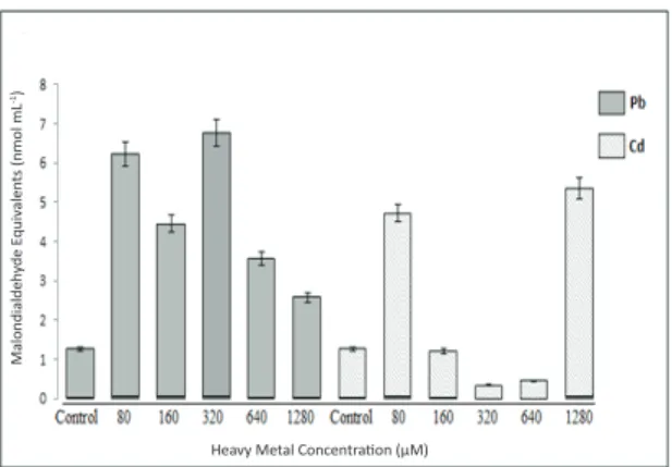
Malondialdehyde (MDA) analysis was used as a marker of oxidative lipid injury, which might have changed in response to Cd+2 and Pb+2 heavy metals
that led to stress in tomato seedlings. Changes in MDA content were observed in the tomato seedlings treated with different concentrations of Pb+2, indicating that the
samples were under stress. It has been demonstrated that increased lipid peroxidation is a characteristic feature of oxidative stress caused by unbalanced equilibrium between ROS production and scavenging or defense mechanisms under heavy metal stress (Smirnoff 1993; Mittler 2002; Lima et al 2006).
Small hydrocarbon fragments, such as ketones and MDAs, which are considered the first evidence of stress in plants, have been used as indicators of lipid peroxidation or membrane damage, (Lyons 1973). In recent studies, increased lipid peroxidation has been observed in plants under heavy metal stress, such as tomato (Krupa & Baszynski 1985; 1989; Quariti et al 1997; Ben Ammar et al 2005), wheat (Malik et al 1992), barley (Vassilev 2004), and mustard (Gaur & Grupa 1994; Nouairi et al 2006). Results of the current study indicate that MDA content substantially increased with all concentrations of Pb+2 contamination. The maximum level of MDA
content was determined in the samples exposed to 320 μM concentrations of Pb+2 contamination while
a significant decrease was measured at 1280 μM among treated samples (Figure 1). The data also show that ROS-induced lipid peroxidation began in a short period of time and that the level of injury was dependent on the exposure concentrations
of Pb+2 contamination. However, MDA contents,
which were obtained in the samples exposed to Cd+2
contamination, showed an increase only with the 80 μM and 1280 μM Cd+2 concentrations. The highest
level of inhibition in MDA content was observed in the treatment with 1280 μM Cd+2. In addition, the
maximum decrease in MDA content was observed in the tomato seedlings exposed to the 320 μM concentration of Cd+2. Surprisingly, lower MDA
levels were observed at 160, 320, and 640 μM of Cd+2
treatment compared to the control samples (Figure1).
Heavy Metal Concentration (μM)
Malondialdeh yde E quiv alen ts (nmol mL -1)
Figure 1- Lipid peroxidation (malondialdehyde, MDA content) in the total tissues of tomato plants exposed to different concentrations of Pb2+ and Cd2+
Şekil 1- Farklı konsantrasyonlarda Pb2+ ve Cd2+’ye
maruz kalan domates bitkisinin tüm dokularındaki lipid peroksidasyonu (malondialdehit, MDA içeriği)
When evaluated SOD enzyme activity and gene expression results of tomato samples exposed to same concentration of Cd+2 contamination in another
study conducted in our laboratory (unpublished data), it was difficult to suggest that prevention of lipid peroxidation in the cells of the tomato samples exposed to 160 μM, 320 μM, and 640 μM Cd+2
concentrations in the current study might result from increased activation of antioxidant mechanisms. Further analysis is necessary to explain this complex connection between lipid peroxidation and antioxidant mechanisms. Results also revealed that lipid peroxidation induced by ROS was observed after contamination of low (80 μM) and high
(1280 μM) Cd+2 concentrations, which generated
an imbalance between production of ROS and antioxidant enzyme activity or gene expression as a component of scavenging and defense mechanisms of ROS.
ROS have a dual action, not only acting as protective or signaling factors, but also as oxidative damaging factors at the cellular level due to an imbalance between the production and removal of ROS under stress conditions (Mittler 2002). To combat the negative effects of this imbalance, an enzymatic antioxidant defense system is activated to protect plants from cellular injury with removing excessively produced H2O2 with the enzymes, such as peroxidase (POD), glutathione reductase (GR), superoxide dismutase (SOD), and catalase (CAT) (Qilin et al 2009). In recent years, genes involved in response to abiotic stress have become the main field for biotechnological studies, as it constitutes the basis for improving the stress tolerance of plants. Many genes play a crucial role in responding to environmental stresses at the post-transcriptional level, and CAT is one of these genes that encode the catalase enzyme. Studies about the protective activity of CAT in response to different abiotic stresses have been demonstrated by previous research studies conducted with Coffea sp., oilseed rape (Brassica
napus L.), pea (Pisum sativum L.), and tomato
(Lycopersicum esculentum L.) plants (Goupil et al 2009; Qilin et al 2009; Fortunato et al 2010).
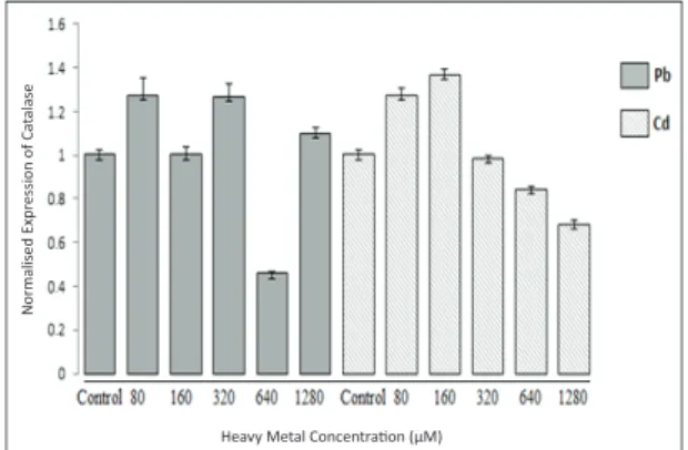
In the current study, exposure of tomato seedlings to Pb+2 and Cd+2 led to significant changes in the
abundance of CAT gene transcripts, suggesting the involvement of the gene in heavy metal tolerance (Figure 2). To evaluate the stability of our results, CAT transcript levels of all samples were measured three times for each stress condition, which indicated steady-state mRNA levels in the cells for both CAT and the reference gene ACT. With regard to the control and to each other, different expression levels were recorded in all stress conditions applied. The highest level of transcript was recorded at 320 μM of Pb+2 and 160 μM of Cd+2 concentrations, and these
results were found to be statistically significant. The lowest levels of mRNA were recorded at the 640 μM of Pb+2 and 1280 μM of Cd+2 concentrations.
Contamination with different concentrations of Pb2+, 80 and 320 μM Pb+2 led to a statistically
significant increase in mRNA level in the tomato seedlings compared to the control, while the 640 μM concentration led to a reduction in the expression of the gene; these results were statistically significant (P1= 0.001; P2= 0.001; P3= 0.000). Changes in the rate of expression level in the catalase gene compared to the control were statistically insignificant upon exposure to 160 and 1280 μM of Pb+2 concentrations. CAT
expression levels decreased significantly in response to 640 μM of Pb+2, which indicates that CAT was strongly
downregulated by this concentration (Figure 2). CAT mRNA levels in the tomato seedlings increased in response to 80 μM and 160 μM of Cd+2 treatments;
these results were statistically significant (P1= 0.000; P2= 0.000) when checked for significance according to one-way ANOVA. The 320, 640, and 1280 μM of Cd+2 treatments led to a decrease in catalase gene
expression level compared to the control, but only the 640 μM and 1280 μM of Cd+2 treatment results were
statistically significant (P1= 0.005; P2= 0.000) (Figure 2). When we analyzed this complex profile of gene expression patterns at the mRNA level and changes in MDA content under different concentrations of Pb+2
and Cd+2 contamination, the results revealed positive
correlations between each other (Figure 1 and 2).
Heavy Metal Concentration (μM)
Normalised Expr
ession of Ca
talase
Figure 2- Catalase mRNA levels in the total tissues of tomato plants exposed to different concentrations of Pb2+ and Cd2+
Şekil 2- Farklı konsantrasyonlarda Pb2+ ve Cd2+’ye
maruz kalan domates bitkisinin tüm dokularındaki katalaz mRNA seviyesi
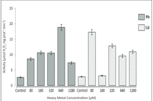
Also, catalase enzyme activity assay revealed different data from gene expression patterns at the mRNA level in tomato seedlings treated with different concentrations of Pb+2 and Cd+2.
In particular, 640 μM Pb+2 and 80 μM Cd+2
contamination levels led to a significant increase in catalase enzyme activity, while gene expression levels under these contamination levels were under the control level or slightly increased (Figure 2 and 3). Increased activity of CAT enzyme indicates the ability of the cell to scavenge increased concentrations of H2O2; this ability was not weakened by Pb+2 or Cd+2 treatment according to
the results of the current study, in accordance with previous reports (Mead 1976). The differences recorded in the enzyme activities of the plants under Pb+2 and Cd+2 stress may be due to the balance of
ROS generation and antioxidant activities that must be achieved to enhance the protection of the cell.
Heavy Metal Concentration (μM)
Activity (μmol H 2 O2 mg pr ot -1 min -1)
Figure 3- Catalase enzyme activity in the total tissues of tomato plants exposed to different concentrations of Pb2+ and Cd2+
Şekil 3- Farklı konsantrasyonlarda Pb2+ ve Cd2+’ye
maruz kalan domates bitkisinin tüm dokularındaki katalaz enzim aktivitesi
The differential recovery in CAT enzyme activity at various concentrations of Pb+2 treatments,
640 Pb+2 and 1280 μM Pb+2, might imply that the
enzyme contributes to cellular defense with differing performances, depending on the concentrations of the heavy metals. In the current study, CAT gene
expression patterns at the mRNA level and changes in CAT enzyme activities under different concentrations of heavy metal treatment revealed no positive correlation or inverse proportion. For example, although the steady-state level of mRNA did not change after the 160 μM Pb+2 treatment (compared
to control), a significant increase was observed in enzyme activity levels at the same concentration. In addition, while the mRNA level of CAT decreased in tomato plants exposed to the 640 μM Pb+2
treatment, enzyme activity significantly increased, and while the mRNA level of CAT increased in tomato plants exposed to the 1280 μM Pb+2 treatment,
enzyme activity slightly increased compared to the control. The results suggest that a translationalpost-translational level of control for CAT gene expression or enzyme activity might be regulated by the reversible modulation of the functionality of its mRNA, depending on the supply of the methyl group donors from glycine and serine and that production of these amino acids is greatly enhanced by photo respiratory carbon flow. Therefore, catalase protein synthesis is associated with the photo respiratory and photosynthetic pathways (Schmidt et al 2002).
4. Conclusions
Results of the current research verified that oxidative stress alerts plant antioxidant defense systems in unsupportable concentrations of heavy metals, and that in most cases, the CAT gene is induced to encode the CAT enzyme to scavenge ROS generated under stress conditions (Lee & An 2005). The data on gene expression and enzyme activity of CAT obtained in the current study could provide a better understanding of the antioxidant mechanisms in tomato plants.
References
Aebi H (1984). Catalase in vitro. Methods in Enzymology
105: 121-126
Asada K & Takahashi M (1987). Production and scavenging of active oxygen in photosynthesis. In: Kyle DJ, Osmond CJ, Artzen CJ, editors. Photoinhibition: Topics in Photosynthesis. Amsterdam, Elsevier, pp. 227-287
Ben Ammar W, Nouairi I, Tray B, Zarrouk M, Jemal F & Ghorbel M H (2005). Cadmium effects on mineral nutrition and lipid contents in tomato leaves (in
French). Journal of Social Biology 199: 157-163
Cervilla L M, Blasco B, Rios J J, Romero L & Ruiz J M (2007). Oxidative stress and antioxidants in tomato (Solanum lycopersicum) plants subjected to boron
toxicity. Annals of Botany 100: 747-756
Cui L, Lee H S, Ndinteh D T, Mbafor J T, Kim Y H, Tv L E, Nguyen P H & Wk O H (2010). New prenylated flavanones from Erythrina abyssinica with protein tyrosine phosphatase 1B (PTP1B) inhibitory activity.
Planta Medica 76: 713-718
El-Beltagi H S, Mohamed A A & Rashed M M (2010). Response of antioxidative enzymes to cadmium stress in leaves and roots of radish (Raphanus sativus L.).
Notulae Scientia Biologicae 2: 76-82
Farrel R E (2007). The regulation of gene expression in plants and animals. In: Bassett CL, editor. Regulation of Gene Expression in Plants: The Role of Transcript Structure and Processing. New York, NY: Springer, pp. 1-34
Fortunato A, Lidon F C, Batista-Santos P, Leitão A E, Pais I P, Ribeiro A I & Ramalho J C (2010). Biochemical and molecular characterization of the antioxidative system of Coffea sp. under cold conditions in genotypes with contrasting tolerance. Journal of
Plant Physiology 167: 333-342
Gaur A & Grupa S K (1994). Lipid components of mustard seeds (Brassica juncea L.) as influenced by cadmium
levels. Plant Food for Human Nutrition 46: 93-102
Goupil P, Souguir D, Ferjani E, Faure O, Hitmi A & Ledoigt G (2009). Expression of stress-related genes in tomato plants exposed to arsenic and chromium in
nutrient solution. Journal of Plant Physiology 166:
1446-1452
Gratao P L, Polle A, Lea P J & Azevedo R A (2005). Making the life of heavy metal-stressed plants a little
easier. Functional Plant Biology 32: 481-494
Henriques F (2003). Gas exchange, chlorophyll a fluorescence kinetics and lipid peroxidation of pecan leaves with varying manganese concentrations. Plant
Science 165: 239-244
Hernandez J A, Olmos E, Corpas F J, Sevilla F & De1 Rio L A (1995). Salt-induced oxidative stress in
chloroplasts of pea plants. Plant Science 105: 151-167
Hodges D M, Delong J M, Forney C F & Prange R K (1999). Improving the thiobarbituric
acid-reactive-substances assay for estimating lipid peroxidation in plant tissues containing anthocyanin and other
interfering compounds. Planta 207: 604-611
Jovanovic Z, Milosevic J & Radovic S (2006). Antioxidative enzymes in the response of buckwheat (Fagopyrum esculentum Moench) to ultraviolet B radiation. Journal of Agriculture and Food Chemistry
54: 9472-9478
Kabata-Pendias A & Pendias H (1989). Trace Elements in the Soil and Plants. Boca Raton, CRC Pres
Krupa Z & Baszynski T (1985). Effects of cadmium on the acyl lipid content and fatty acid composition in thylakoid membranes isolated from tomato leaves.
Acta Physiology Plantarum 7: 55-64
Krupa Z & Baszynski T (1989). Acyl lipid composition of thylakoid membranes of cadmium-treated tomato
plants. Acta Physiology Plantarum 11: 111-116
Lake D L, Kirk P W W & Lester J N (1984). Fractionation, characterization, and speciation of Heavy metals in sewage and sludge amended soil: A review. Jornal of
Environmental Quality 13: 175-183
Lee S H & An C S (2005). Differential expression of three catalase genes in hot pepper (Capsicum annuum L.).
Molecules and Cells 20: 247-255
Lima L, Seabra A, Melo P, Cullimore J & Carvalho H (2006). Phosphorylation and subsequent interaction with 14-3-3 protein regulates plastid glutamine synthetase in
Medicago truncatula. Planta 223: 558-567
Livak K J & Schmittgen T D (2001). Analysis of relative gene expression data using real-time quantitative PCR
and the 2(-delta delta C(T)) method. Methods 25:
402-408
Lyons J M (1973). Chilling injury in plants. Annual
Review of Plant Physiology 24: 445-466
Malik D, Sheoran I S & Singh R (1992). Lipid composition of thylakoid membranes of cadmium treated wheat seedlings. Indian Journal of Biochemistry &
Biophysics 29: 350-354
Matsumura H, Xie Y, Shirakata S, Inoue T, Yoshinaga T, Ueno Y, Izui K & Kai Y (2002). Crystal structures of C4 form maize and quaternary complex of E. coli
phosphoenolpyruvate carboxylases. Structure 10:
1721-1730
Mead J F (1976). Free radical mechanisms of lipid damage and consequences for cellular membranes. In: WA Pryor, editor. Free Radicals in Biology, Vol 1. New York, Academic Press, pp. 51-68
Mittler R (2002). Oxidative stress, antioxidants and stres
tolerance. Trends in Plant Science 7: 405-410
Mittler R, Vanderauwera S, Gollery M & Van Breusegem F (2004). The reactive oxygen gene network in
plants. Trends in Plant Science 9: 490-498
Mittova V, Theodoulou F L, Kiddle G, Gomez L, Volokita M, Tal M, Foyer C H & Guy M (2003). Coordinate induction of glutathione biosynthesis and glutathione-metabolizing enzymes is correlated with salt tolerance
in tomato. FEBS Letter 554: 417-421
NCBI (2013a). Nucleotide. http://www.ncbi.nlm.nih.gov/ nuccore/M93719.1 (Erişim tarihi: 10.01.2014) NCBI (2013b). Nucleotide http://www.ncbi.nlm.nih.
gov/nuccore/NM_001308447.1 (Erişim tarihi:
17.01.2014)
Nouairi I, Ben Ammar W, Ben Youssef N, Ben Mileddaoud D, Habibghorbal M & Zarrouk M (2006). Comparative study of cadmium effects on membrane lipid composition of Brassica juncea and Brassica
napus leaves. Plant Science 170: 511-519
Polle A, Chakrabarti K, Chakrabarti S, Seifert F, Schramel P & Rennenberg H (1992). Antioxidants and manganese deficiency in needles of Norway spruce (Picea abies
L.) trees. Plant Physiology 99: 1084-1089
Qilin D, Jin W, Bin F, Tingting L, Chen C, Honghui L & Shizhang D (2009). Molecular cloning and characterization of a new peroxidase gene (OvRCI) from Orychophragmus violaceus. African Journal of
Biotechnology 8(23): 6511-6517
Quariti O, Boussama N, Zarrouk M, Cherif A & Ghorbalm H (1997). Cadmium and copper-induced changes in
tomato membrane lipids. Phytochemistry 45:
1343-1350
Rao A Q, Hussain S S, Shahzad M S, Bokhari Sy A, Raza M H, Rakha A, Majeed A, Shahid A A, Saleem Z, Husnain T & Riazuddin S (2006). Somatic embryogenesis in wild relatives of cotton (Gossypium spp.). Journal of
Zhejiang University Science B 7(3): 1673-1581
Schmidt M, Dehne S & Feierabend J (2002). Post-transcriptional mechanisms control catalase synthesis during its light-induced turnover in rye leaves through the availability of the hemin cofactor and reversible changes of the translation efficiency of mRNA. Plant
Journal 31: 601-613
Scott D, Keoghan J M & Allen B E (1996). Native and low input grasses-a New Zealand high country perspective. New Zealand Journal of Agricultural
Research 39: 499-512
Sharma R K, Agrawal M & Marshall F (2006). Heavy metals contamination in vegetables grown in wastewater irrigated areas of Varanasi, India. Bulletin
of Environmental Contamination and Toxicology 77:
312-318
Sharma R K, Agrawal M & Marshall F M (2007). Heavy metals contamination of soil and vegetables in suburban areas of Varanasi, India. Ecotoxicology and
Environmental Safety 66: 258-266
Shim I S, Momose Y, Yamamoto A, Kim D W & Usui K (2003). Inhibition of catalase activity by oxidative stress and its relationship to salicylic accumulation in
plants. Plant Growth Regulation 39: 285-292
Sinha S, Saxena R & Singh S (2005). Chromium induced lipid peroxidation in the plants of Pistias tratiotes L., role of antioxidants and antioxidant enzymes.
Chemosphere 58: 595-604
Skorzynska-Polit E, Drazkiewicz M & Krupa Z (2010). Lipid peroxidation and antioxidative response in
Arabidopsis thaliana exposed to cadmium and copper. Acta Physiology Plantarum 32: 169-175
Smirnoff N (1993). The role of active oxygen in the response of plants to water deficit and desiccation.
New Phytology 125: 27-58
Soydam Aydın S, Büyük İ, Çelikkol B P & Aras S (2013). A role of catalase (CAT) in detoxification of reactive oxygen species (ROS) in tomato (Lycopersicum
esculentum) contaminated with manganese (Mn2+).
Biological Diversity and Conservation 6: 140-145
Trichopoulos D (1997). Epidemiology of Cancer. In: DeVita VT editor. Cancer: Principles and Practice of Oncology, Philadelphia, Lippincott Company, pp. 231-258
Türkdoğan M K, Kilicel F, Kara K, Tuncer I & Uygan I (2002). Heavy metals in soil, vegetables and fruits in the endemic upper gastrointestinal cancer region of Turkey. Environmental Toxicology and Pharmacology
13: 175-179
Vassilev A (2004). Cadmium-induced changes in chloroplast lipids and photosystem activities in barley
plants. Biologia Plantarum 48: 153-156
Voutsa D A, Grimanis A & Samara C (1996). Trace elements in vegetables grown in industrial areas in relation to soil and air particulate matter
environ. Environmental Pollution 94: 325-335
Wang D, Heckathorn S A, Barua D, Joshi P, Hamilton E
W & Croix J L A (2008). Effects of elevated CO2 on
the tolerance of photosynthesis to acute heat stress in
C3, C4 and CAM species. American Journal of Botany