GC-MS Analysis of the Antioxidant Active Fractions of
Micromeria juliana with Anticholinesterase Activity
Mehmet Öztürka,b,*, Ufuk Kolakb, Mehmet Emin Durua and Mansur Harmandara
aMugla University, Faculty of Arts and Sciences, Department of Chemistry, 48121 Mugla, Turkey bIstanbul University, Faculty of Pharmacy, Department of General and Analytical Chemistry,
34116 Istanbul, Turkey
[email protected], or [email protected] Received: June 8th, 2009; Accepted: August 20th, 2009
The aerial parts of Micromeria juliana (L.) Bentham ex Reichb. were extracted with light petroleum, acetone and methanol, successively. The antioxidant activity of different concentrations of the extracts was evaluated using different antioxidant tests, namely total antioxidant (lipid peroxidation inhibition activity), DPPH (1,1-diphenyl-2-picrylhydrazyl) radical scavenging, ferric reducing power, and metal chelating. Total antioxidant activity was determined using the β-carotene-linoleic acid assay. Unexpectedly, the light petroleum extract exhibited strong lipid peroxidation inhibition activity. The extract was fractionated on a silica gel column and the antioxidant activity of the fractions was determined by the β-carotene-linoleic assay at 25 μg/mL concentration. The fractions that exhibited more than 50% inhibition activity were analysed by GC and GC/MS; thus, the structure of fourteen compounds were elucidated. In addition, acetyl- and butyryl-cholinesterase inhibitory activities of the extracts were also determined in vitro. The light petroleum and acetone extracts were found to have mild butyrylcholinesterase inhibitory activity.
Keywords: Micromeria juliana, Lamiaceae, antioxidant activity, anticholinesterase activity.
In Turkey, the genus Micromeria, family Lamiaceae, has 14 species and 22 taxa, of which 12 are endemic.
M. juliana (L.) Bentham ex Reichb. grow in open habitats in the Mediterranean region, and in north and west Europe [1].
Several Micromeria species have shown
medicinal value and are used in popular medicine against heart disorders, headache, wound and skin infections [2a,2b], as antispasmodics, stimulants and expectorants [2c] as well as being effective against intestinal colic [2a,2d,2e]. In general, Micromeria species are used for colds and respiratory diseases [2a], and in the perfume industry [3].
The essential oil and/or the activity of nineteen
Micromeria species growing in Turkey have been investigated previously [2d,4a-4d,5a-5f]. Monoterpenoids, such as pulegone, isomenthone,
p-menthone, limonene, linalool, α-pinene, β-pinene,
p-cymene, α-terpinene, γ-terpinene, α-terpineol, camphene, β-bourbonene and borneol were the most encountered components in the essential oils of
Micromeria species.
The chemical composition of the volatile oil of M.
juliana has been determined [4d,6a-6c], as well as antioxidant [6d] and its antimicrobial activities [6a]. The antioxidant activity was only determined using the thiobarbituric acid assay as α-tocopherol equivalents. The aim of this present study was to determine the chemical composition of the antioxidant active fractions of the light petroleum extract of M. juliana by using GC and GC-MS techniques. Since there are some relationships between antioxidant and anticholinesterase activities in the literature [7a,7b], the anti-cholinesterase activity was evaluated as well.
The light petroleum, acetone and methanol extracts of
M. juliana were subjected to antioxidant tests namely, lipid peroxidation inhibition (β-carotene-linoleic acid assay), DPPH free radical scavenging, ferric reducing power, and metal chelating activities. In addition, anticholinesterase activities were performed on the extracts.
Interestingly, the light petroleum extract exhibited stronger lipid peroxidation inhibition activity than
No. 9
1271 - 1276
0 10 20 30 40 50 60 70 80 90 100 Petroleum ether extract Acetone extract Methanol extract α-TOC BHT A n tio xi d an t A ct iv ity (% In h ib it io n ) 10 µg 25 µg 50 µg 100 µg
Figure 1: Inhibition (%) of lipid peroxidation by extracts of M. juliana, BHT,
and α-tocopherol (α-TOC) by the β-carotene bleaching method. Values are mean of n=3. p<0.05, significantly different with Student’s t-test.
0 10 20 30 40 Petroleum ether extract
Acetone extract Methanol extract Quercetin
M et al C h elat in g E ff ect s ( % ) 50 µg/mL 100 µg/mL 200 µg/mL
Figure 2: Metal chelation effect of extracts of M. juliana and quercetin on
ferrous ions. Values are mean of n=3. p<0.05, significantly different with Student’s t-test.
BHT and α-tocopherol, which were used as positive standards (Figure 1). This method reveals the level of inhibition of lipid peroxidation, and it is important to understand the type of antioxidant giving H• radicals to the medium to stop the radical degradation [8]. This method is also important to understand the antioxidants which scavenge singlet oxygen causing radicals in lipids.
The light petroleum extract demonstrated the best metal chelating activity among the others tested, being better than quercetin (Figure 2). Transition ions, such as ferrous, and cupric accelerate lipid oxidation by breaking down hydrogen and lipid peroxides to reactive free radicals via the Fenton reaction [9a,9b].
Fe+2 + H
2O2 Fe+3 +-OH + .OH
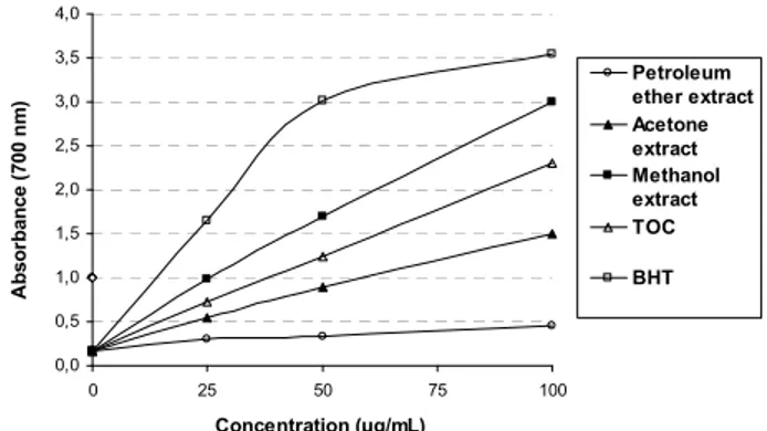
Therefore, chelating agents known as secondary antioxidants are important for antioxidant activity. The light petroleum extract showed almost no activity in the DPPH and reducing power assays. Conversely, the methanol extract was found to be the most active extract in both assays (Figures 3 and 4).
0 10 20 30 40 50 60 70 80 90 100 Petroleum ether extract Acetone extract Methanol extract α-TOC BHT D P P H S ca ve nging E ff ec ts ( % ) 25 µg 50 µg 100 µg
Figure 3: Free radical scavenging activity of extracts of M. juliana, BHT and
α-tocopherol (α-TOC) by DPPH assay. Values are mean of n=3. p<0.05,
significantly different with Student’s t-test.
0,0 0,5 1,0 1,5 2,0 2,5 3,0 3,5 4,0 0 25 50 75 100 Concentration (µg/mL) A b sor b ance ( 700 n m ) Petroleum ether extract Acetone extract Methanol extract TOC BHT
Figure 4: Reductive potential of extracts of M. juliana, BHT and α-tocopherol
(α-TOC) using spectrophotometric detection of the Fe+3-Fe+2 transformations. Values are mean of n=3. p<0.05, significantly different with Student’s t-test.
Table 1: Constituents of fraction 1–3.
Compound No RIa % Identification Method Unidentified – 2.5 MS Unidentified – 58.4 MS, RI Unidentified – 1.1 MS 3- Phenethylphenol (1) 1625 1.2 MS, RI 2,6-Diisopropylnaphthalene (2) 1715 2.4 MS, RI Unidentified – 3.6 MS Unidentified – 3.4 MS Unidentified – 1.3 MS 1,3,3- Trimethyl-1- Phenylindan (3) 1877 1.8 MS, RI Unidentified – 6.1 MS Biformene (4) 1897 1.1 MS, Co-GC, RI Unidentified – 1.8 MS Nordehydroabietane (5) 1919 4.7 MS, RI Abieta-8,11,13-triene (6) 2060 8.6 MS, Co-GC, RI Dehydroabietinal (7) 2181 1.9 MS, Co-GC, RI Total identified: 21.7 a: Kovats index on DB–1 fused silica column; MS: Mass spectrum; Co-GC: Co-injection with authentic compounds; RI: Retention Index literature
comparison.
Based on the results of the antioxidant activity tests on the extracts, the light petroleum extract was studied for its chemical composition by GC and GC-MS. For this purpose, the extract was fractionated on a silica gel column. Fifteen fractions were obtained after similar fractions had been combined. Total antioxidant activity of the fractions at a concentration of 25 μg/mL was also investigated by the β-carotene-linoleic acid assay. Frs 1-3 (78.4%), Frs 4-5 (72.2%), and Frs 8-9 (71.9%) were found to be active, showing
r
-ı
Table 2: Constituents of fraction 4–5. Compound No RIa Content (%) Identification Method Unidentified – 19.3 MS Dihydroactinidiolide (8) 1471 7.7 MS, RI Phytone (9) 1820 60.1 MS, RI Unidentified – 12.0 MS Total identified: 67.7
a: Kovats index on DB–1 fused silica column; MS: Mass spectrum; RI: Retention Index literature comparison.
more than 50% inhibition. Therefore, these three were examined by GC-MS.
The yield of Frs 1-3 in the total light petroleum extract was 0.82% (31.90 mg). The chemical constitution of this fraction is shown in Table 1. Seven compounds were identified by GC and GC-MS analyses, of which major ones were abieta-8,11,13-triene (8.6%), nordehydroabietane (4.7%) and abieta-8,11,13-trien-18-al (1.9%).
Biformene (4) (=labda-8(20),12,14-triene) has been reported as a constituent of Dacrydium and
Helianthus species [10]. Nordehydroabietane (5) (= 18-demethyl triene), triene (6), and dehydroabietinal (7) (= abieta-8,11,13-trien-18-al) are constituents of the oleoresin of Larix and Pinus species [11a-11c]. Compounds 4, 5, 6 and
7 have not been previously reported in Micromeria species.
Frs 4-5 yielded 2.60 mg, corresponding to 0.07% of the light petroleum extract. Only four compounds were detected in this fraction, two of which were identified as dihydroactinidiolide (7.7%) and phytone (= hexahydrofarnesyl acetone) (60.1%), which corresponded to 67.7% of the fraction (Table 2). Dihydroactinidiolide, which showing physiological activity in the cat family, is an important aroma constituent of cigar tobacco and tea [12a]. Phytone, which is an oxidation product of phytol, is widespread in the plant family [12b]. However, both were detected in a Micromeria species for the first time.
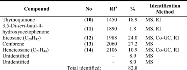
Frs 8-9 yielded 3.00 mg, corresponding to 0.08% of the fraction. Seven compounds were detected, five of which were identified; this corresponded to 82.8% of the fraction. Cembrene (thunbergen) (27.2%) and eicosane (24.0%) were the major compounds (Table 3). Thymoquinone (= 2-isopropyl-5-methyl-1,4-benzo-quinone), a biologically important toxic compound, and cembrene (= thunbergen) are constituents of black cumin and Pinus species, respectively [13-15]. Both compounds were detected in a Micromeria
Table 3: Constituents of Fraction 8–9.
Compound No RIa % Identification Method Thymoquinone (10) 1450 18.9 MS, RI 3,5-Di-tert-butil-4- hydroxyacetophenone (11) 1890 1.8 MS, RI Eicosane (C20H42) (12) 1988 24.0 MS, Co-GC, RI Cembrene (13) 2060 27.2 MS Heneicosane (C21H44) (14) 2106 10.9 MS, Co-GC, RI Unidentified – 8.9 MS Unidentified – 8.0 MS Total identified: 82.8
a: Kovats index on DB–1 fused silica column; MS: Mass spectrum; Co-GC: Co-injection with authentic compounds; RI: Retention Index
literature comparison.
Table 4: Anticholinesterase activity of M. juliana extracts a.
AChE assay BChE assay
Samples 200 µg IC50 (μg/mL) 200 µg IC50 (μg/mL) Light petroleum extract -5.9±4.1 >200 40.9±3.1 >200 Acetone extract 35.3±3.1 >200 52.4±1.8 185.6±1.9 Methanol extract -7.6±6.8 >200 -6.2±2.3 >200 Galantamine b 74.0 ±0.8 5.0±0.1 75.0±0.6 50.8±0.9 a IC
50 values represent the means ± standard deviation of three parallel measurements (p<0.05). b Standard drug (at μM concentration).
species for the first time. In some investigations, radical scavenging activity was related with anticholinesterase activity [7a,7b] and so this was also determined for the extracts (Table 4). The acetone and light petroleum extracts exhibited moderate butyryl-cholinesterase inhibitory activity, whereas no such acetyl-cholinesterase inhibitory activity was observed in either the light petroleum or methanol extracts. Only the acetone extract demonstrated mild acetylcholinesterase inhibitory activity.
In this study, 14 components were identified for the first time from the light petroleum extract of M.
juliana by GC and GC-MS. Interestingly, the volatile oil components, even the major ones previously revealed [4d,6a-6c], were not identified as compounds that showed antioxidant activity.
In conclusion, the identified constituents were found to have cyclic and/or unsaturated structures. These compounds can stop radical degradation or can contribute to antioxidant activity by either scavenging or converting singlet oxygen to triplet oxygen in the medium [15]. Frs 8-9 contained a phenolic, a paraquinoid, a diterpene and two hydrocarbons. However, further studies are needed to understand the origin of the activity. In particularly, other minor and major components need to be tested for their activity, individually, as well as for their possible synergistic effects.
Experimental
General experimental procedures: GC-MS utilized a
Varian Saturn 2100 and were performed at the Department of Chemistry, University of Mugla. Antioxidant activity measurements were recorded on a Shimadzu UV–1601 (Kyoto, Japan), and anticholinesterase activity measurements on a SpectraLab 340PC, Molecular Devices (NY, USA).
Plant material: The aerial parts of Micromeria
juliana were collected from Marmaris-Mugla, Turkey in June 2005 at 250 m altitude and identified by Dr Tuncay Dirmenci. A voucher specimen was deposited in the Herbarium of the Faculty of Arts and Sciences.
Extraction and fractionation: The dried and
powdered aerial parts M. juliana (930 g) were extracted with light petroleum, acetone and methanol, successively, at room temperature (24 h x 3). After filtration, the solvent was evaporated to dryness under vacuum. The crude light petroleum extract (3.88 g) was fractionated on a silica gel column (2.5 x 100 cm) by elution with light petroleum (40-60°), followed by a gradient of dichloromethane up to 100%. Using TLC, 16 fractions, coded Frs 1-3 to Frs 33, were obtained after similar fractions were combined. These 16 fractions were subjected to an antioxidant activity test at 25 μg/mL concentration using the β-carotene-linoleic acid assay. The fractions that exhibited antioxidant activity were studied further. Since the polarity was suitable, these 3 fractions were analyzed by GC and GC-MS. Identification of components 1-14 was based on GC retention indices and computer matching with the Wiley and Nist, 2005 Library, as well as by comparison of the fragmentation patterns of the MS with those reported in the literature and when, ever possible, by co-injection with authentic compounds.
Gas chromatography: GC analyses of the antioxidant
active fractions were performed using a Shimadzu GC-17 AAF, V3, 230V series gas chromatograph equipped with a FID and a DB-1 fused silica capillary column (30 m x 0.25 id., film thickness 0.32 µm); the initial oven temperature was held at 100°C
for 5 min., then programmed to 220°C at 4°C/min
and held isothermal for 15 min; injector and detector
temperatures were 250°C and 270°C, respectively;
carrier gas was He at a flow rate of 1.3 mL/min; sample size, 1.0 µL; split ratio, 1:50. The percentage composition of each fraction was determined with a Class-GC 10 computer programme.
Gas chromatography mass spectrometry: GC-MS
analysis of antioxidant active fractions was performed using a Varian Saturn 2100 (Old York Rd., Ringoes, NJ, USA, Quadrupole, EI-mode, 70 eV) equipped with a DB–1 fused silica capillary column (30 m x 0.25 id., film thickness 0.32 µm). For GC–MS detection, an electron ionisation system with an ionization energy of 70 eV was used. Carrier gas was helium (15 psi) at a flow rate of 1.3 mL/min. The oven temperature was held at 100oC for 5 min, then increased to 220oC with 4oC/min increments and held at this temperature for 15 min. Injector and MS transfer line temperatures were set at 220oC and
290oC, respectively. Ion source temperature was
200°C. The injection volume was 0.5 μL, with a split ratio of 1:30. The mass range was from m/z 28 to 650 amu. Scan time was 0.5 sec, with 0.1 interscan delays. Diluted samples (1/100, v/v, in n-hexane) of 1.0 µL were injected manually in the splitless mode. The relative percentage of each fraction constituent was expressed as percentage.
Antioxidant activity
Chemicals: Potassium ferricyanide, ferrous chloride,
ferric chloride, trichloro acetic acid (TCA), methanol, and quercetin were obtained from E. Merck (Darmstadt, Germany). β-Carotene, linoleic acid, 3- (2-pyridyl)-5,6-bis(4-phenyl-sulphonicacid)-1,2,4-tri-azine (Ferrozine), polyoxyethylene sorbitan mono-palmitate (Tween-40), 1,1-diphenyl-2-picrylhydrazyl
(DPPH), butylated hydroxytoluene (BHT),
α-tocopherol (α-TOC), acetylcholinesterase (AChE),
butyryl-cholinesterase (BChE), 5,5′-dithiobis (2-nitrobenzoic) acid (DTNB), acetylthiocholine iodide, butyrylthiocholine chloride, and galanthamine were obtained from Sigma Chemical Co. (Sigma-Aldrich GmbH, Sternheim, Germany). All other chemicals and solvents were of analytical grade.
Determination of antioxidant activity with the
β-carotene bleaching method: The antioxidant activity
was evaluated using the β-carotene-linoleic acid test
system [16]. β-Carotene (0.5 mg) in 1 mL of
chloroform was added to 25 μL of linoleic acid, and 200 µL of Tween 40 emulsifier mixture. After evaporation of chloroform under vacuum, 100 mL of distilled water, saturated with oxygen, was added by vigorous shaking. Four mL of this mixture was transferred into different test tubes containing different concentrations of the sample. As soon as the emulsion was added to each tube, the zero time absorbance was measured at 470 nm using a spectrophotometer. The emulsion system was
incubated for 2 h at 50 °C. A blank, devoid of β-carotene, was prepared for background subtraction. BHT and α-tocepherol were used as standards.
DPPH free radical scavenging activity: The free
radical scavenging activity was determined by the DPPH assay described by Blois [17,18] with slight modification. DPPH absorbs at 517 nm, but upon reduction by an antioxidant or a radical species its absorption decreases. Briefly, a 0.1 mM solution of DPPH in methanol was prepared and 4 mL of this was added to 1 mL of sample solutions in methanol at different concentrations. Thirty minutes later, the absorbance was measured at 517 nm. The capability to scavenge the DPPH radical was calculated using the following equation:
Antiradical activity (%) = control sample control
A
A
A
−
x 100Reducing power: The reducing power of extracts was
determined according to the iron(III) reductive assay [19]. Sample solutions in different amounts were mixed with 2.5 mL of 0.2 M phosphate buffer (pH 6.6) and 2.5 mL of potassium ferricyanide (1%). After the mixture was incubated at 50oC for 20 min, 2.5 mL of TCA (10%) was added and the mixture centrifuged at 1000 g (MSE Mistral 2000, London, UK) for 10 min. Supernatant (2.5 mL) was mixed with distilled water (2.5 mL) and 0.5 mL of ferric chloride (0.1%), and the absorbance was measured at 700 nm.
Metal chelating activity: The chelating activity of
extract on Fe2+ was measured as reported by Decker and Welch [19,20]. The extract was added to a solution of 2 mM FeCl2 (0.1 mL). The reaction was initiated by the addition of 5 mM ferrozine (0.2 mL). The mixture was shaken vigorously and left standing at room temperature for 10 min. After reaching
equilibrium, the absorbance was determined at 562 nm.
Anticholinesterase activity: Acetyl- and butyryl-cholinesterase inhibitory activities were measured by slightly modifying the spectrophotometric method of Ellman et al [21]. Acetylthiocholine iodide and butyryl-thiocholine chloride were used as substrates of the reaction and DTNB for the measurement of the anticholinesterase activity. Sodium phosphate buffer (pH 8.0; 150 µL of 100 mM solution), 10 µL of test compound solution, and 20 µL AChE or BChE solution were mixed and incubated for 15 min at 25 ºC, and 10 µL of DTNB was added. The reaction was then initiated by the addition of 10 µL of either acetylthiocholine iodide or butyrylthiocholine chloride. The hydrolysis of these substrates, shown by the formation of yellow 5-thio-2-nitrobenzoate anion, was monitored spectrophotometrically at a wavelength of 412 nm. Ethanol was used as a solvent to dissolve the test compounds and controls.
Statistical analysis: All data from all activity tests are
the average of triplicate analyses. The data were recorded as means ± standard deviations. Analysis of variance was performed by ANOVA procedures. Significant differences between means were determined by the Student-t test, with p values of
<0.05 being regarded as significant.
Acknowledgments - This study is a part of M.Ö’s Ph.D. thesis. This study was partly supported by State Planning organization of Turkey (DPT 2003K1208500). We also would like to thank to Dr Tuncay DİRMENCİ, Necati Bey Education Faculty, Department of Biology Education, Balıkesir Üniversity, Turkey, for the identification of the plant sample. GC and GC-MS spectra were performed at the Department of Chemistry, Faculty of Arts and Sciences, University of Muğla.
References
[1] Davis PH. (1982) Micromeria Bentham. In Flora of Turkey and the East Aegean Islands. Davis PH (Ed). Vol.7, Edinburgh University Pres, Edinburg, 335-346.
[2] (a) Ali-Shtayeh MS, Al-Nuri MA, Yaghmour RMR, Faidi YR. (1997) Antimicrobial activity of Micromeria nervosa from the Palestinian area. Journal of Ethnopharmacology, 58, 143–147; (b) Palevitch PD, Yaniv Z. (1991) Medicinal Plants of the Holy
Land. Vols 1 and 2. Tammuz, Tel-Aviv; (c) Formisano C, Mignola E, Rigano D, Senatore F, Bellone G, Bruno M, Rosseli S. (2007)
Chemical composition and antimicrobial activity of the essential oil from aerial parts of Micromeria fruticulosa (Bertol.) Grande (Lamiaceae) growing wild in Southern Italy. Flavour and Fragrance Journal, 22, 289–292; (d) Güllüce M, Sökmen M, Şahin F, Sökmen A, Adıgüzel A, Özer H. (2004) Biological activities of the essential oil and methanolic extract of Micromeria fruticosa (L) Druce spp. serpilifolia (Bieb) PH Davis plants from the eastern Anatolia region of Turkey. Journal of the Science of Food and
Agriculture, 84, 735–741; (e) Ali-Shtayeh MS, Yaghmour RMR, Faidi YR, Salem K, Al-Nuri MA. (1998) Antimicrobial activity of
20 plants used in folkloric medicine in the Palestinian area. Journal of Ethnopharmacology, 60, 265–271.
[3] Puri HS, Jain SP. (1988) Micromeria capitellata Benth.: a new source of pulegone. Parfümerie und Kosmetik, 69: 163, CA 109:43302m.
[4] (a) Aslan I, Çalmaşur Ö, Şahin F, Çağlar Ö. (2005) Insecticidal effects of essential plant oils against Ephestia kuehniella Zell.,
Lasioderma serricorne F. and Sitophillus granarius L. Journal of Plant Diseases and Protection, 112, 257–267; (b) Duru ME,
Öztürk M, Uğur A, Ceylan Ö. (2004) The constituents of essential oil and in vitro antimicrobial activity of Micromeria cilicica from Turkey. Journal of Ethnopharmacology, 94, 43–48; (c) Guarrera PM, Salerno G, Caneva G. (2005) Folk phytotherapeutical plants from Maratea area (Basilicata, Italy). Journal of Ethnopharmacology, 99, 367–378; (d) Başer KHC. (2002) Aromatic biodiversity among the flowering plant taxa of Turkey. Pure and Applied Chemistry, 74, 527–545.
[5] (a) Özcan M. (1999) Antifungal effects of Micromeria myrtifolia Boiss & Hohen in Boiss. and Prangos uechtritzii (Boiss.) Hawsskn decoctions. Acta Alimentaria, 28, 355–360; (b) Harmandar M. (1988) Analysis of essential oil from leaves of Micromeria
fruticosa (L.) Druce subsp. serpillifolia (Bieb.) P.H. Davis. Turkish Journal of Chemistry, 12, 188–194; (c) Kırımer N, Özek T,
Başer KHC. (1991) Composition of the essential oil of Micromeria congesta. Journal of Essential Oil Research, 3, 387–393; (d) Tabanca N, Kırımer N, Demirci B, Demirci F, Başer KHC. (2001) Composition and antimicrobial activity of essential oils of
Micromeria cristata subsp. phrygia and enantiomeric distribution of borneol. Journal of Agricultural and Food Chemistry, 49,
4300–4303; (e) Başer KHC, Demirçakmak B, Duman H. (1997) Composition of the essential oil of Micromeria cremnophila Boiss. et Heldr. subsp. amana (Rech.fil) P.H.Davis. Journal of Essential Oil Research, 9, 725–726; (f) Başer KHC, Kırımer N, Özek T., Tümen G. (1995) Essential oil of Micromeria carminea P.H.Davis. Journal of Essential Oil Research, 7, 457–458.
[6] (a) Stojanovic G, Palic I, Ursic-Jankovic J. (2006) Composition and antimicrobial activity of the essential oil of Micromeria cristata and Micromeria juliana. Flavour and Fragrance Journal, 21, 77–79; (b) Mastelic J, Jerkovic I, Kustrac D. (2005) Aromatic compounds of Micromeria juliana (L.). Bentham ex Reichenb. from Croatia(a). Journal of Essential Oil Research, 17, 516–518; (c) Slavkovska V, Couladis M, Bojovic S, Tzakou O, Pavlovic M, Lakusic B, Jancic R. (2005) Essential oil and its systematic significance in species of Micromeria Bentham from Serbia & Montenegro. Plant Systematics and Evolution, 255, 1–15; (d) Couladis M, Tzakou O, Verykokidou E, Harvala C. (2003) Screening of some Greek aromatic plants for antioxidant activity. [7] (a) Agrawal R, Tyagi E, Shukla R, Nath C. (2009) A study of brain insulin receptors, AChE activity and oxidative stress in rat
model of ICV STZ induced dementia. Neuropharmacology, 56, 779–787; (b) Papandreou MA, Dimakopoulou A, Linardaki ZI, Cordopatis P, Klimis-Zacas D, Margarity M, Lamari FN. (2009) Effect of a polyphenol-rich wild blueberry extract on cognitive performance of mice, brain antioxidant markers and acetylcholinesterase activity. Behavioural Brain Research, 198, 352–358. [8] Huang D, Ou B, Prior RL. (2005) The chemistry behind antioxidant capacity assays. Journal of Agricultural Food Chemistry, 53,
1841–1856.
[9] (a) Halliwell B, Gutteridge JM. (1984) Oxygen toxicology, oxygen radicals, transition metals and disease. Biochemical Journal,
219, 1-4; (b) Aruoma OI, Halliwell B, Dizdaroğlu M. (1989) Iron ion-depended modification of bases in DNA by the superoxide
radical-generating system hypoxanthine/xanthine oxidase. Journal of Biological Chemistry, 264, 13024–13028.
[10] Bohlmann F, Jakupovic J, King RM, Robinson H. (1980) Neue ent-atisiren- und ent-kaurensäure-derivate aus Helianthus-arten.
Phytochemistry, 19, 863–868.
[11] (a) Bol'shakova VI, Demenkova LI, Khan VA, Dubovenko ZhV, Shmidt ÉN, Pentegova VA. (1985) Chemical composition of the oleoresin of Larix kamtschatica. Chemistry of Natural Compounds, 21, 749–752; (b) Vlad PF, Russo AG, Koltsa MN, Paukov VN. (1971) Dehydroabietane from the oleoresin of Pinus pallasiana. Chemistry of Natural Compounds,7, 16–18; (c) Russo AG, Vlad PF, Lazur'evskii GV. (1968) The neutral substances of the oleoresin of Pinus pallasiana. Chemistry of Natural Compounds, 4, 167–168.
[12] (a) Isoe S, Hyeon SB, Katsumura S, Sakan T. (1972) Photo-oxygenation of carotenoids. II. The absolute configuration of loliolide and dihydroactinidiolide. Tetrahedron Letters, 13, 2517–2520; (b) Ogunwande IA, Walker TM, Setzer WN. (2007) A review of aromatic herbal plants of medicinal importance from Nigeria. Natural Product Communications, 2, 1311–1316.
[13] El-Dakhakhny M. (1963) Studies on the chemical constitution of Egyptian Nigella sativa L. seeds. II. The essential oil. Planta
Medica, 11, 465–470.
[14] Song ZQ, Liang ZQ, Liu X. (1995) Chemical characteristics of oleoresins from Chinese pine species. Biochemical Systematics and
Ecology, 23, 517–522.
[15] Wienkotter N, Hopner D, Schutte U, Bauer K, Begrow F, El-Dakhakhny M, Verspoh EJ. (2008) The effect of nigellone and thymoquinone on inhibiting trachea contraction and mucociliary clearance. Planta Medica, 74, 105-108.
[16] Miller HE. (1971) A simplified method for the evaluation of antioxidants. Journal of the American Oil Chemists’ Society, 48, 91. [17] Blois MS. (1958) Antioxidant determinations by the use of a stable free radical. Nature, 181, 1199–1200.
[18] Vera N, Zampini C, Isla MI, Bardón A. (2007) Antioxidant and XOD inhibitory coumarins from Pterocaulon polystachyum DC.
Natural Product Communications, 2, 551–556.
[19] Dorman HJD, Kosar M, Baser KHC, Hiltunen R. (2009) Phenolic profile and antioxidant evaluation of Mentha x piperita L. (peppermint) extracts. Natural Product Communications, 4, 535–542.
[20] Decker EA, Welch B. (1990) Role of ferritin as a lipid oxidation catalyst in muscle food. Journal of Agricultural and Food
Chemistry, 38, 674-677.
[21] Ellman GL, Courtney KD, Andres V, Featherstone RM (1961) A new and rapid colorimetric determination of acetylcholinesterase activity. Biochemistry and Pharmacology and Behavior, 7, 88–95.