ORIGINAL PAPER
Factors associated with prolonged wound drainage
after hemiarthroplasty for hip fractures in elderly
Umut Canbek1 &Ulas Akgun1 &Nevres Hurriyet Aydogan1 &Tugba Dubektas Canbek2 &Ali Turgut3 & Oguzhan Samil Erciyes1
Received: 24 February 2020 / Accepted: 14 July 2020 # SICOT aisbl 2020
Abstract
Purpose The aim of this study was to determine the incidence of prolonged wound drainage (PWD) and the amount of drainage fluid after hip hemiarthroplasty (HA) and to investigate the risk factors for the development of PWD associated with the patient, fracture and surgical treatment.
Methods Data from 313 patients who underwent HA were prospectively analysed. The mean drainage time and drainage amount of patients with PWD were calculated. Patient demographic data, pre-operative ASA scores and anticoagulation status, presence of diabetes, fracture type, surgical approach, femoral stem type, cable usage, amount of drain output, blood transfusion quantity, time from injury to surgery, time from surgery to discharge and patient blood tests were investigated.
Results The incidence of PWD after HA was 8.9% (28 patients). The mean drainage time in patients with PWD was 4.9 ± 1.85 (3–9) days, and the mean collected total fluid volume was 51.1 ± 26.9 (21–132) mL. PWD was more commonly observed in the lateral approach group (p< 0.001) and morbidly obese patients (p < 0.001). In the PWD group, the mean post-operative first-day haemoglobin value was lower (p< 0.001), more blood transfusions were required (p < 0.001) and the amount of drainage output from the closed suction drain (CSD) was higher (p < 0.001). The duration of hospitalization was longer in patients with PWD (p < 0.001). Lateral approach, morbid obesity and increased drainage output were found to be associated with PWD in logistic regression analysis.
Conclusion Lateral approach, morbid obesity and increased drainage output were found to be risk factors for the occurrence of PWD.
Keywords Drainage time . Drainage volume . Hemiarthroplasty . Prolonged wound drainage
Introduction
Prolonged wound drainage (PWD) is considered one of the few factors that cause early periprosthetic joint infection (PJI) following arthroplasty [1–5]. It is not clear whether patients
with PWD develop surgical site infections (SSIs) or heal with-out complications [5,6]. Due to the apprehension of PJI com-plications, increased hospitalization time is enforced, and sometimes unnecessary irrigation and debridement surgeries
* Umut Canbek [email protected] Ulas Akgun
[email protected] Nevres Hurriyet Aydogan [email protected] Tugba Dubektas Canbek [email protected] Ali Turgut
Oguzhan Samil Erciyes [email protected]
1 Department of Orthopaedics and Traumatology, Faculty of Medicine,
Mugla Sitki Kocman University, Mugla, Turkey
2
Department of Internal Medicine, Mugla Sitki Kocman University Training and Research Hospital, Mugla, Turkey
3 Department of Orthopaedics and Traumatology, Tepecik Training
and Research Hospital, Izmir, Turkey
https://doi.org/10.1007/s00264-020-04738-z
are performed [5,7]. Such factors increase costs for the patient and healthcare systems [5,8].
Although there is no clear consensus in the literature, con-tinuous wet spots larger than 2 × 2 cm on the dressings cov-ering the surgical area that remain after the third day of arthroplasty surgery are defined as PWD [7,9–11]. The PWD incidence rate following total joint arthroplasty (TJA) is reported to be between 0.2 and 21%, and PWD is thought to be more frequent following revision arthroplasty surgeries [5,
12–15].
Low-molecular-weight heparin (LMWH), obesity and ex-cessive drain output after surgery are some of the identified risk factors for PWD after TJA surgery [14]. The studies about the incidence rate and possible risk factors for PWD were all conducted with patients undergoing TJA interventions, and to the best of our knowledge, there are no similar studies in patients with hip fractures treated with HA [6, 12–15]. Additionally, we could not find a study in the literature that reports the collection of the leaking fluid and calculation of the amount of leakage in patients with PWD.
The main objective of our study is to evaluate the incidence of and risk factors for PWD in patients with a hip fracture who are treated with HA. In addition, we also aimed to quantita-tively measure the drainage volume and duration by collecting the fluid using a simple method that we developed.
Patients and methods
Study population
The study prospectively collected data from 448 consecutive proximal femur fracture patients over 65 years of age who were treated in our clinic from January 2017 to January 2020. Local ethics committee approval was obtained prior to study initiation, and all patients gave their informed consent before being included in the study. After excluding patients who do not meet the inclusion criteria, the study was per-formed using data from 313 patients. The flowchart of the study is shown in Fig.1.
Decision process and surgical procedure
Clinical and radiological evaluation of all trauma patients in our clinic is performed by a team of four orthopaedic surgeons with ten to 15 years of clinical experience and a senior ortho-paedic surgeon with over 35 years of clinical experience. The orthopaedic council determines the type of surgery (open re-duction and internal fixation vs arthroplasty) to be performed in elderly patients with hip fractures with consideration of the patient’s age, comorbid diseases, activity level prior to frac-ture and fracfrac-ture type.
The surgical approach type (lateral or posterior) and femo-ral stem type (cemented or uncemented) to be used are chosen by the surgeon who will perform the surgery in patients un-dergoing HA. Three surgeons in our clinic used the posterior approach, and 1 surgeon preferred the lateral approach for HA surgery. All femoral stems and modular bipolar head implants used for HA were obtained from two local orthopaedic supply producers (Hipokrat, Izmir, Turkey, and Tipmed, Izmir, Turkey).
Poly-methyl methacrylate (PMMA) (Biofix-Synimed) ce-ment was used for fixation of polished femoral stems, and no additional antibiotics were added. A distal centralizer and a bone plug were used with all cemented femoral implants. Proximal 1/3 hydroxyapatite-coated implants were used for uncemented femoral stem applications. Displaced trochanter major and minor fragments in trochanteric fractures were fixed to the femoral stem with 1 to 3 multifilament metallic cable systems according to the size of the fragments. After stability control of the hip joint, a closed suction drainage (CSD) system was placed 3–5 cm distal to the incision. A polyglactin suture material was used for fascia and subcuta-neous tissues with intermittent fashion. A polypropylene su-ture material was used for skin closure.
Peri- and post-operative follow-up protocol
and collection of drainage fluid
An antibiotic prophylaxis was administered by 2-g cefazolin in patients without penicillin allergy, and patients with peni-cillin allergy were given 600-mg clindamycin. Antibiotic pro-phylaxis was continued for 24 hours following the surgery (cefazolin 3 × 1 g or clindamycin 2 × 600 mg) [14]. Low-molecular-weight heparin (enoxaparin sodium– 4000 IU) was administered as deep vein thrombosis (DVT) prophylaxis.
The amount of accumulated fluid in the negative pressure CSD system was recorded. All drains were removed under sterile conditions after 48 hours. Following CSD system remov-al, a sterile disposable paediatric urine collection bag with a 100-cc volume was placed on the drain exit point with the collection part stuck to the drainage point using sterile tech-niques (Fig. 2). The fluid volume collected in the collection bag via passive drainage was measured daily and was replaced daily as the drainage continued. Patients with daily fluid collec-tion > 2 ml for three consecutive days after the placement of the collection bag were considered PWD cases. Collection bags were removed in patients with no fluid collection for 24 hours, and the drain exit point was covered using sterile gauze. Patients with dry dressings 24 hours after fluid collection procedure completion were discharged if there was no contraindication. Drainage amounts in all patients were recorded daily. Patients with cemented prosthesis without any drainage were mobilized with full weight-bearing, whereas patients with uncemented prostheses were mobilized with partial weight-bearing.
Patients with persistent drainage were not mobilized and only lower extremity isometric exercises were given.
Patients were divided into two groups according to the pres-ence of PWD. Mean age, sex, body mass index (BMI) values, pre-operative ASA scores and anticoagulation status, presence of diabetes, hospitalization time from initial referral to the sur-gery, period from the surgery to discharge, fracture region (fem-oral neck or trochanter), surgical approach used (lateral or pos-terior), cement usage (cemented vs uncemented) and cable us-age (yes/no) were recorded. Patients were categorized as normal (≤ 24.9 kg/m2
), overweight (25.0 to 29.9 kg/m2), class I obese (30 to 34.9 kg/m2), class II obese (35 to 39.9 kg/m2) or morbid obese (≥ 40 kg/m2
) according to their BMI. Finally, pre-operative blood glucose levels, haemoglobin (Hgb), lymphocyte number and percentage, total protein and albumin values prior to surgery, 24 hours after surgery and at discharge, total blood transfusion requirement amounts and total fluid amounts drained from the CSD system following surgery were also compared.
448 23 425 50 62 9 6 10 6 9 22 313 28 285
Fig. 1 The flowchart of the study
Fig. 2 A sterile disposable paediatric urine collection bag with 100-cc capacity was used for collection of draining fluid from drain exit point
Statistical analysis
The IBM SPSS Statistics version 22 (IBM Corp, Armonk NY, USA) was used for statistical analysis. Continuous variables are expressed as the mean, standard deviation and max-min values, and categorical variables are expressed as percentages. The normality of the groups was evaluated with the Shapiro-Wilk test. The Mann-Whitney U test was used for the non-normally distribut-ed variables, and the t test was usdistribut-ed to compare normally
distributed variables. The chi-square tests were used to compare categorical variables. Variables with p < 0.25 were included in the multivariate logistic regression anal-ysis. The age-corrected probability rate (PR) and 95% confidence interval were set using the maximum proba-bility method. Sub-group analysis was performed about variables that are statistically significant in logistic re-gression to investigate the effect of the distribution of the fracture type. p < 0.05 values were deemed statisti-cally significant. 0,0 0,2 0,4 0,6 0,8 1,0 0 1 2 3 4 5 6 7 8 9 10
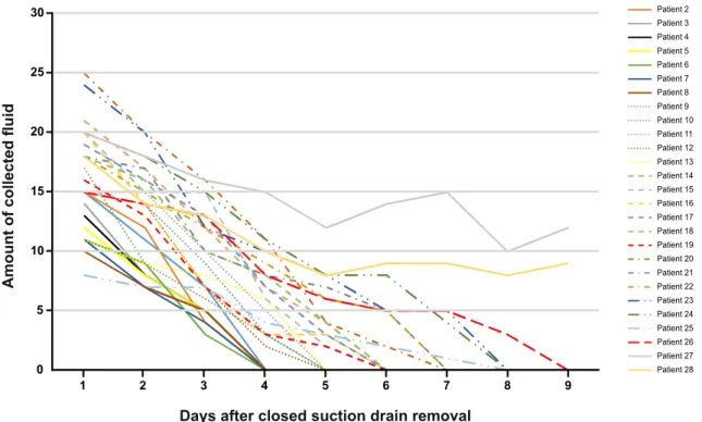
Fig. 3 The Kaplan-Meier curve for the duration of prolonged wound drainage within all study group after closed suction drain removal 0 5 10 15 20 25 30 1 2 3 4 5 6 7 8 9
Days after closed suction drain removal
Amount of collected fluid Patient 1 Patient 2 Patient 3 Patient 4 Patient 5 Patient 6 Patient 7 Patient 8 Patient 9 Patient 10 Patient 11 Patient 12 Patient 13 Patient 14 Patient 15 Patient 16 Patient 17 Patient 18 Patient 19 Patient 20 Patient 21 Patient 22 Patient 23 Patient 24 Patient 25 Patient 26 Patient 27 Patient 28
Results
One hundred fourteen (36.4%) patients were male, whereas 199 (63.6%) were female. The mean age of the patients was
81.2 ± 8.3 (range 65 to 99) years. Fifty-two (16.7%) patients showed no collection of fluid in the collection bag following drain removal. One hundred twenty-two (38.9%) patients showed fluid collection on the first day, and 111 (35.5%)
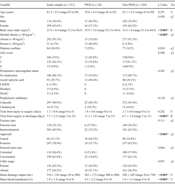
Table 1 The comparison of demographic and surgical data
Variable Entire sample (n = 313) PWD (n = 28) Non-PWD (n = 285) p Value Test
Age (years) 81.2 ± 8.3 (range 65 to 99) 79.6 ± 8.4 (range 65 to 95) 81.3 ± 8.3 (range 65 to 99) 0.251 U
Sex 0.458 χ2
Male 114 (36.4%) 12 (42.9%) 102 (35.8%)
Female 199 (63.6%) 16 (57.1%) 183 (64.2%)
Body mass index (kg/m2) 31.8 ± 4.4 (range 21.5 to 44.5) 35.9 ± 5.8 (range 25.2 to 44.4) 31.4 ± 4 (range 21.5 to 44.5) < 0.001* U
Morbid obesity (≥ 40 kg/m2) < 0.001* χ2 Absent (< 40 kg/m2) 292 (93.3%) 15 (53.6%) 277 (97.2%) Present (≥ 40 kg/m2) 21 (6.7%) 13 (46.4%) 8 (2.8%) Diabetes mellitus 84 (26.8%) 7 (25%) 77 (%27) 0.818 χ2 ASA score 0.440 χ2 2 166 (53%) 12 (42.8%) 154(54%) 3 132 (42.2%) 15 (53.6%) 117(41.1%) 4 15 (4.8%) 1 (3.6%) 14(4.9%)
Preoperative anticoagulant status 0.283 χ2
No medication 188 (60.1%) 15 (53.6%) 173 (60.7%)
Acetyl salicylic acid 93 (29.7%) 13 (46.4%) 80 (28.1%)
LMWH 6 (1.9%) 0 6 (2.1%) Warfarin 15 (4.8%) 0 15 (5.3%) NOAC 11 (3.5%) 0 11 (3.8%) Prophylactic antibiotics 0.158 χ2 Cefazolin 297 (94.9%) 25 (89.3%) 272 (95.4%) Clindamycin 16 (5.1%) 3 (10.7%) 13 (4.6%)
Time from injury to surgery (days) 1.7 ± 0.8 (range 0 to 5) 1.8 ± 0.6 (range 0 to 5) 1.7 ± 0.8 (range 0 to 5) 0.282 U
Time from surgery to discharge (days) 7.7 ± 2.2 (range 5 to 22) 11.3 ± 3.8 (range 7 to 21) 6.7 ± 1.4 (range 3 to 11) < 0.001* U
Fracture type 0.111 χ2 Femoral neck 110 (35.1%) 6 (27.8%) 104 (36.5%) Intertrochanteric 203 (65.9%) 22 (72.2%) 181 (63.5%) Approach < 0.001* χ2 Lateral 66 (21.1%) 18 (64.3%) 48 (16.8%) Posterior 247 (78.9%) 10 (35.7%) 237 (83.2%)
Femoral stem type 0.084 χ2
Cemented 114 (36.4%) 6 (21.4%) 108 (37.9%)
Uncemented 199 (63.6%) 22 (78.6%) 177 (62.1%)
Cable usage 0.947 χ2
Present 136 (43.5%) 12 (42.9%) 124 (43.5%)
Absent 177 (56.5%) 16 (57.1%) 161 (56.5%)
Mean drainage output (mL) 318 ± 136 (range 20 to 900) 563 ± 173 (range 300 to 900) 294 ± 105 (range 20 to 750) < 0.001* U
Mean blood transfusion (U) 1.9 ± 1.8 (range 0 to 9) 4.5 ± 2.2 (range 0 to 9) 1.6 ± 1.5 (range 0 to 9) < 0.001* U
χ2 chi-square test
U Mann-Whitney U test
ASA score American Society of Anesthesiologists physical status classification score LMWH low-molecular-weight heparin
patients showed fluid collection on two consecutive days. Twenty-eight (8.9%) patients showed fluid drainage for three or more consecutive days after drain removal and these pa-tients are accepted as PWD. The Kaplan-Meier curve for the proportion of patients’ remaining drainage by days after closed suction drain removal is shown in Fig.3. The mean drainage time after CSD removal in the PWD group was 4.9 ± 1.9 (range 3 to 9) days, and the mean collected drainage vol-ume was 51.1 ± 26.9 (range 21 to 132) mL. The daily changes of collected fluid volume in PWD patients following CSD removal is shown in Fig.4. Drainage stopped on its own within seven days of CSD removal in 26 (92.9%) PWD pa-tients. Two (7.1%) patients in the PWD group required irriga-tion and debridement on the ninth day (11th day following surgery) due to continuous drainage and no reduction in drain-age volume with time. These patients did not experience prolonged drainage again or surgical field infection following irrigation.
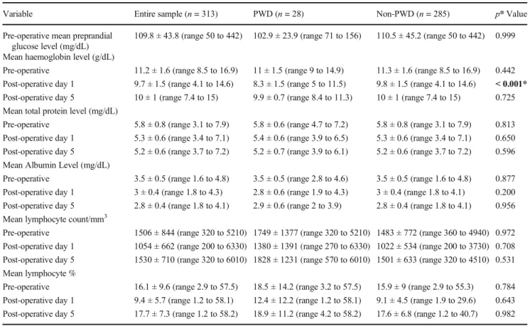
PWD was more frequently observed in HA patients in whom the lateral approach was used (p < 0.001) and morbidly obese patients (p < 0.001). In the PWD group, the mean Hgb level on the first day after surgery was lower (p < 0.001)), blood transfusion levels were higher (p < 0.001) and drain output from CSD was higher (p < 0.001). The hospitalization period of PWD patients was significantly longer (p < 0.001). No significant difference was seen between the groups in
other variables. Table1shows the comparison of demograph-ic and surgdemograph-ical data, and Table 2 shows the comparison of laboratory results between the groups.
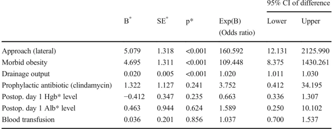
The multivariate logistic regression analysis showed that the most important risk factors for PWD were lateral ap-proach, morbid obesity and increased CSD output volume (Table3). There was no significant difference between frac-ture type groups in approach type, morbid obesity and CSD output volumes (Table4).
Discussion
The reported incidence rate of PWD in the literature varies from 0.2 to 21%, and PWD is more frequent following revi-sion arthroplasty [5,12–15]. Maathuis et al. [15] reported the PWD incidence rate in 366 THA patients as 11% when there was no protocol defined for and as 18% when a protocol was defined based on C-reactive protein and wound status. Aggarwal et al. [16] reported a PWD rate of 21% in their study of 263 THA patients. The mentioned studies are conducted using total joint arthroplasty patient groups, and knee and hip arthroplasties were assessed together in most of these studies [6,12–15]. In our study, the incidence of PWD after HA was 8.9%. The mean drainage time in patients with PWD was about five days, and the mean total fluid volume was 51.1
Table 2 Comparison of laboratory results between the groups
Variable Entire sample (n = 313) PWD (n = 28) Non-PWD (n = 285) p* Value
Pre-operative mean preprandial glucose level (mg/dL)
109.8 ± 43.8 (range 50 to 442) 102.9 ± 23.9 (range 71 to 156) 110.5 ± 45.2 (range 50 to 442) 0.999
Mean haemoglobin level (g/dL)
Pre-operative 11.2 ± 1.6 (range 8.5 to 16.9) 11 ± 1.5 (range 9 to 14.9) 11.3 ± 1.6 (range 8.5 to 16.9) 0.442
Post-operative day 1 9.7 ± 1.5 (range 4.1 to 14.6) 8.3 ± 1.5 (range 5 to 11.5) 9.8 ± 1.5 (range 4.1 to 14.6) < 0.001*
Post-operative day 5 10 ± 1 (range 7.4 to 15) 9.9 ± 0.7 (range 8.4 to 11.3) 10 ± 1 (range 7.4 to 15) 0.725
Mean total protein level (mg/dL)
Pre-operative 5.8 ± 0.8 (range 3.1 to 7.9) 5.8 ± 0.6 (range 4.7 to 7.2) 5.8 ± 0.8 (range 3.1 to 7.9) 0.813
Post-operative day 1 5.3 ± 0.6 (range 3.4 to 7.1) 5.4 ± 0.6 (range 3.9 to 6.5) 5.3 ± 0.6 (range 3.4 to 7.1) 0.650
Post-operative day 5 5.2 ± 0.6 (range 3.7 to 7.2) 5.2 ± 0.7 (range 3.9 to 6.1) 5.2 ± 0.6 (range 3.7 to 7.2) 0.596
Mean Albumin Level (mg/dL)
Pre-operative 3.5 ± 0.5 (range 1.6 to 4.8) 3.5 ± 0.5 (range 2.8 to 4.6) 3.5 ± 0.5 (range 1.6 to 4.8) 0.877
Post-operative day 1 3 ± 0.4 (range 1.8 to 4.3) 2.8 ± 0.6 (range 1.9 to 4.3) 3 ± 0.4 (range 1.8 to 4.1) 0.200
Post-operative day 5 2.8 ± 0.4 (range 1.8 to 4.1) 2.9 ± 0.6 (range 2 to 3.9) 2.8 ± 0.4 (range 1.8 to 4.1) 0.956
Mean lymphocyte count/mm3
Pre-operative 1506 ± 844 (range 320 to 5210) 1749 ± 1377 (range 320 to 5210) 1483 ± 772 (range 360 to 4940) 0.972
Post-operative day 1 1054 ± 662 (range 200 to 6330) 1380 ± 1391 (range 270 to 6330) 1022 ± 534 (range 200 to 3730) 0.708
Post-operative day 5 1530 ± 710 (range 320 to 6010) 1828 ± 1231 (range 570 to 6010) 1501 ± 633 (range 320 to 4510) 0.531
Mean lymphocyte %
Pre-operative 16.1 ± 9.6 (range 2.9 to 57.5) 18.5 ± 14.2 (range 3.2 to 57.5) 15.9 ± 9 (range 2.9 to 55.3) 0.784
Post-operative day 1 9.4 ± 5.7 (range 1.2 to 58.1) 12.4 ± 12.2 (range 1.2 to 58.1) 9.1 ± 4.5 (range 1.9 to 29.6) 0.643
mL. There is no similar method in the literature for quantita-tive measurement of PWD fluid amount.
To the best of our knowledge, there are no particular stud-ies about the etiologic factors of PWD following hip HA in elderly patients. We found that PWD risk was higher in pa-tients in whom the lateral approach was used and in those with higher drainage output from the CSD. Outcomes of surgical approaches used for HA in the treatment of hip fractures were evaluated in a meta-analysis performed by van der Sijp et al. and shorter operation time and less blood loss was observed with posterior approaches compared with lateral [17]. Biber et al. [18] reported that patients treated with HA for femoral neck fractures using the lateral approach showed more post-operative haemorrhage and revision surgery rates due to haematoma formation than patients treated using the posterior approach. Patel et al. [14] reported a significant relationship between drainage output volume and PWD. Ahmed et al. [19] observed more frequent PWD after THA in hypertensive pa-tients compared with the normotensive ones and prolonged bleeding was accused. Although the main cause of PWD is still unclear, it is thought to be caused by haemorrhage from the veins in the surgical field [6,19–21]. As our study results showed that patients in the PWD group required more blood transfusions, had more drainage output volumes and underwent surgery using the lateral approach, we consider that our results support this theory.
There are various advantages of application of CSD, such as reduction of hematoma formation, decreased probability of prolonged wound drainage and lower rate of infection [22]. However, using CSD is no longer recommended after primary total hip arthroplasty [23]. On the other hand, the utilization of CSD in revision cases and after proximal femur fractures is believed to be beneficial due to susceptibility to haematoma formation. Also, the use of closed drain may be a cause of
prolonged drainage. Strahovnik et al. found that the presence of closed suction drainage was related with prolonged wound drainage in total joint arthroplasty cases [22]. However, in that study, an increased pain and swelling on the thigh was en-countered much more in the group where CSD was not used. Since CSD was utilized for all patients in this study, we cannot comment on its effect on PWD, but we think that the use of CSD is helpful in reducing haematoma formation.
Patel et al. [14] showed a relationship between morbid obesity and PWD formation risk following hip arthroplasty and explained this by fat necrosis and the requirement for a larger incision. The BMI value was found to be higher in the PWD group compared with non-PWD in our study. Furthermore, when patients with morbid obesity were strati-fied and logistic regression was performed, morbid obesity was found to be highly related to PWD. More bleeding due to the requirement of a larger surgical incision and the fact that there is more dead space following surgery in those patients are thought to be the reasons for increased PWD frequency in morbidly obese patients [24–26].
It is usually recommended to perform irrigation and de-bridement in patients with prolonged drainage for more than five to seven days following TJA. Patel et al. [14] reported that PJI risk increased by 42% each day with prolonged drainage after the fifth day of THA and recommended open debride-ment for patients with prolonged drainage lasting longer than seven days. In our study, patients with decreased daily drain output were conservatively managed, whereas two patients with constant daily drainage required irrigation and debride-ment. We think that a personalized approach can be utilized to evaluate the daily drainage output. Patients with a gradual decrease in drainage output can be conservatively managed; on the other hand, surgical management would be reasonable in patients with consistent drainage amounts.
Table 3 Multivariate logistic regression analysis revealed the most important risk factors for PWD 95% CI of difference B* SE* p* Exp(B) (Odds ratio) Lower Upper Approach (lateral) 5.079 1.318 <0.001 160.592 12.131 2125.990 Morbid obesity 4.695 1.311 <0.001 109.448 8.375 1430.261 Drainage output 0.020 0.005 <0.001 1.020 1.011 1.030
Prophylactic antibiotic (clindamycin) 1.322 1.127 0.241 3.752 0.412 34.195
Postop. day 1 Hgb* level −0.412 0.347 0.235 0.663 0.336 1.307
Postop. day 1 Alb* level 0.463 0.944 0.624 1.589 0.250 10.102
Blood transfusion 0.036 0.201 0.856 1.037 0.700 1.537
*Postop. day 1 Hgb postoperative day 1 haemoglobin level Postop. day 1 Alb postoperative day 1 serum albumin level B regression coefficient
SE standard error p statistical significance
Our study has several limitations. The fact that surgical interventions were performed by different surgeons and the lack of randomization in treatment modalities are foremost limitations of our study. Not measuring the depth of the inci-sion, not including the patients who were taken to the inten-sive care unit following surgery and not measuring the drain-age volume in those patients are the other limitations of our study.
Conclusion
In conclusion, 8.9% of elderly patients treated with HA fol-lowing hip fractures experienced prolonged wound drainage in our study group. Lateral surgical approach, morbid obesity and increased closed suction drainage volume were all found to be related to prolonged wound drainage. We believe that evaluating the course of daily collected drainage amounts may help to apply a more personalized management in patients with prolonged wound drainage.
Funding information This research did not receive any specific grant from funding agencies in the public, commercial or not-for-profit sectors.
Compliance with ethical standards
Conflict of interest The authors declare that they have no conflict of
interest.
Ethical approval All procedures performed in studies involving human
participants were in accordance with the ethical standards of the institu-tional and/or nainstitu-tional research committee and with the 1964 Helsinki declaration and its later amendments or comparable ethical standards.
Informed consent Informed consent was obtained from all individual
participants included in the study.
References
1. Cordero-Ampuero J, de Dios M (2010) What are the risk factors for
infection in hemiarthroplasties and total hip arthroplasties? Clin
Orthop Relat Res 468:3268–3277. https://doi.org/10.1007/
s11999-010-1411-8
2. Florschutz AV, Fagan RP, Matar WY, Sawyer RG, Berrios-Torres
SI (2015) Surgical site infection risk factors and risk stratification. J
Am Acad Orthop Surg 23(Suppl):S8–s11.https://doi.org/10.5435/
jaaos-d-14-00447
3. Noailles T, Brulefert K, Chalopin A, Longis PM, Gouin F (2016)
What are the risk factors for post-operative infection after hip hemiarthroplasty? Systematic review of literature. Int Orthop 40:
1843–1848.https://doi.org/10.1007/s00264-015-3033-y
4. Pulido L, Ghanem E, Joshi A, Purtill JJ, Parvizi J (2008)
Periprosthetic joint infection: the incidence, timing, and predispos-ing factors. Clin Orthop Relat Res 466:1710–1715.https://doi.org/ 10.1007/s11999-008-0209-4
5. Wagenaar FBM, Lowik CAM, Zahar A, Jutte PC, Gehrke T,
Parvizi J (2019) Persistent wound drainage after total joint
arthroplasty: a narrative review. J Arthroplasty 34:175–182.
https://doi.org/10.1016/j.arth.2018.08.034
6. CAM L, Wagenaar FC, van der Weegen W, Poolman RW,
Nelissen R, Bulstra SK, Pronk Y, Vermeulen KM, Wouthuyzen-Bakker M, van den Akker-Scheek I, Stevens M, Jutte PC, group Ls (2017) LEAK study: design of a nationwide randomised controlled trial to find the best way to treat wound leakage after primary hip
and knee arthroplasty. BMJ Open 7:e018673.https://doi.org/10.
1136/bmjopen-2017-018673
7. Ghanem E, Heppert V, Spangehl M, Abraham J, Azzam K, Barnes
L, Burgo FJ, Ebeid W, Goyal N, Guerra E, Hitt K, Kallel S, Klein G, Kosashvili Y, Levine B, Matsen L, Morris MJ, Purtill JJ, Ranawat C, Sharkey PF, Sierra R, Stefansdottir A (2014) Wound
management. J Orthop Res 32(Suppl 1):S108–S119.https://doi.
org/10.1002/jor.22554
8. Kurtz SM, Lau E, Watson H, Schmier JK, Parvizi J (2012)
Economic burden of periprosthetic joint infection in the United States. J Arthroplasty 27:61-65.e61. doi:https://doi.org/10.1016/j. arth.2012.02.022
9. Al-Houraibi RK, Aalirezaie A, Adib F, Anoushiravani A,
Bhashyam A, Binlaksar R, Blevins K, Bonanzinga T, Chih-Kuo F, Cordova M, Deirmengian GK, Fillingham Y, Frenkel T, Gomez J, Gundtoft P, Harris MA, Harris M, Heller S, Jennings JA, Jiménez-Garrido C, Karam JA, Khlopas A, Klement MR, Komnos G, Krebs V, Lachiewicz P, Miller AO, Mont MA, Montañez E, Romero CA, Schwarzkopf R, Shaffer A, Sharkey PF, Smith BM, Sodhi N, Thienpont E, Villanueva AO, Yazdi H (2019) General assembly, prevention, wound management: pro-ceedings of international consensus on orthopedic infections. J
Arthroplasty 34:S157–s168. https://doi.org/10.1016/j.arth.2018.
09.066
10. Ng VY, Lustenberger D, Hoang K, Urchek R, Beal M, Calhoun JH,
Glassman AH (2013) Preoperative risk stratification and risk Table 4 Subgroup analysis
according to fracture type Variable Femoral neck fracture (n =
110) Intertrochanteric fracture (n = 203) p Value Test Approach 0.132 χ2 Lateral 18 (16.4%) 48 (23.6%) Posterior 92 (83.6%) 155 (76.4%) Morbid obesity (≥ 40 kg/m2) 0.26 χ2 Absent (< 40 kg/m2) 105 (95.5%) 187 (92.1%) Present (≥ 40 kg/m2 ) 5 (4.5%) 16 (7.9%)
Mean drainage output (mL)
reduction for total joint reconstruction: AAOS exhibit selection. J Bone Joint Surg Am 95:e191–e115.https://doi.org/10.2106/jbjs.l. 00603
11. Parvizi J, Gehrke T, Chen A (2013) Proceedings of the international consensus on periprosthetic joint infection. Bone Joint J 95:1450–
1452.https://doi.org/10.1302/0301-620X.95B11.33135
12. Adelani M, Johnson S, Keeney J, Nunley R, Barrack R (2014)
Clinical outcomes following re-admission for non-infectious wound complications after primary total knee replacement. Bone
Joint J 96:619–621. https://doi.org/10.1302/0301-620X.96B5.
33479
13. Jaberi FM, Parvizi J, Haytmanek CT, Joshi A, Purtill J (2008)
Procrastination of wound drainage and malnutrition affect the out-come of joint arthroplasty. Clin Orthop Relat Res 466:1368–1371. https://doi.org/10.1007/s11999-008-0214-7
14. Patel VP, Walsh M, Sehgal B, Preston C, DeWal H, Di Cesare PE
(2007) Factors associated with prolonged wound drainage after primary total hip and knee arthroplasty. J Bone Joint Surg Am 89: 33–38.https://doi.org/10.2106/JBJS.F.00163
15. Maathuis P, de Hartog B, Bulstra S (2009) Timing of open
debride-ment for suspected infection of joint prosthesis: a report on 551
patients. Curr Orthop Pract 20:541–545.https://doi.org/10.1097/
BCO.0b013e3181a0a7fb
16. Aggarwal VK, Elbuluk A, Dundon J, Herrero C, Hernandez C,
Vigdorchik JM, Schwarzkopf R, Iorio R, Long WJ (2019) Surgical approach significantly affects the complication rates asso-ciated with total hip arthroplasty. Bone Joint J 101-b:646-651. doi: https://doi.org/10.1302/0301-620x.101b6.bjj-2018-1474.r1
17. van der Sijp MPL, van Delft D, Krijnen P, Niggebrugge AHP,
Schipper IB (2018) Surgical approaches and hemiarthroplasty out-comes for femoral neck fractures: a meta-analysis. J Arthroplasty
33:1617-1627.e1619. doi:https://doi.org/10.1016/j.arth.2017.12.
029
18. Biber R, Brem M, Singler K, Moellers M, Sieber C, Bail HJ (2012)
Dorsal versus transgluteal approach for hip hemiarthroplasty: an analysis of early complications in seven hundred and four
consec-utive cases. Int Orthop 36:2219–2223.https://doi.org/10.1007/
s00264-012-1624-4
19. Ahmed AA, Mooar PA, Kleiner M, Torg JS, Miyamoto CT (2011)
Hypertensive patients show delayed wound healing following total
hip arthroplasty. PLoS One 6:e23224.https://doi.org/10.1371/
journal.pone.0023224
20. Kilpadi DV, Cunningham MR (2011) Evaluation of closed incision
management with negative pressure wound therapy (CIM): hematoma/seroma and involvement of the lymphatic system.
Wound Repair Regen 19:588–596.
https://doi.org/10.1111/j.1524-475X.2011.00714.x
21. Pauser J, Nordmeyer M, Biber R, Jantsch J, Kopschina C, Bail HJ,
Brem MH (2016) Incisional negative pressure wound therapy after hemiarthroplasty for femoral neck fractures - reduction of wound
complications. Int Wound J 13:663–667.https://doi.org/10.1111/
iwj.12344
22. Strahovnik A, Fokter SK, Kotnik M (2010) Comparison of
drain-age techniques on prolonged serous draindrain-age after total hip arthroplasty. J Arthroplasty 25:244–248.https://doi.org/10.1016/j. arth.2008.08.014
23. Abdel MP, Barreira P, Battenberg A, Berry DJ, Blevins K,
Font-Vizcarra L, Frommelt L, Goswami K, Greiner J, Janz V, Kendoff DO, Limberg AK, Manrique J, Moretti B, Murylev V, O'Byrne J, Petrie MJ, Porteous A, Saleri S, Sandiford NA, Sharma V, Shubnyakov I, Sporer S, Squire MW, Stockley I, Tibbo ME, Turgeon T, Varshneya A, Wellman S, Zahar A (2019) Hip and knee section, treatment, two-stage exchange spacer-related: proceedings of international consensus on orthopedic infections. J Arthroplasty 34:S427–s438.https://doi.org/10.1016/j.arth.2018.09.027
24. Kapadia BH, Berg RA, Daley JA, Fritz J, Bhave A, Mont MA
(2016) Periprosthetic joint infection. Lancet 387:386–394.https:// doi.org/10.1016/S0140-6736(14)61798-0
25. Toma O, Suntrup P, Stefanescu A, London A, Mutch M, Kharasch
E (2011) Pharmacokinetics and tissue penetration of cefoxitin in obesity: implications for risk of surgical site infection. Anesth A n a l g 1 1 3 : 7 3 0–737. h t t p s : / / d o i . o r g / 1 0 . 1 2 1 3 / A N E . 0b013e31821fff74
26. D’Apuzzo MR, Novicoff WM, Browne JA (2015) The John Insall
award: morbid obesity independently impacts complications, mor-tality, and resource use after TKA. Clin Orthop Relat Res 473:57– 63.https://doi.org/10.1007/s11999-014-3668-9
Publisher’s note Springer Nature remains neutral with regard to jurisdic-tional claims in published maps and institujurisdic-tional affiliations.