© Turkish Society of Radiology 2011
195
C
omputed tomographic angiography (CTA) is an excellent meth-od, with low morbidity and great availability, for the evaluation of stroke patients. High-quality images are essential for a correct diagnosis. To obtain sufficient images, techniques are used to eliminate artifacts and ensure proper image post-processing (1–3).Greater density in the neck veins may lead to streak artifacts and may ultimately be the cause of incorrect diagnoses, such as thrombosis or dis-section within the adjacent arteries. Increased densities may also prevent high-quality reconstructions. Contrast material reflux into the veins leads to a higher density in the veins and may be seen in daily practice for no apparent reason. A few studies have suggested that when inject-ing contrast material into the right arm, there is a significant reduction in jugular vein reflux and perivenous artifacts. It has been reported that brachiocephalic vein compression and left jugular vein stasis might be related to decreased retrosternal distance and may be prevented with left arm injection(3–6).
We evaluated all of the parameters that have been considered in previ-ous studies related to the injection side and those issues in CTA perform-ance that may lead to misdiagnoses with nondiagnostic images.
In this study, artifacts that may be related to right or left arm injec-tions, as well as artifacts that may reduce image quality of CTA, were investigated in a systematic fashion.
Materials and methods Patient population
CTAs of 90 patients with clinically suspected cervical or intracra-nial vascular disease were retrospectively reviewed. CTAs performed on patients with heart failure, vascular anomalies, or occlusions were not included. All patients were informed of the study before the CTA examination.
The first group of 44 patients, which consisted of 21 men and 23 wom-en, with a mean age of 64±13.2 years and an age range of 31 to 85 years, received contrast material in their right arms. The second group, consist-ing of 18 men and 28 women, with ages rangconsist-ing from 31 to 87 years and a mean age of 60.76±13.23 years, received contrast material injections in their left arms.
CT protocol
All CTA scans were obtained using a four-detector row CT scanner (HiSpeed; GE Medical Systems, Milwaukee, Wisconsin, USA). The pa-tients assumed a supine position with their arms at their sides. Prior to the procedure, the patients were instructed to remove any movable dental implants, to breathe quietly without swallowing, and to remain still during the scan. A lateral scout view including the thorax, neck,
N E U R O R A D I O L O G Y
O R I G I N A L A R T I C L ECarotid CT angiography: comparison of image quality for left
versus right arm injections
Gülen Demirpolat, Mürvet Yüksel, Gülgün Kavukçu, Deniz Tuncel
From the Department of Radiology (G.D. gulendemirpolat@
hotmail.com), Balıkesir University School of Medicine, Balıkesir,
Turkey; the Departments of Radiology (M.Y.), and Neurology (D.T.), Kahramanmaraş Sütçü İmam University School of Medicine, Kahramanmaraş, Turkey; and the Department of Radiology (G.K.), Ege University School of Medicine, İzmir, Turkey.
Received 18 January 2010; revision requested 6 April 2010; revision received 26 May 2010; accepted 29 May 2010.
Published online 9 August 2010 DOI 10.4261/1305-3825.DIR.3290-10.1
PURPOSE
To evaluate the differences in image quality of carotid com-puted tomographic angiography (CTA) of patients injected with contrast material in their right arms versus patients in-jected with contrast material in their left arms.
MATERIALS AND METHODS
Patients who had cerebrovascular accidents and subsequently underwent CTA were included in the study. Contrast material was injected into the right arms of 44 patients and into the left arms of 46 patients. Source images of a total of 90 CTAs were retrospectively evaluated for perivenous streak artifacts and contrast material reflux into the veins of the neck and upper thorax. After adjusting for differences in gender, the relation-ship between the injection site and the intensity of perivenous streak artifacts and venous reflux was determined.
RESULTS
Perivenous streak artifacts and venous reflux were demon-strated in patients who underwent either right or left arm in-jections. However, the intensity of perivenous streak artifacts was stronger in patients who were injected with contrast ma-terial in the left arm. Venous reflux into the neck and upper thorax veins was also more severe and more frequent with left arm injections. A decreased retrosternal distance facilitated reversed flow into the veins when the left arm injection was used.
CONCLUSION
Perivenous beam hardening streak artifacts and venous re-flux could not be prevented with right or left arm injections. However, patients who were injected with contrast material in their right arms showed fewer artifacts, thus allowing for better quality images on CTA.
Key words: • tomography, X-ray computed • angiography • artifacts • veins
Demirpolat et al.
196•September 2011•Diagnostic and Interventional Radiology
and (c) reflux into all three branch veins. The relationships between these three groups and the intensity of the perivenous streak artifacts and injec-tion site were evaluated.
Perivenous artifacts were evaluated on axial images and were scored as fol-lows: 0, no artifact, clear visualization of the perivenous tissues; 1, artifacts that may lead to false positive steno-sis or dissection in the arteries near the veins; and 2, profuse artifacts that make the image unusable for assessment. The relationship between artifact scores and right and left arm injections and venous reflux was evaluated.
Statistical analysis
Software (SPSS 10.0 for Windows; SPSS Inc., Chicago, USA) was used for the statistical analysis. The dif-ferences between right and left arm injection in regards to age, gender, and length of IJV reflux were evalu-ated using a 2 test. Fisher’s exact test was used to evaluate the relationship between IJV reflux frequency and right and left arm injections. The dif-ference in the retrosternal distance in patients with and without IJV reflux was determined with the Mann-Whit-ney U test. The relationship between perivenous artifact scores and gender, injection site, presence of branch vein reflux, and the presence and length of IJV reflux was evaluated using a 2 test. A value of P < 0.05 was consid-ered significant.
Results
The mean age and gender differences between right and left arm injections were not statistically significant (Table 1) (P > 0.05). IJV reflux was observed in 54.4% (49/90) of the patients. The frequency and length were statistically higher in left arm injections as com-pared to right arm injections (Tables 1 and 2). IJV reflux was not affected by gender.
The mean shortest retrosternal dis-tance was 9.3±5.4 mm for IJV-reflux-negative left arm injections. This value was smaller in the patients with IJV re-flux (6.87±3.98) (Fig. 1a, b).
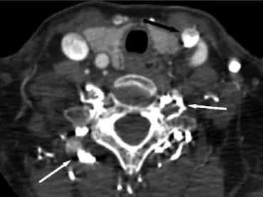
Branch vein reflux (Fig. 2) was ob-served in 25 patients. It was more fre-quent with left arm injections than with the right arm. It was detected in 50% of patients in whom a left arm injection was performed and 4.5% of patients in whom a right arm injection was performed. Severe branch vein re-flux, defined as having reflux into two or three branch veins, was associated with left arm injections, and it was not detected with right arm injections. The artifact score was not correlated with the severity of the branch vein reflux.
Streak artifacts were more frequent when the left arm was used for con-trast material injection. The severity of perivenous streak artifacts (Fig. 3) was independent of the injection site, the presence of branch vein reflux, and the presence and extension of IJV reflux (Table 3) (P > 0.05).
and skull was acquired. The CTA scan range reached from the aortic arch to the vertex. The scan parameters were as follows: the section thickness was 2.5 mm, the table feed for rotation was 2.5 mm, the pitch was at unity, the voltage was set at 120 kV, and the current was 250 mA. The scanning range was planned in a caudocranial direction. Nonionic contrast material, with an iodine concentration of 300 mg I/mL (iohexol [Omnipaque 300]; Amersham Health, Cork, Ireland), was injected with a power injector through an 18- to 20-gauge IV cannu-la (depending on the size of the vein) in an antecubital vein. All patients received 120 mL of contrast mate-rial, at an injection rate of 3.5 mL/s. Saline was not injected following the administration of the contrast mate-rial. Synchronization between the passage of contrast material and data acquisition was achieved with a bo-lus tracking technique. The CTA scan was triggered automatically by means of a threshold, measured in a region of interest (ROI) set in the common carotid artery. The threshold was set at 100 HU.
Slice thicknesses and intervals of 1.25 mm were used during post-processing and reconstruction.
Data collection and image analysis The age and gender of each patient were noted. Subsequently, the patients were grouped according to the con-trast material injection site. For each patient, axial and reformatted images were analyzed for perivenous streak artifacts and reflux. The shortest retro-sternal distance between the sternum or sternoclavicular joint and the aortic arch or its branches was measured in axial images in patients with left arm injections.
To evaluate the relationship between the injection side and the internal jugular vein (IJV) reflux severity, the length of the IJV reflux was measured in millimeters on sagittal or coronal maximum-intensity-projection imag-es. The length was compared between right and left arm injections.
Reflux in the branches of the subclavi-an vein (SCV) subclavi-and brachiocephalic vein (BCV), involving veins other than the IJV, was grouped as follows: (a) reflux into one of the branch veins (external jugular, paravertebral or mediastinal veins), (b) reflux into two branch veins,
Table 1. The relationship of age, gender, internal jugular vein reflux and contrast material injection arm Parameter Right arm (n=44) Left arm (n=46) P Age 62.36±1.95 62.12±1.95 0.25 Male/female 21/23 18/28 0.411
IJV reflux length (cm) 6.5±11.73 32.93±6.57 0.00
Table 2. The relationship between IJV reflux frequency and right and left arm injections
Injection site IJV reflux P Reflux (-) n % Reflux (+) n % Left arm 16 34.8 30 65.2 0.03 Right arm 25 56.8 19 43.2
Carotid CT angiography image quality for left versus right arm injections•197
Volume 17•Issue 3
Figure 1. a, b. Axial CT image (a) demonstrating brachiocephalic vein compression (arrow). Sagittal reformatted image (b) showing reflux of contrast medium into the left IJV (arrows) due to constitutional retrosternal narrowing.
b
a
Figure 2. Reflux into the branching veins (paravertebral, external jugular veins) (white arrows) and the IJV (black arrow) are clearly shown on an axial source image.
Figure 3. Transverse CT image showing prominent perivenous streak artifacts (arrow) due to a higher density in the brachiocephalic vein. The artifacts obscure the supraaortic branches.
Table 3. Relationship of perivenous artifacts and injection site, IJV reflux, length of IJV reflux, gender, and branching vein reflux
Parameter Artifact score P 0 1 2 n % n % n % Injection site Left arm 3 6.5 12 26.1 31 67.4 0.22 Right arm 3 6.8 19 43.2 22 50.4 IJV reflux (+) 1 2.0 16 32.7 32 65.3 0.12 (-) 5 12.2 15 36.6 21 51.2
IJV reflux length
0–10 mm 5 8.5 23 39.0 31 52.5 0.22 >10 mm 1 3.2 8 25.8 22 71.0 Branching vein reflux
(+) 1 1.1 6 6.6 18 20 0.29 (-) 5 5.5 25 27.7 35 38.8 Age 74.16±10.6 (n=6) 66.39±10.36 (n=31) 58.64±13.7 (n=53) Gender Female 2 3.9 18 35.3 31 60.8 0.49 Male 4 10.3 13 33.3 22 56.4 IJV, internal jugular vein
Demirpolat et al.
198•September 2011•Diagnostic and Interventional Radiology
Discussion
Adequate homogenous luminal con-trast is required for optimal visualiza-tion of an artery with CTA. The attenu-ation of the surrounding structures should be less than the arteries to ob-tain good quality reconstructions and to avoid streak artifacts. Venous con-tamination and venous stasis lead to higher attenuation in veins and distort images of adjacent arteries. Common reasons for venous stasis and venous hyperdensity include venous reflux, severe heart failure, expiration, medi-astinal masses, aortic aneurysms, bra-chiocephalic vein stenosis and superior vena cava syndrome (7–10).
Barmeir et al. (6) described a technical artifact that resulted in nondiagnostic examinations in 3 of 54 carotid CTAs. They changed the technical parameters and the volume of contrast material used, but could not improve the image quality. They reported that they did not see IJV reflux in 1812 carotid CTAs ob-tained after changing the injection side from the left to right arm (6). In our study, reflux into the IJV was found in 49 patients (54.4%). This finding signif-icantly increased when the left antecu-bital vein was used for injection. In 30 of the 49 patients with reflux (61.2%), the contrast material was injected into the left side. We also found that a decrease in the retrosternal distance predisposed patients to reflux when the left arm was used for injection. The length of the re-flux was also significantly higher in left arm injections. The results of our study are consistent with the study performed by Tseng et al. (4), who measured the retrosternal distance in nine patients in whom CTAs had been repeated with contrast material injections to the right and left arms. Their work showed that the retrosternal distance was 6.5±0.17 mm for four patients who had obvi-ous reflux after left arm injection and 13.2±0.22 mm for five patients who had no reflux (4).
Branching vein reflux, involving veins other than the IJV, is another artifact source and prevents the ac-quisition of high-quality carotid CTA reconstructions. This was found in 25 of 90 patients (27.7%) and was signifi-cantly less frequent than IJV reflux.
It was more frequent with left arm injections. Our results are concord-ant with the study of You et al. (5), who evaluated image quality differ-ences between right and left arm in-jections. They administered the con-trast material to the right (n=30) or the left (n=30) antecubital vein while obtaining head and neck CT and com-pared venous reflux into the branch-ing veins and perivenous artifacts be-tween the two groups. They reported that the scores of the perivenous ar-tifacts for the right arm injections were lower than the scores for the left arm injections. They suggested that the venous system is longer in the left arm and that this might have a role in the severity of artifacts (5). The authors did not investigate a pos-sible relationship between branching vein reflux and perivenous artifacts. Our results show that the severity of the perivenous streak artifacts is inde-pendent of branching vein reflux.
The factors that predispose certain patients to IJV reflux may lead to re-flux in the branching veins. However, a small diameter and different angles with the brachiocephalic and subcla-vian veins with respect to the IJV may reduce reflux.
Various methods were evaluated for eliminating the perivenous streak arti-facts. Dilution of the contrast material is one of these methods. However, it is not suitable for CTA because highly concentrated (greater amount of io-dine) contrast medium is preferable in CTA (11). The addition of a chaser bolus is another possible method that may be used to reduce streak artifacts in CTA. This method requires a dual-barrel power injector, and it is not use-ful for carotid CTA because the scan delay is shorter in carotid CTA and scanning should be started before the injection of contrastmaterial is com-plete (12, 13).
In conclusion, the injection site of the contrast medium is important to the image quality of carotid CTA. Injec-tion into the right arm decreases reflux into the IJV, BCV and SCV branches. Use of the right arm does not prevent perivenous streak artifacts but does re-duce their frequency.
References
1. Gupta R, Jones SE, Mooyaart EAQ, Pomerantz SR. Computed tomographic an-giography in stroke imaging: fundamental principles, pathologic findings, and com-mon pitfalls. Semin Ultrasound CT MRI 2006; 27:221–242.
2. Monye C, Weert TT, Zaalberg W, et al. Optimization of CT angiography of the carotid artery with a 16-MDCT scanner: craniocaudal scan direction reduces con-trast material–related perivenous artifacts. AJR Am J Roentgenol 2006; 186:1737– 1745.
3. Takhtani D. CT neuroangiography: a glance at the common pitfalls and their prevention AJR Am J Roentgenol 2005; 185:772–783.
4. Tseng Y, Hsu H, Lee T, Chen C. Venous reflux on carotid computed tomography angiography: relationship with left-arm injection. J Comput Assist Tomogr 2007; 31:360–364.
5. You SY, Yoon DY, Choi CS, et al. Effects of right- versus left-arm injections of contrast material on computed tomography of the head and neck. J Comput Assist Tomogr 2007; 31:677–681.
6. Barmeir E, Tann M, Zur S, Braun J. Improving CT angiography of the carotid artery using the “right” arm. AJR Am J Roentgenol 1998; 170:1657–1658. 7. Tanaka T, Uemura, K, Takahashi M, et al.
Compression of the left brachiocephalic vein: cause of high signal intensity of the left sigmold sinus and internal jugular vein on MR images. Radiology 1993; 188:355– 361.
8. Friedman BH, Lovegrove FT, Wagner HN Jr. An unusual variant in cerebral circulation studies. J Nucl Med 1974; 15:363–364. 9. Bok B, Marsault C, Aubin ML, et al. Jugular
venous reflux in cerebral radionuclide an-giography: an explanation. Eur J Nucl Med 1978; 3:63–65.
10. Paksoy Y, Genc BO, Genc E. Retrograde flow in the left inferior petrosal sinus and blood steal of the cavernous sinus associ-ated with central vein stenosis: MR angi-ographic findings. AJNR Am J Neuroradiol 2003; 24:1364–1368.
11. Lell MM, Anders K, Uder M, et al. New tech-niques in CT angiography. Radiographics 2006; 26:45–62.
12. Haage P, Schmitz-Rode T, Hübner D, Piroth W, Günther RW. Reduction of contrast material dose and artifacts by a saline flush using a double power injector in helical CT of the thorax. AJR Am J Roentgenol 2000; 174:1049–1053.
13. de Monye C, de Weert TT, Zaalberg W, et al. Optimization of CT angiography of the carotid artery with a 16-MDCT scanner: Craniocaudal scan direction reduces con-trast material-related perivenous artifacts. AJR Am J Roentgenol 2006; 186:1737– 1745.

