Diagn Interv Radiol 2013; 19:187–190 © Turkish Society of Radiology 2013
Gadolinium leakage into subarachnoid space and cystic metastases
Adalet Elçin Yıldız, Eray Atlı, Kader Karlı Oğuz
NEURORADIOLOGY
CASE REPORTABSTRACT
Subarachnoid space (SAS) and cystic metastatic lesions of brain parenchyma appear hypointense on fluid-attenuated inversion-recovery (FLAIR) and T1-weighted magnetic reso-nance imaging (MRI) unless there is a hemorrhage or ele-vated protein content. Otherwise, delayed enhancement and accumulation of contrast media in SAS or cyst of metastases should be considered. We present hyperintense SAS and cys-tic brain metastases of lung cancer on FLAIR and T1-weight-ed MRI, respectively, in a patient who had been previously given contrast media for imaging of spinal metastases and had mildly impaired renal functions, and discuss the relevant literature.
G
adolinium (Gd) enhanced magnetic resonance imaging (MRI) is routinely performed for diagnosis and monitoring of various cen-tral nervous system diseases. On noncontrast T1-weighted imaging, most lesions are hypointense in the absence of blood products and diverse paramagnetic substances, fat or elevated protein content. Blood-brain bar-rier (BBB) damage and the related leakage of contrast media into the extra-cellular space from the vascular system result in abnormal enhancement following intravenous administration of Gd-based paramagnetic contrast materials. Therefore, numerous lesions of infectious, inflammatory, de-myelinating, and malignant diseases are enhanced after contrast medium administration in the brain parenchyma (1). Gd accumulation within the cystic fluid of metastases causing a potential diagnostic misunderstanding has not been reported in the literature, to our knowledge.On normal fluid-attenuated inversion-recovery (FLAIR) imaging, the inversion-recovery pulse nulls the signal from subarachnoid space (SAS), and it is hypointense unless there is accompanying blood, tu-mor, inflammation or infection, as well as vascular engorgement. These conditions include subarachnoid hemorrhage, meningitis, meningeal carcinomatosis, leptomeningeal metastases, subacute infarct, subdural hematoma, adjacent neoplasms, dural venous thrombosis, and status epilepticus (2, 3). Rarely, oxygen supplementation during MRI and pre-vious Gd-based contrast media administration may result in hyperin-tense SAS. Gd accumulation in SAS may occur in patients with or with-out renal insufficiency, and in conditions with or withwith-out BBB damage (2–5). Herein, we present a case with Gd leakage into SAS and cystic fluid of metastases with a brief review of the literature.
Case report
A 48-year-old male with a metastatic lung cancer (squamous cell carci-noma) was admitted to our hospital with complaints of lower back pain and lower limb weakness for 20 days. He had received his last chemother-apy and radiotherchemother-apy treatment five months prior. On admission, neuro-logical examination revealed lower limb paresis and paresthesia at T11– L1 dermatomes, and the patient reported urine and fecal incontinence. In laboratory tests, blood urea concentration was 23.98 mg/dL (normal range, 6–20 mg/dL), blood creatinine concentration was 1.39 mg/dL (nor-mal range, 0.7–1.2 mg/dL), and glomerular filtration rate (GFR) was 54.31 mL/min/1.73 m² (estimated Modification of Diet in Renal Disease Formu-la GFR, <60 mL/min/1.73 m²). Contrast enhanced whole spine MRI was performed using 0.01 mmol/kg gadopentetate dimeglumine for a suspect-ed diagnosis of vertebral metastases and/or pathological fracture between T11 and L1 levels.
From the Department of Radiology (A.E.Y. [email protected], E.A., K.K.O.), Hacettepe University School of Medicine, Ankara, Turkey; National Magnetic Resonance Research Centre (UMRAM) (K.K.O.), Bilkent University, Ankara, Turkey.
Received 4 September 2012; accepted 14 October 2012. Published online 21 January 2013
DOI 10.5152.dir.2013.040
188 •May–June 2013 • Diagnostic and Interventional Radiology Yıldız et al.
MRI studies were performed on a 1.5 Tesla MR scanner (Symphony, Tim Sys-tems, Siemens Medical SysSys-tems, Erlan-gen, Germany). Spine MRI demonstrat-ed multiple bone metastases that were hypointense on precontrast T1-weight-ed imaging (Fig. 1a) and enhancT1-weight-ed on postcontrast images throughout the spine. There were also enhancing pial nodular metastases adjacent to the spinal cord and at the cauda equina on postcontrast T1-weighted imaging. Marked edema of the spinal cord was observed between C7 and T4 levels without a remarkable intramedullary enhancement. At the time of spinal im-aging, because extensive spinal intradu-ral extramedullary metastases and en-hancing metastases in the cerebellum
were detected (Fig. 1a), additional post-contrast T1-weighted imaging of the brain was also obtained. It revealed lep-tomeningeal carcinomatosis and cystic metastases in the right cerebral and left cerebellar hemispheres with a peripher-al enhancement and hypointense cystic fluid (Fig. 1b).
The patient was admitted to the medi-cal oncology clinic with these findings. Approximately 24 hours after the first MRI, his physician, who was unaware of the postcontrast T1-weighted imag-ing of the brain obtained durimag-ing the spine imaging session, requested a new brain contrast-enhanced MRI. . At this second MRI, SAS was hypointense but the cystic metastatic lesions were hy-perintense on precontrast T1-weighted
imaging (Fig. 1c). Additionally, SAS was diffusely hyperintense (Fig. 2a) and cystic metastases were bright on FLAIR imaging. Following intravenous administration of same contrast media with the previous exam, leptomenin-geal carcinomatosis and widespread parenchymal micronodular metasta-ses were also observed. Together, the findings from both imaging sessions suggested leakage of Gd from the vas-cular system into the central cystic fluid of parenchymal metastases and SAS. Two additional separate MRI, in-cluding T1-weighted imaging, FLAIR, and gradient echo (GRE) images, were performed to follow Gd clearance from cystic fluid of parenchymal metastases and SAS, and to rule out hemorrhage
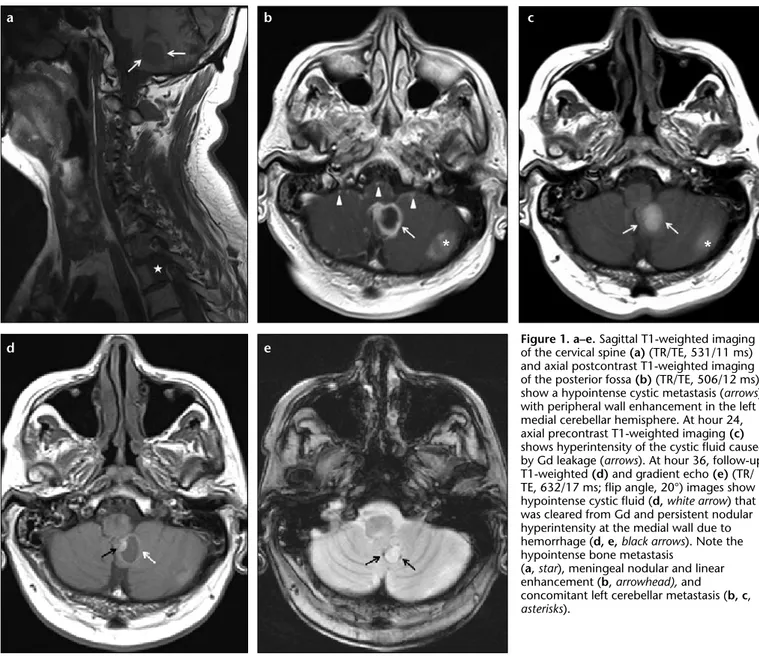
Figure 1. a–e. Sagittal T1-weighted imaging of the cervical spine (a) (TR/TE, 531/11 ms) and axial postcontrast T1-weighted imaging of the posterior fossa (b) (TR/TE, 506/12 ms) show a hypointense cystic metastasis (arrows) with peripheral wall enhancement in the left medial cerebellar hemisphere. At hour 24, axial precontrast T1-weighted imaging (c) shows hyperintensity of the cystic fluid caused by Gd leakage (arrows). At hour 36, follow-up T1-weighted (d) and gradient echo (e) (TR/ TE, 632/17 ms; flip angle, 20°) images show hypointense cystic fluid (d, white arrow) that was cleared from Gd and persistent nodular hyperintensity at the medial wall due to hemorrhage (d, e, black arrows). Note the hypointense bone metastasis
(a, star), meningeal nodular and linear enhancement (b, arrowhead), and concomitant left cerebellar metastasis (b, c, asterisks). b e a d c
Volume 19 • Issue 3 Gadolinium leakage into subarachnoid space and cystic metastases • 189
that might have also caused T1 short-ening. The scans were performed at ap-proximately 36 and 160hours after ad-ministration of the contrast material. At 36 hours, cystic fluids of the parenchy-mal metastases were hypointense with continued enhancement at the periph-ery of the cyst on T1-weighted imag-ing (Fig. 1d) that may in part be due to hemorrhage observed on GRE imaging (Fig. 1e); SAS and cyst fluid, however, were still hyperintense on FLAIR imag-ing (not shown). On GRE imagimag-ing, SAS had no evidence of susceptibility (Fig. 2b). After 160 hours, SAS and cyst flu-id were cleared of Gd on FLAIR images (Fig. 2c), but subtle T1 hyperintensity at the periphery of the cyst remained on T1-weighted imaging images.
The total dose of Gd given to the pa-tient in the two scanning sessions was 30 mL (gadopentetate dimeglumine, 0.01 mmol/kg). During the hospital stay, re-nal function parameters of the patient were mildly impaired.
Discussion
Increased signal intensity in the SAS has been described in numerous pathologic changes of the meninges and brain tissue on FLAIR imaging, including subarachnoid hemorrhage, meningitis, meningeal carcinomatosis, leptomeningeal metastases, subacute infarct, subdural hematoma, adjacent neoplasms, dural venous thrombosis, and status epilepticus (2, 3). Addition-ally, supplemental oxygen during MRI and previous contrast media adminis-tration can also cause artifactual SAS
hyperintensity (1, 4–6). According to Braga et al. (7), supplemental oxygen at 100% is a main cause of artifactual SAS hyperintensity on FLAIR imaging, regardless of the anesthetic drug used. This artifact does not develop when 50% oxygen is administered. Another cause for artifactual SAS hyperintensi-ty is previous contrast media adminis-tration. In a large series of 33 patients, Bozzao et al. (8) demonstrated hyper-intense SAS on FLAIR imaging due to Gd leakage in 2–24 hours following Gd administration in pathologic condi-tions with BBB disruption and neovas-cularization.
Although BBB disruption and neo-vascularization result in leakage of contrast media into the extracellular space from vascular system, there is no evidence from a histopatholog-ic study showing that damaged pial vessels could allow leakage of Gd into the SAS. The exact mechanism of lep-tomeningeal and SAS enhancement re-mains unclear (1, 8). Notably, Morris and Miller (3) showed that presence of abnormalities known to disrupt the BBB and renal insufficiency are not mandatory for SAS hyperintensity on FLAIR caused by previous Gd admin-istration. Their findings also suggested that Gd chelates might move across an osmotic gradient at the circumventric-ular organs with elevated plasma con-centrations where there is no BBB, as in normal dura mater. An in vitro and animal model study by Mamourian et al. (9) with healthy dogs also showed that intravenously administered Gd
could cross into the SAS in sufficient concentrations to alter the appearance of the SAS on FLAIR. They emphasized that SAS concentrations of Gd were proportional to the administered Gd dose, and that a dose three times high-er than standard amounts (0.03 mmol/ kg) was essential to produce detectable changes (9). Therefore, signal changes related to leakage of Gd into SAS re-quire sufficient Gd concentration in the plasma and/or BBB disruption or neovascularization.
Not only is the total dose adminis-tered to the patient important in af-fecting the detection of Gd in the SAS and persistence of the hyperintensity on images, but also the persistence of Gd in plasma caused by renal func-tional impairment plays a role (10). Gd chelates are cleared from plasma via glomerular filtration, with a nor-mal plasma half-life of 1.6 hours. In patients with renal insufficiency, the plasma half-life of Gd chelates may be prolonged up to 30 hours (11, 12). Hence, either renal insufficiency or an overdose administered to patients may be responsible for hyperintense SAS on FLAIR imaging.
In our patient, resolution of cystic flu-id hyperintensity on T1-weighted imag-es occurred at 36 hours, and both cystic fluid and SAS hyperintensity on FLAIR images was observed at 160 hours. In the literature, resolution of the hy-perintensity on FLAIR or T1-weighted images, which developed following ad-ministration of gadopentetate dimeglu-mine and gadodiamide (5, 8, 9) ranged Figure 2. a–c. Axial FLAIR (TR/TE/TI, 8000/127/2183 ms) image at hour 24 (a) shows diffuse SAS hyperintensity due to Gd leakage. On the gradient echo image (b), there is no evidence of hemorrhage. At hour 160, the follow-up FLAIR image (c) shows hypointense SAS with total clearance from Gd.
b
190 •May–June 2013 • Diagnostic and Interventional Radiology Yıldız et al.
between 48 hours to two weeks (3, 8-10). Although Bozzao et al. (8) stat-ed that resorption time of Gd from SAS is 48 hours on FLAIR, no other study has investigated the exact resolution time of hyperintensity in varying con-ditions, including total dose or renal function degree. In the current patient, BBB disruption and abnormal plasma clearance due to mild renal function-al impairment might have resulted in accumulation of Gd in cystic metasta-ses, and SAS on T1-weighted and FLAIR images. Two administrations of Gd (15 cc each, for a total of 30 cc) within ap-proximately 30 hours also might have prolonged the clearance of the Gd from the plasma and resulted in continued hyperintensity up to the 160 hours on FLAIR images.
In our patient, SAS hyperintensi-ty on FLAIR could not be attributed to subarachnoid hemorrhage, as evi-denced from GRE. However, the cystic metastasis located in the left medial cerebellar hemisphere had micronodu-lar and peripheral susceptibility due to blood, and this may explain the lack of apparent signal alteration in the lesion wall on T1-weighted images from 36 to 160 hours. Still, the cystic fluid did not show hemorrhage on GRE, supporting our Gd-leakage hypothesis.
We also observed hyperintense cys-tic metastases on T1-weighted images caused by previous Gd administration. Breast cancer, adenocarcinoma of lung cancer, thymic squamous cell carcino-ma metastases can be cystic/necrotic (13). Abnormally proliferating vessels of high-grade tumors have intercellu-lar gaps and discontinuous basement
membrane, causing both intravascular (flow related) and interstitial (perme-ability related) enhancement (1). Boz-zao et al. (8) showed FLAIR hyperin-tensity of cystic cavities in a necrotic brain mass and a surgical cavity due to Gd leakage. Given that FLAIR is much more sensitive to changes in Gd con-centration (9), we further demonstrat-ed this finding on T1-weightdemonstrat-ed imag-ing.
In conclusion, radiologists should be aware of delayed or persistent Gd enhancement that can occur in cysts of the metastases (and possibly of other necrotic tumors), and SAS on T1-weighted and FLAIR imaging. In such situations, radiologists should check history of previous Gd admin-istration in the patients, especially in those without renal impairment. Conflict of interest disclosure
The authors declared no conflicts of interest. References
1. Smirniotopoulos JG, Murphy FM, Rushing EJ, Rees JH, Schroeder JW. Patterns of con-trast enhancement in the brain and me-ninges. Radiographics 2007; 27:525–551.
[CrossRef]
2. Maeda M, Yagishita A, Yamamoto T, Saku-ma H, Takeda K. AbnorSaku-mal hyperintensity within the subarachnoid space evaluated by fluid-attenuated inversion-recovery MR imaging: a spectrum of central nervous sys-tem diseases. Eur Radiol 2003; 13:192–201.
[CrossRef]
3. Morris JM, Miller GM. Increased signal in the subarachnoid space on fluid-attenuated inversion recovery imaging associated with the clearance dynamics of gadolinium che-late: a potential diagnostic pitfall. AJNR Am J Neuroradiol 2007; 28:1964–1967. [CrossRef]
4. Deliganis AV, Fisher DJ, Lam AM, Maravil-la KR. Cerebrospinal fluid signal intensity increase on FLAIR MR images in patients under general anesthesia: the role of supple-mental O2. Radiology 2001; 218:152–156. 5. Zatman P, Hourihan MD. Contrast
enhance-ment of cerebrospinal fluid on delayed MRI in a patient with an epidural abscess and re-nal failure. Br J Radiol 2002; 75:474–477. 6. Stuckey SL, Goh TD, Heffernan T, Rowan D.
Hyperintensity in the subarachnoid space on FLAIR MRI. AJR Am J Roentgenol 2007; 189:913–921. [CrossRef]
7. Braga FT, da Rocha AJ, Hernandez Filho G, Arikawa RK, Ribeiro IM, Fonseca RB. Rela-tionship between the concentration of sup-plemental oxygen and signal intensity of CSF depicted by fluid-attenuated inversion recovery imaging. AJNR Am J Neuroradiol 2003; 24:1863–1868.
8. Bozzao A, Floris R, Fasoli F, Fantozzi LM, Col-onnese C, Simonetti G. Cerebrospinal fluid changes after intravenous injection of gad-olinium chelate: assessment by FLAIR MR imaging. Eur Radiol 2003; 13:592–597. 9. Mamourian AC, Hoopes PJ, Lewis LD.
Visu-alization of intravenously administered con-trast material in the CSF on fluid-attenuated inversion-recovery MR images: an in vitro and animal-model investigation. AJNR Am J Neuroradiol 2000; 21:105–111.
10. Rai AT, Hogg JP. Persistence of gadolinium in CSF: a diagnostic pitfall in patients with end-stage renal disease. AJNR Am J Neurora-diol 2001; 22:1357–1361.
11. Joffe P, Thomsen HS, Meusel M. Pharmaco-kinetics of gadodiamide injection in patients with severe renal insufficiency and patients undergoing hemodialysis or continous am-bulatory peritoneal dialysis. Acad Radiol 1998; 5:491–502. [CrossRef]
12. Shellock FG, Kanal E. Safety of magnetic resonance imaging contrast agents. J Magn Reson Imaging 1999; 10:477–484. [CrossRef]
13. Troiani C, Lopes CC, Scardovelli CA, Nai GA. Cystic brain metastases radiologically simulating neurocysticercosis. Sao Paulo Med J 2011; 129:352–356. [CrossRef]