Cukurova Medical Journal
Cukurova Med J 2018;43(2):495-497 ÇUKUROVA ÜNİVERSİTESİ TIP FAKÜLTESİ DOI: 10.17826/cumj.342717Yazışma Adresi/Address for Correspondence: Dr. Hüseyin Onur Aydın, 1Baskent University Faculty of Medicine,
Department of General Surgery, Ankara, Turkey E-mail: dronuraydı[email protected] Geliş tarihi/Received: 05.01.2017 Kabul tarihi/Accepted: 15.03.2017
EDİTÖRE MEKTUP / LETTER TO THE EDITOR
Retroperitoneal cystic lesion mimicking malign sarcoma as a
complication of lost gallstones during laparoscopic cholecystectomy
Laparoskopik kolesistektomi sırasında kaybolan safra taşı komplikasyonu sonrası
malign sarkomu taklit eden retroperitoneal kistik lezyon
Hüseyin Onur Aydın
1, Ebru Hatice Ayvazoğlu Soy
1, Tevfik Avcı
1, Sedat Yıldırım
11Baskent University Faculty of Medicine, Department of General Surgery, Ankara, Turkey
Cukurova Medical Journal 2018;43(2):495-497
Dear Editor,
The gold standard treatment of symptomatic gallstone disease is prefered to be laparoscopic cholecystectomy (LC) due to shorter hospital stay and reduced postoperative pain. Biliary tract injury or lost gallstones are important complications of LC¹. Biliary tract injury is reduced in the last two decades with expertised surgeons. However, lost gallstones are still one of the major problems following LC.
The incidence of perforated gallbladder, diagnosed during surgery is about 18%². In literature there are many reports about the complications of lost or undetected gallstones. Among them the incidence of intraabdominal abcess is reported to be 0.3 %³. Abcess or intraabdominal lesions due to lost gallstones are frequently seen in subhepatic recess or retroperitoneum⁴. Diagnosis and management of these confusing complications are still major problems following LC. Especially, cases presenting with intraabdominal suspicious tumoral mass are serious social, psychological and medico-legal concerns.
A 60 year old man admitted to our hospital with abdominal pain. Abdominal computed tomography scans showed a 88x64x40 mm probably malign appearing mass in right hepatic recess. Calcified statellite lesions were accompanying the infiltrating mass radiologically (Figure 1). He had LC due to symptomatic gallstones in our center 10 months ago . The pathology report of the gallbladder did not
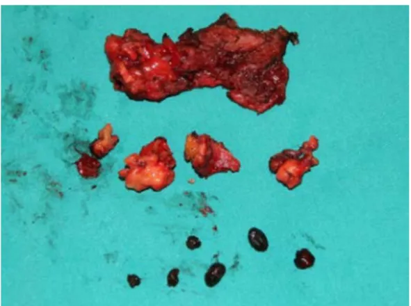
mention about perforation and he had no co morbid disease. We performed true-cut biopsy due to suspicious findings of the lesion, but as specimen was not enough for definitive diagnosis we decided to perform laparatomy. Intraoperative findings revealed a 10cm mass with firm adhesions to liver and surrounding tissue (Figure 2). During dissection, the mass was perforated and purulent discharge was drained. In further dissection, gallstones were seen at the core of the mass (Figure 3). Intraoperative frozen sections were reported as chronic inflammation and necrosis without any malignancy. The retroperitoneal mass of spilled gallstones was excised. The definite pathology was reported as granulomatous inflammation and necrosis. The patient was discharged after three days without any complication.
Cilt/Volume 43 Yıl/Year 2018 Retroperitoneal cystic lesion mimicking malign sarcoma
496
With increased number of LC procedures, the variety of the complications are also increased. The complications due to lost gallstones are reported to be 0.1-0.3%⁴. The predisposing factors for undetected spilled gallstones are male gender, older age, pigment stones, over 15 stones, over 1,5 cm sized stones and perihepatic location of lost stones⁵. These lost gallstones can be presented as abcess or retroperitoneal mass due to induced inflammation⁶.
Figure 2. Intraoperative view
Retroperitoneal mass is diagnosed with clinical and radiological findings. Spicular growing to surrounding tissue is one of the criteria of malignancy⁷. Malign retroperitoneal mass needs agressive treatment. In literature there are few reports about lost gallstones presenting as suspicious retroperitoneal mass. Leland and Dawson showed adhesions caused by inserted sterile gallstones in rats. So during LC all spilled gallstones should be removed instantly⁸.
Abcess formation after lost gallstones are reported to be 4 months-10 years after LC⁹. Perihepatic abcess and granulation can be radiologically misdiagnosed with retroperitoneal mass. Intra abdominal foreign bodies cause adhesion, encapsulation and finally granuloma formation10.
Kim et al. reported subhepatic abcess due to gallstones mimicking retroperitoneal sarcoma at the 5th month of LC¹¹. Dasari et al. presented the case of peritoneal masses of gallstones mimicking metastatic peritoneal lesions 2 years after LC¹². In our case lost spilled gall stones were presented with a suspicious retroperitoneal mass at the 10th month of LC. Computed tomography (CT) and magnetic resonance imaging (MRI) are capable of diagnosis of calcified gallstone⁷. However, like our patient, pure
cholesterol stones can not be seen in CT scans or MRI and they are represented as intraabdominal or retroperitoneal masses.
Figure 3. Gallstones and mass core
Spilled gallstones may cause severe complications so they should be removed at hte same operation. For prevention of spilled and lost gallstones, perforation of gallbladder should be avoided during dissection; both proximal and distal ends of cystic canal should be ligated safetly and endobag usage may be useful for extraction of dissected gallbladder. Lost gallstones mimicking malign or suspicious mass are rare in literature. Although the known history of LC, retroperitoneal or intraabdominal mass should be investigated for lost gallstones and managed carefully.
REFERENCES
1. Luu MB, Deziel DJ. Unusual Complications of Gallstones. Surg Clin North Am. 2014;94:377-94. 2. Woodfield J, Rodgers M, Windsor J. Peritoneal
gallstones following laparoscopic cholecystectomy. Surg Endosc. 2004;18:1200-7
3. Noda Y, Kanematsu M, Goshima S et al. Peritoneal chronic inflammatory mass formation due to gallstones lost during laparoscopic cholecystectomy. Clin Imaging. 2014;38:758-61.
4. Zehetner J, Shamiyeh A, Wayand W. Lost gallstones in laparoscopic cholecystectomy: all possible complications. Am J Surg. 2007;193:73-8
5. Brockmann J, Kocher T, Senninger N, Schürmann G. Complications due to gallstones lost during laparoscopic cholecystectomy. Surg Endosc. 2002;16:1226-32.
6. Koç E, Suher M, Öztugut ŞU, Ensari C, Karakurt M, Özlem N. Retroperitoneal abscess as a late complication following laparoscopic cholecystectomy. Med Sci Monit. 2004;10:CS27-9.
Aydun et al. Cukurova Medical Journal
497 7. Bennett A, Gilkeson R, Haaga J, Makkar V, Onders
R. Complications of “dropped” gallstones after laparoscopic cholecystectomy: technical considerations and imaging findings. Abdom Imaging. 2000;25:190-3.
8. Leland D, Dawson D. Adhesions and experimental intraperitoneal gallstones. Contemp Surg. 1993;42:273-6.
9. Morrin M, Kruskal J, Hochman M, Saldinger P, Kane R. Radiologic features of complications arising from dropped gallstones in laparoscopic
cholecystectomy patients. AJR Am J Roentgenol. 2000;174:1441-5.
10. Yildirim T, Parlakgumus A, Yildirim S. Diagnosis and management of retained foreign objects. J Coll Physicians Surg Pak. 2015;25:367-71.
11. Kim B-S, Joo S-H, Kim H-C. Spilled gallstones mimicking a retroperitoneal sarcoma following laparoscopic cholecystectomy. World J Gastroenterol. 2016;22:4421-6.
12. Dasari B, Loan W, Carey DP. Spilled gallstones mimicking peritoneal metastases. JSLS 2009;13:73-6