Kardiochirurgia i Torakochirurgia Polska 2015; 12 (3) 246
Address for correspondence: Faruk Cingoz, MD, Gülhane Askeri Tıp Akademisi, Gn. Dr. Tevfik Sağlam Cad., 06010 Etlik, Ankara, Turkey, phone: +90 312 3045220, fax: +90 312 3045200, e-mail: [email protected]
Streszczenie
W pracy opisano przypadek kobiety, lat 55, bez choroby wień-cowej w wywiadzie, chorującej na nadciśnienie od 17 lat, któ-ra została przyjęta ze spoczynkowym bólem klatki piersiowej. Badanie elektrokardiograficzne wykazało ujemny załamek T w odprowadzeniach przedsercowych. Angiografia naczyń wieńcowych uwidoczniła nietypową anatomię naczyniową w postaci wspólnego odejścia prawej i lewej tętnicy wieńcowej od zatoki Valsalvy stanowiącej wspólny pień wieńcowy. War-to zwrócić uwagę, że nieprawidłowości załamka T i bóle klatki piersiowej mogą wynikać z tej wrodzonej anomalii wieńcowej. Słowa kluczowe: anomalia wieńcowa, choroba niedokrwienna serca.
Case report
Abstract
A 55-year-old female without a history of coronary artery dis-ease, hypertensive for the past 17 years, was admitted with resting chest pain. Electrocardiography revealed a negative T-wave in anterior chest leads. Coronary angiography visualised anomalous coronary anatomy, with a common origin of the right coronary artery and the left main coronary artery in the right sinus of Valsalva serving as a common coronary trunk. It should be emphasised that T-wave abnormalities and chest angina may be related to this congenital coronary anomaly. Key words: coronary anomaly, ischaemic heart disease.
Novel variant of dual left anterior descending artery
arising from single right coronary artery anomaly
presenting with angina inversa
Faruk Cingoz1, Gokhan Arslan1, Atilla Iyisoy2, Hakan Bingol3
1Department of Cardiovascular Surgery, Gulhane Military Medical Academy, Etlik, Ankara, Turkey 2Department of Cardiology, Gulhane Military Medical Academy, Etlik, Ankara, Turkey
3Department of Cardiovascular Surgery, Baskent University, Konya, Turkey
Kardiochirurgia i Torakochirurgia Polska 2015; 12 (3): 246-247
DOI: 10.5114/kitp.2015.54462
Case report
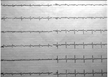
A 55-yeold female without a history of coronary ar-tery disease, hypertensive for 17 years duration was ad-mitted with chest pain at rest. Her electrocardiography re-vealed negative T-wave in anterior chest leads (Fig. 1), and T waves were returned to normal fashion without medi-cal treatment within 10 minutes (Fig. 2). Echocardiogram showed normal left ventricle systolic function and ejection fraction (> 60%). Her biochemical parameters were within normal limits and her stress test was normal as well. She was subjected to coronary angiography to rule out any is-chaemic aetiology. At coronary angiography, there was no obstructive coronary artery disease; however, the coronary angiogram demonstrated an anomalous coronary anatomy, with the origin of the right coronary artery and left main stem from the right sinus of Valsalva as a common
coro-nary trunk (single corocoro-nary artery) (Fig. 3). Circumflex and left anterior descending arteries were arising from the left main stem. Right coronary artery was normal. Meanwhile, a second left anterior descending artery was seen aris-ing from the proximal right coronary artery (Fig. 3 and 4). This was consistent with the novel variant of dual left ante-rior descending artery maybe subgroup of type IV accord-ing to Spinaldo-Franco Classification [1]. It emphasises that T wave abnormalities and chest angina may be related this congenital coronary anomaly.
Reference
1. Spinaldo-Franco H, Grose R, Solomon N. Dual left anterior descending ar-tery: angiographic description of important variants and surgical implica-tions. Am Heart J 1983; 105: 445-448.
Kardiochirurgia i Torakochirurgia Polska 2015; 12 (3) 247
CASE REPORT
Fig. 1. 12-lead electrocardiogram showing negative T waves in an-terior chest leads during chest pain at rest
Fig. 2. 12-lead electrocardiogram showing normal T-waves in ante-rior chest leads after chest pain
Fig. 4. Multidetector computed tomography, 3-dimensional vo-lume-rendered reconstructed image of the coronary system. LAD2 (arrow) arises from the right coronary artery (arrow) and then turns sharply down the anterior interventricular sulcus and extends to the apex of the heart. Left anterior descending artery (arrow) forms a short vessel
RCA – right coronary artery, LAD – left anterior descending artery Fig. 3. Single coronary artery and double LAD arteries. Coronary
angiography in a caudal right anterior oblique view shows all co-ronary arteries arising from right sinus of Valsalva with a common trunk. LAD2 (arrows) arises from the RCA (arrow) and then turns sharply down the anterior interventricular sulcus. LAD (arrow) forms a short vessel, which produces both septal perforators and diagonal branches.
LMCA – left main coronary artery, Cx – circumflex artery, LAD – left anterior descending artery, RCA – right coronary artery