Turk Kardiyol Dern Ars 2017;45(1):73-76 doi: 10.5543/tkda.2016.44449
Complicated left-sided infective endocarditis in chronic
hemodialysis patients: a case report
Kronik hemodiyaliz uygulanan hastalarda komplike sol taraf
enfektif endokarditi: Olgu sunumu
Department of Cardiology, Başkent University İstanbul Medical and Research Center Hospital, İstanbul, Turkey
#Department of Infectious Disease, Başkent University İstanbul Medical and Research Center Hospital, İstanbul, Turkey
Öykü Gülmez, M.D., Mehtap Aydın, M.D.#
Özet– Enfektif endokardit (EE) son dönem böbrek hastala-rında (SDBH) yüksek mortalite ve morbiditesi olan ciddi bir enfeksiyon hastalığıdır. Özellikle çift lümenli damar kate-teri olanlarda tekrarlayan bakkate-teriyemilerle ilişkilidir. En sık etken Staphylococcus aureus ve en çok etkilenen kapak sıklıkla kireçlenmiş mitral kapaktır. Yaşları 56 ve 88 olan ve hemodiyaliz kateterinden hemodiyalize giren iki hasta ateş ve troponin yüksekliği ile başvurdu. Kan kültürlerinde
Staphylococcus aureus üredi ve iyi kalitede çekilen
trans-torasik ekokardiyografi’de (TTE) kireçlenmiş mitral ve aort kapak saptandı; vejetasyon veya apse oluşumu izlenmedi. Kateter enfeksiyonuna ikincil miyokart nekrozu düşünüldü. Her ikisinde de antibiyotik tedavisi başlandıktan 3–5 gün sonra alınan kültürlerde tekrar üreme saptandı. Bu nedenle transözofajiyal ekokardiyografi (TEE) uygulandı. Yaşlı has-tada perivalvüler apse, genç hashas-tada hareketli vejetasyon saptandı. Yaşlı hasta cerrahiyi reddetti ve dirençli şoka ikin-cil kaybedildi. Genç hastada mitral kapak cerrahisi yapıl-dı ancak takibinde sol kalp yetersizliği ve kanama gelişti; dirençli şok nedeniyle bu hasta da kaybedildi. Hastaları-mızın değerlendirilmesinde birincil odak olarak kateter en-feksiyonunun bulunması ve TTE ile vejetasyon veya anüler apse saptanmadığı için hastalıkların seyri şanssız oldu. Unutulmaması gereken nokta, SDBH’de Duke kriterlerine göre EE tanısını koymak güçtür ve bu nedenle iyi kalitede TTE’de vejetasyon veya apse saptanmasa bile TEE plan-lanması gereklidir.
Summary– Infective endocarditis (IE) is a serious infectious condition with high morbidity and mortality in patients with end-stage renal disease (ESRD). It has been particularly as-sociated with recurrent bacteremia due to vascular access via lumen catheters. The most common pathogen is
Staphy-lococcus (S.) aureus, and most affected valve is mitral valve,
which frequently calcified. Two patients with ESRD who re-ceived hemodialysis treatment via tunneled catheters, aged 56 and 88 years, were admitted with fever and high troponin level. Blood cultures revealed growth of S. aureus. Good quality transthoracic echocardiography (TTE) displayed cal-cified mitral and aortic valves with no vegetation or abscess formation. Myocardial necrosis as result of catheter infec-tion was considered. Both patients had persistent positive blood cultures 3 and 5 days after initiation of antibiotic treat-ment. Therefore, transesophageal echocardiogram (TEE) was scheduled. Results revealed perivalvular abscess in the older patient, and highly mobile vegetation in the younger patient. The older patient refused surgery and died soon af-ter due to refractory shock. Mitral valve surgery was planned for the other patient; however, she developed left ventricular failure and bleeding, and also subsequently died as result of refractory shock. Patient evaluations were particularly unfa-vorable: they had catheter infection as primary focus, and TTE did not detect vegetation or annular abscess. Diagnosis of IE in patients with ESRD using Duke criteria is problem-atic; we have to keep use of TEE in mind to detect vegetation or abscess formation when there is clinical suspicion regard-ing ESRD patients even after good quality TTE.
73
D
espite developments in the diagnosis andman-agement of infective endocarditis (IE), it is still associated with significant mortality and morbidity, most often due to cardiovascular failure. Incidence of
bacteremia is elevated in patients with end-stage re-nal disease (ESRD) as result of frequent vascular ac-cess, arteriovenous fistula, and comorbid conditions, such as malnutrition, uremia, or diabetes. Incidence
Received:March 29, 2016 Accepted: June 06, 2016
Correspondence: Dr. Öykü Gülmez. Başkent Üniversitesi İstanbul Uygulama ve Araştırma Merkezi Hast., Kardiyoloji Anabilim Dalı, Oymacı Sokak, No: 7, Altunizade, İstanbul, Turkey.
Tel: +90 216 - 554 15 00 e-mail: [email protected] © 2017 Turkish Society of Cardiology
ranges from 12% to 22%, with mortal-ity rate ranging from 30% to 56%.[1–3]
Pres-ently described are 2 cases of left-sided IE in patients with
ESRD with indwelling catheter who were diagnosed based on results of transesophageal echocardiography (TEE), despite good quality transthoracic echocar-diography (TTE) image, complicated by cardiogenic shock and death.
CASE REPORT
Case 1– An 88-year old male with history of ESRD was admitted with fever, melena, and high troponin level. Physical examination was remarkable for fever of 38°C, heart rate of 110 beats/min, blood pressure of 100/50 mmHg. Indwelling catheter had been inserted as primary vascular access for hemodialysis (HD) 5 years previously. Good quality TTE demonstrated cal-cific aortic valve with mean gradient of 15 mmHg and moderate aortic and tricuspid regurgitation with ejec-tion fracejec-tion of 50%. Two sets of blood samples from
permanent catheter and 1 sample from peripheral vein were obtained simultaneously for blood cultures. All blood cultures showed growth of methicillin-resistant coagulase-negative Staphylococci (MRCNS). Intra-venous vancomycin was initiated (1.5 g every 72 h), with dose adjusted based on renal function. Therapy for melena was also implemented. After 3 days, TEE was scheduled, as new blood cultures derived from permanent catheter and peripheral vein still showed growth of MRCNS. TEE revealed aortic annular ab-scess with connection to left ventricular (LV) cavity (Figure 1). Patient and his relatives declined surgery due to high surgical risk. His hemodynamic status progressively deteriorated, resulting in need for ino-tropic and vasopressor support. Unfortunately, he died 6 days after diagnosis of IE due to refractory shock. Case 2– A 56 year-old woman with medical history of ESRD due to systemic lupus erythematosus, renal transplantation and rejection 5 years earlier, atrial fi-brillation, and coronary artery bypass graft (CABG) operation 6 years prior, was admitted with chest pain and fever of 38.7ºC during HD and high troponin lev-el. She had been using indwelling catheter as primary vascular access for HD for 2 years.
Electrocardiogra-Turk Kardiyol Dern Ars 74
Abbreviations:
ESRD End-stage renal disease HD Hemodialysis IE Infective endocarditis LV Left ventricle
TEE Transesophageal echocardiogram TTE Transthoracic echocardiography
Figure 1. Aortic annular abscess formation, short-axis and 4-chamber view.
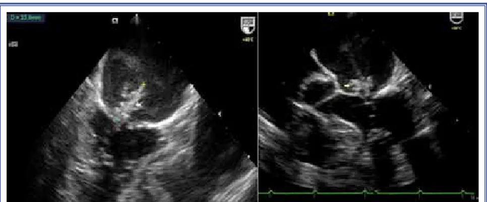
Figure 2. Vegetation 23.8×13.0 mm in size on the mitral valve, 2- and 4-chamber views.
Complicated left-sided infective endocarditis in chronic hemodialysis patients 75
phy revealed atrial fibrillation, heart rate of 120 beats/ min and diffuse ST segment depression in inferolat-eral leads. A good quality TTE yielded results simi-lar to previous echocardiograms: mild calcific mitral stenosis (valve area of 2.6 cm²), mild mitral regurgita-tion, mitral annular calcificaregurgita-tion, moderate tricuspid regurgitation, and normal LV function. Four sets of blood samples (2 sets from vascular access and 2 sets from peripheral vein) were taken. Empiric treatment of intravenous vancomycin was initiated (1 g every 72 h), with dose adjusted based on renal function, as she had history of positive blood culture with growth of methicillin-resistant S. aureus (MRSA) 2 months ear-lier. Medical therapy for ischemic heart disease was also implemented. Vancomycin was replaced with cefozolin and TEE was scheduled, as all new blood cultures showed growth of methicillin-sensitive S.
aureus. TEE revealed mobile vegetation 23.8×13.0
mm in size on the mitral valve (Figure 2). Coronary angiography was performed. Patient underwent mitral valve surgery and single-vessel CABG. Postoperative blood cultures were negative, but she developed LV failure, which resulted in need for use of inotrope and vasopressor, as well as bleeding, which required re-peated blood transfusions. She died on 10th postop-erative day due to refractory shock.
DISCUSSION
Outcome for 2 patients with IE in cases presently de-scribed was death due to heart failure, refractory in-fection, bleeding, and cardiorespiratory failure. The risk of mortality was high for both patients at admis-sion. Moreover, patient evaluations were particularly unfavorable; they had vascular access for HD as pri-mary focus for catheter infection, and TTE did not de-tect vegetation or annular abscess. In this report, we would like to discuss the epidemiology, microbiology, clinical features, diagnosis, and management of IE in patients with ESRF.
Epidemiological studies have reported incidence of IE in patients with ESRD is 15 to 60 times higher
than overall incidence of IE in general population.[3]
Moreover, it is second most frequently seen cardio-vascular disease as leading cause of death in patients with ESRD. The in-hospital mortality rate approaches 50%.[1,2] Heart failure and cardiogenic shock are
in-dependent predictors of mortality.[4] Mitral valve
in-volvement, septic embolism, vegetation size >2 cm³
on TEE, IE related to drug-resistant organism (espe-cially MRSA and vancomycin-resistant
Enterococ-cus), age over 65 years, and cerebrovascular accident/
transient ischemic attack are also associated with poor
overall patient survival.[5] Moreover, patients with
elevated troponin level are at high risk for poor out-come, including higher mortality and surgery rates, central nervous system events, and cardiac abscess.
[6] Elevated troponin level is associated with local
in-vasion of the myocardium, coronary embolism, more extensive infection, and sepsis; it can identify
high-risk patients who need more aggressive treatment.[6]
Despite improvements in medical and surgical thera-py for IE, survival rates of ESRD patients with endo-carditis have changed only a little.[5]
Patients with ESRD are prone to metastatic blood-stream infections due to tunneled catheters.[1]
Recur-rent bacteremia as result of vascular access infection acquired during HD treatment occurs at rate of 1 epi-sode per 100 patient-months, and IE develops in 1% to 12%. Bacteremia risk is due to impaired immune system caused by malnutrition or underlying systemic disease, such as diabetes mellitus, or uremia.[5] Since
premature degenerative heart valve disease, such as calcific aortic stenosis and regurgitation, or mitral annular calcification with or without calcific mitral stenosis and MR are frequent in ESRD patients, left-sided endocarditis occurs twice as often as right-left-sided endocarditis. Mitral valve is involved in more than half of cases, while multiple valve infections occur in 20% of cases.[1,5]
Previous studies have demonstrated that primary causative organisms are predominantly
Staphylo-coccus species (up to 75%), which are particularly
virulent.[1,2,5,7] Nasal carriage of S. aureus is
associ-ated with recurrent blood stream infections due to in-creased vascular access manipulation.
Diagnosis of IE in patients with ESRD remains a clinical dilemma, as clinical presentation usually re-sembles an access infection. Moreover, using Duke criteria in diagnosis has several limitations. Vascular access as primary focus of infection (one of the major Duke criteria is positive blood culture for typical IE organisms in the absence of primary focus), and less frequently, presentation with fever (one of the minor Duke criteria), make diagnosis of IE in patients with
Any ESRD patient with suspicion of IE should be screened with TTE. Moreover, use of TEE should be added after TTE in patients with high clinical suspi-cion, such as new-onset heart failure; other stigmata of endocarditis; development of HD-related hypoten-sion, especially in previously hypertensive patient; history of IE or prior valvular surgery; or bacteremia with typical organism for IE. TEE is rarely necessary in absence of high clinical suspicion with negative, good quality TTE.[5]
Treatment of IE requires appropriate antibiotic therapy and duration, as well as surgery, in selected cases. The 2015 European Society of Cardiology guidelines report that IE pathogen-specific antimicro-bial therapy and duration of therapy recommended for IE could be applied for patients both with and with-out ESRD.[8] Candidates for surgery are patients with
heart failure refractory to medical therapy, recurrent embolic events, vegetation of more than 10 mm in size, or mechanical complications, such as new heart block, perivalvular or aortic abscess, or valvular per-foration. Data related to decision to remove HD cath-eter in patients with IE are limited. Limited number of reports suggest removal and transfer of the patient to peritoneal dialysis or replacement of infected cath-eter with new cathcath-eter. However, effectiveness is not clearly defined and data from controlled trials are scarce.[5,9,10]
Conclusion
Consistent with previous reports, we report left-sided endocarditis with Staphylococcal infection and high mortality rate due to high-risk criteria present on ad-mission. Diagnosis of IE in patients with ESRD us-ing Duke criteria is problematic; we have to keep use of TEE in mind to detect vegetation even after good quality TTE for ESRD patients with clinical suspi-cion.
Conflict-of-interest issues regarding the authorship or article: None declared.
REFERENCES
1. Lewis SS, Sexton DJ. Metastatic complications of blood-stream infections in hemodialysis patients. Semin Dial 2013;26:47–53. Crossref
2. Kamalakannan D, Pai RM, Johnson LB, Gardin JM, Sara-volatz LD. Epidemiology and clinical outcomes of infec-tive endocarditis in hemodialysis patients. Ann Thorac Surg 2007;83:2081–6. Crossref
3. Chou MT, Wang JJ, Wu WS, Weng SF, Ho CH, Lin ZZ, et al. Epidemiologic features and long-term outcome of dialysis patients with infective endocarditis in Taiwan. Int J Cardiol 2015;179:465–9. Crossref
4. Nadji G, Rusinaru D, Rémadi JP, Jeu A, Sorel C, Tribouilloy C. Heart failure in left-sided native valve infective endocar-ditis: characteristics, prognosis, and results of surgical treat-ment. Eur J Heart Fail 2009;11:668–75. Crossref
5. Nucifora G, Badano LP, Viale P, Gianfagna P, Allocca G, Montanaro D, et al. Infective endocarditis in chronic haemo-dialysis patients: an increasing clinical challenge. Eur Heart J 2007;28:2307–12. Crossref
6. Gucuk Ipek E, Guray Y, Acar B, Kafes H, Dinc Asarcikli L, et al. The usefulness of serum troponin levels to predict 1-year survival rates in infective endocarditis. Int J Infect Dis 2015;34:71–5. Crossref
7. Rekik S, Trabelsi I, Hentati M, Hammami A, Jemaa MB, Hachicha J, et al. Infective endocarditis in hemodialysis patients: clinical features, echocardiographic data and out-come: a 10-year descriptive analysis. Clin Exp Nephrol 2009;13:350–4. Crossref
8. Habib G1, Lancellotti P1, Antunes MJ, Bongiorni MG, Casa-lta JP, Del Zotti F, et al. 2015 ESC Guidelines for the manage-ment of infective endocarditis: The Task Force for the Man-agement of Infective Endocarditis of the European Society of Cardiology (ESC). Eur Heart J 2015;36:3075-128. Crossref
9. Fernández-Cean J, Alvarez A, Burguez S, Baldovinos G, Larre-Borges P, Cha M. Infective endocarditis in chronic hae-modialysis: two treatment strategies. Nephrol Dial Transplant 2002;17:2226–30. Crossref
10. Maraj S, Jacobs LE, Maraj R, Kotler MN. Bacteremia and infective endocarditis in patients on hemodialysis. Am J Med Sci 2004;327:242–9. Crossref
Turk Kardiyol Dern Ars 76
Keywords: Catheter infection; end-stage renal disease; hemodialy-sis; infective endocarditis.
Anahtar sözcükler: Kateter enfeksiyonu; son dönem böbrek hasta-lığı; hemodiyaliz; infektif endokardit.