© TÜBİTAK
Isolation, Characterization and Identification of
Thiram-degrading Microorganisms from Soil
Enrichment Cultures
Nurettin ŞAHİN
Muğla University, Faculty of Arts and Sciences, Department of Biology, 48187, Kötekli, Muğla-TURKEY
A. Üsame TAMER
Celal Bayar University, Faculty of Arts and Sciences, Department of Biology, 45040, Manisa-TURKEY
Received: 10.02.1999
Abstract: Mixed microbial cultures were obtained by enrichment from soil samples collected from the Gediz basin. A number of species of bacteria and fungi were isolated and partially characterized from enrichment clutures containing the fungicide thiram. According to their responses in morphological and biochemical tests fungi were identified as Aspergillus niger, A. flavus and Penicilium steckii. Bacterial isolates were assigned to the genera: Bacillus, Arthrobacter, Moraxella-like Acinetobacter and Streptomyces. Islates grew in a mineral salts medium containing 0.2 mg/ml thiram and no growth factors were required.
Key Words: Pesticide, fungicide, thiram (TMTD), biodegradation
Zenginleştirilmiş Toprak Kültüründen Thiramı Parçalayan Mikroorganizmaların İzolasyonu, Karakterizasyonu ve Tanımlanması
Özet: Karışık mikrobiyal kültürler Gediz havzasından alınan toprak örneklerinden zenginleştirme ile elde edildi. Fungusid thiram içeren zenginleştirilmiş kültürlerden fungus ve bakteri türleri izole edildi ve kısmen tanımlandı. Morfolojik ve biyokimyasal testlere verdikleri cevaplara göre funguslar; Aspergillus niger, A. flavus ve Penicillium steckii, olarak tanımlandı. Bakteriyal izolatlar; Bacillus, Arthrobacter, Moraxella-like Acinetobacter ve Streptomyces genuslarına dahil edildi. İzolatlar 0.2 mg/ml thiram içeren mineral besiyerinde geliştiler ve büyüme faktörlerine ihtiyaç duymazlar. Anahtar Sözcükler: Pestisid, fungusid, thiram (TMTD), biyolojik parçalanma.
Introduction
Thiram [tetramethylthiuram disulfide (TMTD)] is one of the most widely applied dithiocarbamate fungicides in modern agriculture for the control of Botrytis spp., Uredinales on ornamentals and Venturia pirina. As a seed treatment, sometimes insecticide or other fungicide is also added, to control damping-off disease (Phytophthora and Pythium spp.) of maize,
orna-mentals and vegetables. High doses of TMTD are used to repel birds, rodents and deer in fields and orchards (1). High concentrations of TMTD in seed beds of nursery soil have been shown to be quite persistent (2) and may affect the Rhizobium-leguminose symbiosis. In the last few decades, the microbial decomposition of TMTD has been investigated by several authors. In the experiments of Richardson, it was found that more than 95% of the TMTD was completely converted to metabolites by soil microorganisms after 55 days of incubation (3). TMTD decomposition by microorganisms was reported in a recent study (4), in which it was observed that exposure of sewage, fresh water, sea water and field soils to annual applications of TMTD and disposition of the soils degrade TMTD more rapidly. Pseudomonas aeruginosa was isolated from soil which utilizes TMTD as a major source of carbon and energy (5). Other studies also reported the same phenomenon in field and laboratory experiments (6, 7) but the isolation and characterization of other TMTD-degrading microbial species has not been investigated.
The aim of the present work the isolation and primary characterization of microbial species obtained from enrichment soil cultures capable of utilizing TMTD.
Materials and Methods
Chemicals
Analytical grade (97.5 % active substance) Tetramethylthiuram disulfide (TMTD) was obtained from KORUMA® Industrial Products Co. (Gebze, Turkey). Other chemicals, also of
analytical grade, were purchased from Merck. Isolation of TMTD-Utilizers
TMTD-degrading microorganisms were isolated by the enrichment and adaptation techniques of Klages and Linges (8). Soil samples (150 g) were obtained from fifteen agricultural soils (Gediz, Turkey) that had been exposed to TMTD for a duration of one to five years (400 g commercial formulation ha-1), and were stored frozen. When required, soil was
held at ambient temperature for a week and then sieved (<2mm). This soil had pHs of 7.39-8.00 and 0.46-1.62 % of organic matter. Ten grams (oven dry basis) of soil were added to flasks containing 150 ml of the modified mineral salts medium (MSM) (9). It contained the following in one liter of distilled water: K2HPO4, 0.8 g; KH2PO4, 0.2 g; MgSO4. 7H2O, 0.2 g;
CaSO4, 0.1 g (NH4)6Mo7O24.4H2O, 0.001 g; (NH4)SO4, 5 g; yeast extract, 0.1 g; 1 ml. of a stock
solution of trace elements prepared in 100 ml distilled water which contained ZnSO4. 7H2O, 10
mg; MnCl2. 4H2O, 30 mg; H3BO3, 30 mg; CaCl2. 6H2O, 20 mg; CuCl2. 2H2O, 10 mg; NiCl2.
6H2O, 20 mg; NaMoO4. 2H2O, 30 mg; and supplemented with 200 mg thiram I-1from a sterile
stock solution prepared by passing on aqueous TMTD solution through a 4.5 µm Millipore filter. Flasks we incubated on a rotary shaker at 27 °C and the pH: ±7.2.
After 7 days of incubation, 1 ml soil suspension was transferred from each flask to flasks in the same medium. After another 7 days of incubation at 27°C, microorganisms were isolated by spread plating them into the same medium but solidified. Isolation plates were incubated at 27°C. Culture purity was verified by an absence of growth on nutrient agar plates and by microscopic examination.
Characterization of cultures
Preliminary taxonomic classification of the bacterial isolates was performed using classical biochemical and staining properties to provide a basis for differentiating strains which were otherwise distinct on the basis of colony morphology. Each bacterial isolate which showed different degrees of TMTD-degrading, was characterized by Gram staining and determining their biochemical properties including catalase production, oxidase activity, growth on Simmons Citrate agar, McConkey agar, and growth at selected temperatures and pH values. Auxanographic features were determined with the API 20 E System (Api Biomérieux SA, France). Growth (at 27°C) was checked after 24-48 h incubation under water saturated air.
Susceptibility towards antibiotics of gram negative isolates were tested with impregnated discs (Oxoid) by the Kirby-Bauer method. Inhibition zones were assessed on Mueller-Hinton agar and evaluated after 18 and 24 hours of incubation at 30°C.
The Streptomyces isolates were grown on Inorganic Salts Starch (ISP 4; International Streptomyces Project, Difco) and Pepton Iron agars (Difco 00891) to determine spore chain morphology and their ability to produce soluble and melanin pigments. Isolates were grown on Oatmeal agar (ISP 3; Difco) to determine aerial and substrate mycelium colour at 25°C for 7-14 days. Their morphological characteristics were examined by eye and with Hund Wetzlar microscope objectives at 100x and 400x magnification. The occurrence of LL-A2pm
(diaminopimelic acid) was ascertained by TLC of whole cell hydrolysates (10).
Other tests used for the taxonomical characterization of isolates were performed as described in the literature (11). Fungal cultures were identified in routine morphological tests (12-14).
Growth Experiments
Growth experiments with TMTD as the sole carbon source were performed in 1000 ml Erlenmayer flasks containing 250 ml of media added with 0.2 mg/ml TMTD. The cultures were incubated for two weeks on a rotary shaker at 27°C.
Bacterial biomass was measured in MSM as dry weight by filtration through a 0.2 µm pore size polycarbonate membrane filter (Nuclepore, U.S.A) cells collected on filters were washed with distilled water and filters were dried at 100°C to constant weight, optical density was determined at OD436nm;
Streptomyces isolates were measured as dry weight in yeast malt extract broth (ISP 2), whereas fungi were measured as colony diameter in Czapek-Dox agar (Oxoid). These experiments were repeated twice.
Degradation Studies
Isolates were tested for their ability to degrade TMTD. By the following protocol bacteria and Streptomyces isolates were grown in triplicate in flasks containing 150 ml MSM supplemented with 0.2 mg/ml TMTD. After 80 h at 27°C bacteria and Streptomyces cultures were centrifuged at 4000 g for 15 min and TMTD in the supernatant was extracted with
chloroform for spectrophotometrical analysis at 440 nm. Fungal isolates were grown in triplicate in 100 ml of Czapek-Dox broth (Oxoid) supplemented with 0.2 mg/ml TMTD. Each bacterial and fungal isolate consisted of the described media supplemented with TMTD but without any organisms for controls. After 7 days of incubation at 25°C, fungal cultures were filtered through Whatman no. 1 paper. The TMTD in the filtrate was extracted with chloroform for spectrophotometric analysis. TMTD was determined according to the method described by the AOAC (15).
Effects of TMTD on Microbial Growth
Selected bacterial isolates were tested for sensitivity to TMTD. Technical grade formulation of TMTD was dissolved in chloroform at concentrations of 100 ppm (w/v). Sterile paper disks (13 mm diam.) were saturated in TMTD solutions, air dried and placed on nutrient agar which had been previously seeded with a bacterial isolate. After 48 h of incubation at 27°C, the plates were examined for inhibition zones of bacterial growth around each disk (16).
Data Processing
Numerical analysis of the phenetic data was performed with the simple matching coefficient (SSM; 17) and the coefficient of Jaccard (Sj; 18, 19). A phenogram was constructed from the
matrix of distance with the single linkage clustering method (20). Statistical tests were done with the StatView SE+GraphicsTM program (Apple Computer Inc., California, USA). All counts
reported were the means of three determinations. The differences between the means were detected by a one-way analysis of variance. Probabilities lower than or equal to 0.05 were considered significant.
Results and Discussion
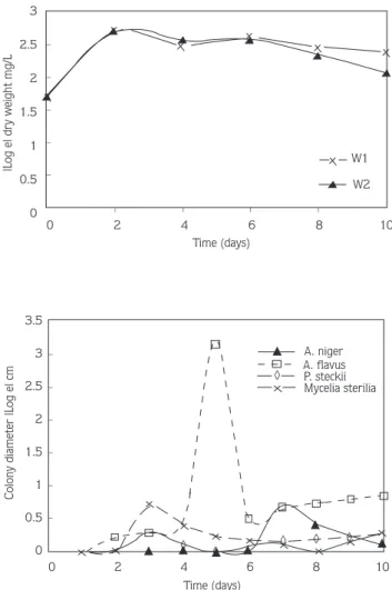
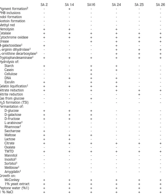
Highly enriched (>25 serial transfers) bacterial and fungi isolates were obtained from the soils of the Gediz basin without any growth factors. All isolates were capable of growth at the expense of TMTD (0.2 mg/ml). Their biomass increased relatively during growth in MSM which contained only TMTD as the carbon source (Fig. 1). Eight out of the twenty selected different type isolates were tested for their ability to degrade TMTD (Table 1).
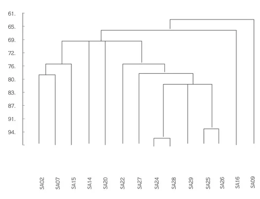
Table 2 lists the various phenotyphical characteristics of the selected TMTD-degrading bacteria strains. The phenogram in figure 2 was constructed from these tests using the simple matching coefficient (SSM) by means of the single linkage clustering method.
All isolates were catalase positive. The optimum temperature for growth was 28-30°C. The pH optimum for growth with TMTD was between 6.5 and 7.0, for growth on complex medium 6.8-7.3. The maximum specific growth rate, µmax, observed in batch culture for growth with
TMTD as the source of carbon was between 0.17 and 0.19 h-1. Four isolates (SAO2, SA16,
SA20, SA27) were observed to be motile under oil immersion and Simms motility test medium. The isolates designated as SAO2, SAO7, SA16, SA20, SA22, SA27 were Gram-positive, aerobic, endospore forming rods. According to their properties the organisms were assigned to
Time (days) 0 2 4 6 8 10 _ _ _ _ _ _ _ _ _ 3 2.5 2 1.5 1 0.5 0
|Log e| dry weight mg/L W1
W2 3.5 3 2.5 2 1.5 1 0.5 0
Colony diameter |Log e| cm
_ _ _ _ _ _ 0 2 4 6 8 10 Time (days) A. niger A. flavus P. steckii Mycelia sterilia ◊ ◊ ◊ ◊ ◊ ◊
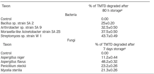
Figure 1. Growth Rates of TMTD Degrading Fungi and Steptomyces in MSM.
the genus Bacillus. The properties distinguish it from other Bacillaceae (all endosporeformers) are their aerobic nature, which may be strict or facultative, rod shape, and catalase production. The other genera of sporeformers include Sporolactibacillus, which is microaerophilic and catalase-negative; Clostridium, anaerobic but does not reduce sulfate; Desulfotomaculum, anaerobic but does reduce sulfate; Sporosarcina, a coccus; and Thermoactinomycetes, which while forming endospores displays typical actinomycete characteristics. Culture of SAO2 is rhizoid, root-like structures from the point of inoculation, identified as Bacillus mycoides, and the two isolates designated as SAO9, SA14 were Gram-positive, pigmented, endospore-free
forming rods, growth on methyl red (150 µg/ml) positive and β-galactosidase negative. They had a characteristic rod-coccus growth cycle during the mid-log phase of growth. Due to this special characteristic they were identified as Arthrobacter spp. Five other isolates (SA24, SA25, SA26, SA28, SA29) showed smooth, opaque colonies on complex media, nonmotile, Gram-negative, oxidase and catalase positive, nonpigmented rods. According to these properties they were assigned to the genus Moraxella. All Gram-negative isolates were resistant to Streptomycine 10U and Cefalexine 30µg (Table 3).
Table 4 gives some morphological and biochemical characteristics of soil Streptomyces spp. isolates. The Streptomyces strains were characterized by the presence of LL-A2pm and gray
spores in the rectus-flexibilis. Actinomycete Promicromonospora citrea and Streptomyces sp. have been reported to utilize carbofuran and butylate as a sole source of carbon and energy for growth (9). We were able to isolate two Streptomyces strains designed as W1 and W2 which are shown in Table 4. Both isolates exhibited a similar morphology on TMTD agar plates. The colonies were opaque, soft in texture, with a regular shape and smooth surface. Our Streptomyces isolates included substrate and aerial mycelium, nonmotile arthrospore, considering at the same time the fact that they form long chains of spores on aerial mycelium (hyphae), it was determined that they were subsumed under the genus of Streptomyces.
Furthermore, our isolates were separated from Kineosporia, Streptoverticillium and Sporichtya, due to the existence of sporangium and motile spores within the substrate mycelium, the spore chains being rectus-flexibilis and the existence of substrate mycelium respectively.
We were also able to determine the four fungal TMTD degrading isolates Aspergillus niger, A. flavus, Penicillium steckii and Mycelia sterilia. Streptomyces and fungal isolates did not require
Table 1. Degradation of TMTD by selected strains
Taxon % of TMTD degraded after
80 h storagea
Bacteria
Control 0.00
Bacillus sp. strain SA 2 25±0.20
Arthrobacter sp. strain SA 9 32.5±0.50
Moraxella-like Acinetobacter strain SA 25 37.5±0.50
Streptomyces sp. strain W 1 43.7±0.49
Fungi
Taxon % of TMTD degraded after 7 days storagea Control 0.00 Aspergillus niger 11.2±0.44 Aspergillus flavus 48.2±0.32 Penicillium steckii 23.2±0.26 Mycelia sterilia 21.3±0.26 aexpressed as mean ± S. D.
Table 2. Overview of the Morphological, Physiological and Biochemical Characteristics of Selected Bacterial Isolates with TMTD as Sole Carbon Source
SA 2 SA 14 SA16 SA 24 SA 25 SA 26 Pigment formation2 - + - - - -PHB inclusions - - - -Indol formation - - - -Acetoin formation - - - -Methyl red - - - -Hemolysin + + - + - -Catalase + + + + + + Cytochrome oxidase + + + + + + Urease - - + + + + β-galactosidase1 + - - + - -L-arginin dihydrolase1 - - - + + + L-ornithine decarboxylase1 - - - + + + Tryptophandesaminase1 + + + + + + Hydrolysis of: Starch + - + + - -Casein - - + + - -Cellulose - - + - - -DNA - + + + - -Esculin + - + + - -Gelatin liquification1 + + + + - -Nitrate reduction + - + + + + Nitrite reduction - - - - +
-Gas from glucose - - + - -
-H2S formation (TSI) - - - -Fermantation of: D-glucose + + + - - -D-galactose + + + - - -D-fructose + + + - - -L-arabinose1 - - - - - -Rhamnose1 - - - - - -Saccharose + - - - - -Maltose + + + - - -Lactose + + - - - -Citrate + + + + + + Oxalate - - - -TMTD + + + + + + Mannitol - - - -Inositol1 - - - - - -Sorbitol1 - - - - - -Melibiose1 - - - - - -Amygdalin1 - - - - - -Growth on: McConkey + + + + + + 1% yeast extract + + + + + + Peptone water (%1) + + + + + + 1 % NaCl + + + + + +
1Tested with API 20E with cells grown aerobically on TMTD agar plates. 2Tested on King’s medium A and B.
Table 3. Antibiotic sensivity of Gram-negative strains. (C: Chloramphenicol 30µg, NA: Nalidixic acid 30µg, NV: Novobiocine 30µg, S: Streptomycine 10 U, CL: Cefalexine 30 µg, CN: Gentamycine 10µg, AMP: Ampicillin 10µg.
Figure 2. Phenogram Based on the Phenotyphic Tests Listed in Table 2. The scale shows the similarity values in (%)
C NA NV S CL CN AMP SA24 S S R R R R R SA25 S S S R R R S SA26 S S S R R S S SA28 S S R R R S S SA29 R S R S R S R _ _ _ _ _ _ _ _ _ _ _ 61. 65. 69. 72. 76. 80. 83. 87. 91. 94.
growth factors. The optimum pH and temperature were 6.8 and 27°C respectively.
Selected isolates (W 1, SA 2, SA 14, SA 16, SA 24, SA 25) were not inhibited by the presence of 100 ppm (w/v) of thiram (data not shown).
Many microorganisms that are capable of degrading fungicides have been described (21). Bacillus spp. and Arthrobacter spp. were isolated which are capable of hydrolyzing the insecticide parathion. One of these Arthrobacter strains is also able to utilize p-nitrophenol as a sole carbon source (22). Recently, Bacillus sp. and Arthrobacter sp. having the ability to degrade endosulfan and carbaryl were isolated from soil (23).
Our isolates SA 2, SA 14 and SA 16, had similar characteristics to the isolates mentioned above. Furthermore, SA 25 and SA 26 had the same physiological and nutritional characteristics of Moraxella-like Acinetobacters (24). The results of morphological and physiological tests suggested that SA 25 and SA 26 most closely resemble species belonging to the genus Moraxella-like Acinetobacter, while SA 14 appears to belong to the genus Arthrobacter.
Fusarium spp., Penicillium spp., Trichoderma spp., Paecilomyces spp., Epicoccum sp., Diheterospora spp. and Verticillium spp. were capable of hydrolyzing EPTC from a soil previously exposed to the herbicide EPTC (25). Aspergillus sp., Fusarium sp., Mortierella sp. and Penicillium sp. were able to defluorinate activity 1080 from soils previously exposed to Na-monofluoracetate (1080) (26). Mucor sp., Fusarium sp., Monilia sp. and Cephalotrichum sp. suppressed by TMTD in a parallel experiment (3), could not be isolated in our study. Of our isolates, Aspergillus flavus, showed better growth (p<0.05) in 2 mg/ml TMTD than Penicillium steckii, Aspergillus niger and Mycelia sterilia (Fig. 1). From the isolation studies we obtained recently, no Mycelia sterilia isolates have been reported yet.
Isolates of Streptomyces sp. (strain W1) and Aspergillus flavus were the most effective degraders even after 80 h storage (Table 1). Streptomyces may be the primary degrading
Table 4. Some Properties of Soil Streptomyces Isolates
Strain W1 Strain W2
Spore chain morphology Rectus-flexibilis Rectus-flexibilis
Color of aerial mycelium gray gray
Color of substrad mycelium white gray
Color of soluble pigment - gray-brown to black
A2pm (Diaminopimelic acid) LL LL Production of: Melanin - -Urease + + α-amylase + + Catalase + + Degredation of: Oxalate - -Esculin + +
organisms of TMTD and the others are opportunistic strains growing on the excretion or lysis products of TMTD.
TMTD increased the biomass (dry weight) of Aspergillus flavus at 5 d and Streptomyces isolates at 2 d (Fig. 1). However, at 6 d growth of Streptomyces and Aspergillus flavus isolates on TMTD-amended medium were significantly less on the medium, possibly indicating a decrease in the rate of pesticide metabolism due to depletion of pesticide substrate or accumulation toxic by-products. In conclusion, the present results illustrate the minor role soil bacteria play in TMTD degradation. Based on the results of these pure culture experiments, bacteria metabolized TMTD relatively slowly possibly due to the loss of TMTD-utilizing ability.
One explanation for the loss of TMTD-utilizing ability in pure culture by microorganisms from soils (3-5) is that the genes for degrading TMTD may be located on plasmids which were lost during prolonged storage and sub-culturing. This phenomenon was also noticed by workers handling EPTC-degrading microorganisms (24). Further studies are required to identify the possible pathways for the microbial degradation of thiram and presence of plasmid DNA. Recent evidence suggests that possible pathways for he handling of thiram by yeast are considered with respect to the activity of glutathione reductase of the cells (27). One of the possible pathways for the microbial degradation of thiram could be hydrolysis with amidases or esterases, since amidases and esterases are very common in soil microorganisms. These aspects are currently being studied.
Acknowledgements
This work was funded by the Scientific and Technical Research Council of Turkey (TÜBİTAK), project no: TBAG/AY-44. The authors are indebted to Prof. İsmet HASENEKOĞLU (Atatürk Üniv.) and Dr. A. Akın DENİZCİ (Ege Üniv.) for their help with this work. We are indebted to Prof. Joh. WÖSTEMEYER (Universität Jena, Germany) for critical reading of the manuscript.
References
1. Worthing, C. R., Hance, R. J.: The pesticide manual, 9th Ed. Published by the British Crop Protection Council, 1991, 822-823.
2. Duffield, W. J., Eide, R. R., Application of rabbit repellent to coniferous planting stock in the Pacific Northwest. J. Forest 60: 109-111, 1962.
3. Richardson, L. T., The persistance of thiram in soil and its relationship to the microbiological balance and damping -off control. Canad. J. Bot. 32: 335-346, 1954.
4. Raghu, K., Murthy, N. B. K., Kumaraswamy, R., Rao, R. S., Sane, P. V.: Proceedings of the joint FAO/IAEA. Division of atomic energy in food and agriculture, Vienna, 1975, 137.
5. Shirkot, C. K.: In studies on the interaction of soil microorganisms and dithiocarbamates. Ph. D. thesis, Punjab University, Chandigarh, India, 1983, 48-60.
6. Shirkot, C. K., Gupta, K. G., Accelerated tetramethylthiuram disulfide (TMTD) degradation in soil by inoculation with TMTD-utilizing bacteria. Bull. Environ. Contam. Toxicol. 35: 354-361, 1985.
tetramethylthiuram disufide (TMTD) from contaminated soil. Bull. Environ. Contam. Toxicol. 44: 317-324, 1990.
8. Klages, U., Lingens, F., Degradation of 4-chlorobenzoic acid by a Pseudomonas sp. Zenterblatt. Bakt. Hyg., I. Abt. Orig. C 1: 215-223, 1980.
9. Edwards, D. E., Kremer, R. J., Keaster, A. J., Characterization and growth response of bacteria in soil following application of carbofuran. J. Environmen. Sci. Health. B 27: (2), 139-154, 1992.
10. Steneck, J. L., Roberts, G. D., Simplified approach to identification of aerobic actinomycetes by thin-layer chromatography. Appl. Microbiol. 28: 226-231, 1974.
11. Staley, J. T., Bryant, M. P., Pfenning, N., Hotl, J. G.: Bergey’s manual of systematic bacteriology. vol. 1-4. Williams & Wilkins, Baltimore, Md., 1984.
12. Ellis, M. B.: Dematiaceaus hyphomycetes. Kew, Commonwealth mycological Institute, 1971.
13. Domsch, K. H., Gams, W., Anderson, T. H.: Compendium of soil fungi. London, 1980, Academic press. 14. Arx, J. A.: The genera of fungi sporulating in pure culture. 1981, Vaduz J. Cramer, 1-335.
15. AOAC : Official Methods of Analysis of the Association of Official Analytical Chemists. (Ed. Williams S.), Arlington, Virginia, 1990, Association Official Analytical Chemists, Inc.
16. Wollum, A. G.: Cultural methods for soil microorganisms, In: Page, A. L (ed) Methods of soil analysis, part 2, 1982, Madison, Wisconsin, American Society for Agronomy, 781.
17. Sokal, R. R., Michener, C. D., A statistical method for evaluating systematic relationships. Univ. Kansas Sci. Bull., 38: 1409-1438, 1958.
18. Jaccard, P., Nouvelles recherches sur la distribution florale. Bull. Soc. Vaud. Sci. Nat. 44: 226-270, 1908. 19. Sneath, P. H. A., Some thoughts on bacterial classification. J. Gen. Microbiol., 17: 184-200, 1957. 20. Sneath, P. H. A., The application of computers to taxonomy. J. Gen. Microbiol., 17: 206-226, 1957.
21. Wallnöfer, P. R., Engelhardt, G.: Microbial degradation of pesticides. Chemistry of plant protection 2, Berlin-Heidelberg, 1989, Spinger-Verlag, 1-115.
22. Nelson, L. M., Biologycally-induced hydrolysis of parathion in soil: isolation of hydrolyzing bacteria. Soil Biol. Biochem. 14: 219-222, 1982.
23. Barlas (Emir), N., The effects of commercial and microorganism-degraded solutions of endosulfan and carbaryl on albino mice. Tr. J. of Zoology. 18: 221-226, 1994.
24. Chen, T. C., Levin, R. E., Taxonomic significance of phenethyl alcohol production by Achromobacter isolated from fishery sources. Appl. Microbiol. 28: 681-687, 1974.
25. Lee, A., EPTC (S-ethyl N, N-dipropylthiocarbamate) degrading microorganisms isolated from a soil previously exposed to EPTC. Soil Biol. Biochem., 16: (5), 529-531, 1984.
26. Wong, D. H., Kirkpatrick, W. E., King, D. R., Kinnear, J. E., Defluorination of sodium monofluoroacetate (1080) by microorganisms isolated from western Australian soils. Soil Biol. Biochem. 24: (9), 833-838, 1992. 27. Elskens, M. T., Penninckx, M. J., Thiram and dimethyldithiocarbamic acid interconversion in Saccharomyces
cerevisiae: a possible metabolic pathway under the control of the glutathione redox cycle. Appl. Environ. Microbiol. 63: 7, 2857-2862, 1997.


