doi: 10.3389/fgene.2021.585556
Edited by: Dimitrios P. Vlachakis, Agricultural University of Athens, Greece Reviewed by: Jovanny Zabaleta, Louisiana State University, United States Gianvito Pio, University of Bari Aldo Moro, Italy Tatjana Loncar-Turukalo, University of Novi Sad Faculty of Technical Sciences, Serbia *Correspondence: Tu ˘gba Önal-Süzek [email protected]
Specialty section: This article was submitted to Nutrigenomics, a section of the journal Frontiers in Genetics Received: 21 July 2020 Accepted: 12 February 2021 Published: 04 March 2021 Citation: Saliho ˘glu R and Önal-Süzek T (2021) Tissue Microbiome Associated With Human Diseases by Whole Transcriptome Sequencing and 16S Metagenomics. Front. Genet. 12:585556. doi: 10.3389/fgene.2021.585556
Tissue Microbiome Associated With
Human Diseases by Whole
Transcriptome Sequencing and 16S
Metagenomics
Rana Saliho ˘glu
1and Tu ˘gba Önal-Süzek
1,2*
1Bioinformatics Department, Graduate School of Natural and Applied Sciences, Mu ˘gla Sıtkı Koçman University, Mu ˘gla, Turkey,2Computer Engineering Department, Faculty of Engineering, Mu ˘gla Sıtkı Koçman University, Mu ˘gla, Turkey
In recent years, a substantial number of tissue microbiome studies have been published,
mainly due to the recent improvements in the minimization of microbial contamination
during whole transcriptome analysis. Another reason for this trend is due to the capability
of next-generation sequencing (NGS) to detect microbiome composition even in low
biomass samples. Several recent studies demonstrate a significant role for the tissue
microbiome in the development and progression of cancer and other diseases. For
example, the increase of the abundance of Proteobacteria in tumor tissues of the breast
has been revealed by gene expression analysis. The link between human papillomavirus
infection and cervical cancer has been known for some time, but the relationship
between the microbiome and breast cancer (BC) is more novel. There are also recent
attempts to investigate the possible link between the brain microbiome and the cognitive
dysfunction caused by neurological diseases. Such studies pointing to the role of the
brain microbiome in Huntington’s disease (HD) and Alzheimer’s disease (AD) suggest
that microbial colonization is a risk factor. In this review, we aim to summarize the
studies that associate the tissue microbiome, rather than gut microbiome, with cancer
and other diseases using whole-transcriptome analysis, along with 16S rRNA analysis.
After providing several case studies for each relationship, we will discuss the potential
role of transcriptome analysis on the broader portrayal of the pathophysiology of the
breast, brain, and vaginal microbiome.
Keywords: neurodegenerative, vagina, tissue microbiome, whole transcriptome, RNA-seq, breast cancer, brain microbiome, 16S RNA analysis
INTRODUCTION
The human body hosts a microbiome of microbes, bacteria, and viruses (
Morgan and Huttenhower,
2012
) that reside in human tissues and biofluids accompanied by various anatomical sites (e.g.,
mammary glands, placenta, and ovarian follicles;
Marchesi and Ravel, 2015
). The majority of
the studies on the human microbiome are focused on microbial diversity and interactions
only with the surface or the epithelial layer. Sequencing analyses of microbiomes have mostly
focused on taxonomy profiling using 16S-rRNA amplicon sequencing, which efficiently covers the
biodiversity of the samples using minimal sequencing by directly characterizing the microbiome
taxonomy (
Shakya et al., 2019
). The whole-transcriptome
analysis offers an alternative to 16S-rRNA sequencing by
detecting and quantifying the low expression levels including
non-coding RNAs (
Shakya et al., 2019
).
The link between the human microbiome and some specific
diseases/cancer types has been tackled by several comprehensive
reviews (e.g., vaginal microbiome,
Ma et al., 2012
;
Muls et al.,
2017
;
Champer et al., 2018
;
Xu et al., 2020
; breast microbiome,
Chadha et al., 2020
; and brain microbiome,
Piacentini et al., 2014
;
Harris and Harris, 2018
).
In this review, rather than the commonly studied gut
microbiome, we summarize the recent but less frequent
microbiome studies analyzing the important role of the
whole
tissue microbiome not limited to but including the epithelial
microbiome, providing a more wholesome picture of the
association of dysbiosis with cancer, neurodegenerative, and
inflammatory diseases.
Various
omics
technologies
such
as
transcriptomics,
proteomics,
metabolomics,
metagenomics,
and
their
combinations provide new insights into the understanding
of the human microbiome and its role in cancer/disease
development (
Komorowski and Pezo, 2020
). As most of the
microorganisms can not be practically cloned and cultured
by the conventional methods, most of the recent microbiome
studies utilize 16S/18S/ITS Amplicon sequencing or whole
transcriptome analysis. Among these techniques, our
mini-review focuses on 16S-rRNA and whole transcriptome analysis;
both of which are demonstrated to be equally sensitive in
bacterial genus detection (
Razzauti et al., 2015
). Of 16S-rRNA
and whole transcriptome analysis, whole transcriptome analysis
is more robust and cost-effective in both capturing the coding
and non-coding RNA and quantifying the heterogeneity in gene
expressions of cells, tissues, and organs (
Ozsolak and Milos,
2011
;
Sancesario and Bernardini, 2018
). The advantages of the
whole transcriptome analysis compared to other omic methods
are vast, the most fundamental one being its contribution
to the determination of new strategies for drug discoveries
and therapeutic interventions (
Ozsolak and Milos, 2011
;
Jiang et al., 2015
).
This mini-review aims to provide a summary of the recent
studies related to the role of the issue microbiome changes
in cancer/unhealthy tissues of the brain, breast, and vagina
using 16S-RNA and whole transcriptome analysis. The studies
considering the tissue microbiome as originating not only from
the surface but all parts of the aforementioned tissues are
included within the scope of this article.
BREAST INTRACELLULAR
MICROBIOME
Breast cancer (BC) microbiome (Figure 1) is hypothesized to
be affected by bacteria-related inflammation in the mammary
ducts and glands disrupting the hierarchy of stem cells. Several
recent studies focus on the intra-tissue microbial and the viral
composition in BC [e.g.,
human papillomavirus (HPV);
Nejman
et al., 2020
;
Sher et al., 2020
]. HPVs are more abundant in BCs
compared to benign breast and normal breast controls (
Lawson
et al., 2016
). Some studies claim that HPV may play a role in the
formation of breast ductal carcinoma, together with the ability to
immortalize human epithelial cells (
Di Lonardo et al., 1992
;
Kan
et al., 2005
;
Heng et al., 2009
). In addition to the epithelial tissue
microbiome studies, several recent studies point to the presence
of intra-tissue bacteria in healthy controls (
Nejman et al., 2020
).
Some studies have shown that pregnancy and breastfeeding
might reduce the risk of BC due to the protective behavior of
the lactose fermenting bacterial flora in the mammary ducts
(
Marwaha et al., 2020
). Another important finding related to BC
is that some patients with hormone receptor-positive (HR+) tend
to have more aggressive BC possibly due to the dysbiosis that
triggers the early inflammation in the mammary gland during the
progression of HR+ breast tumor by disrupting the mammary
tissue homeostasis (
Rosean et al., 2019
).
The presence of mucosal-associated invariant T (MAIT)
cells in the breast ducts intervenes T-helper 17 cell responses
that might be regulated during breast carcinogenesis by
the indications of breast microbiome and the expression of
stress-related ligands by neoplastic breast duct epithelial cells
(
Zumwalde et al., 2018
).
Live and metabolically active
Proteobacteria, Firmicutes, and
Actinobacteria are discovered in breast tumors (
Nejman et al.,
2020
).
Thompson et al. (2017)
pointed to the increase of
Proteobacteria in tumor tissues and Actinobacteria in normal
tissues. Additionally, the correlation of expression profiles with
the microbiome data indicates that
H. influenza is significantly
correlated with genes in the G2M checkpoint, E2F transcription,
and mitotic spindle assembly pathways (
Thompson et al.,
2017
) (Table 1).
Although the microbiome profiles of malignant and benign
breast tumors are different (
Costantini et al., 2018
;
Meng et al.,
2018
), there are some significant similarities in the profiles
of normal and tumor breast tissues revealed by 16S rRNA
amplicon sequencing (
Urbaniak et al., 2016
) (Table 1). The genus
Propionicimonas and families Micrococcaceae, Caulobacteraceae,
Rhodobacteraceae, Nocardioidaceae, and Methylobacteriaceae in
malignant tissues are the enriched microbial biomarkers, and
the development of malignancy results in the decrease in
Bacteroidaceae and the increase in Agrococcus (
Meng et al., 2018
)
(Table 1). Additionally, the genus
Fusobacterium, Bacteroides,
and
Allistipes are especially related to BC (
Philley et al., 2019
.).
Another important finding is the racial differences in the
microbiome of breast tissue first identified by
Smith et al. (2019)
,
i.e., relatively higher abundance of the genera Ralstonia in
non-Hispanic Black women (Table 1).
The effect of HPV on BC has been investigated for years
(
Di Lonardo et al., 1992
;
Widschwendter et al., 2004
;
Sher
et al., 2020
). The presence of HPV in BC tissue has resulted
in increased histopathological activity in the tumor (
Al-Badry
et al., 2019
). This situation may also be presented with the
pathologic nipple discharge (PND;
Balcı et al., 2019
). Also, the
serum derived-extracellular vesicles (EVs) from the BC patients
containing HPV DNA reveal the role of HPV as a potential
trigger for aggressive BC (
De Carolis et al., 2019
). The expressions
of
P53, RB, BRCA1, and BRCA2 in HPV positive BC patients
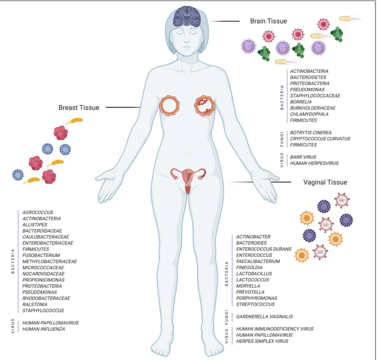
FIGURE 1 | Illustration of different tissues and their microorganisms (bacteria, fungi, and viruses) in normal and disease cases (Created with BioRender.com).
are reduced compared to those in HPV negative ones, possibly
indicating the positive relationship between the increase in
the inflammatory cytokines (e.g., IL-1 and IL-6) and tumor
progression (
Khodabandehlou et al., 2019
).
Chemotherapy, unfortunately, has significantly exacerbated
the disease progression by shifting the microbiome of breast
tumors and increasing the
Pseudomonas spp. (
Chiba et al., 2020
)
(Table 1). There is a need for further studies that cover a larger
and more racially diverse data population, and any findings will
provide new insights into the role of the microbiome in the
therapy stage and the investigation of novel bacterial biomarkers
(
Chiba et al., 2020
). In summary, the recent developments related
to the role of the microbiome in BC reveal the importance of this
relationship for the investigation of BC (
Sher et al., 2020
).
BRAIN MICROBIOME
An interesting indication of tissue microbiome is its association
with neurodegenerative diseases. The polymicrobial infections
are comprised of fungi and bacteria found in the brain tissues
of AD patients (
Pisa et al., 2015, 2017
). Many other studies
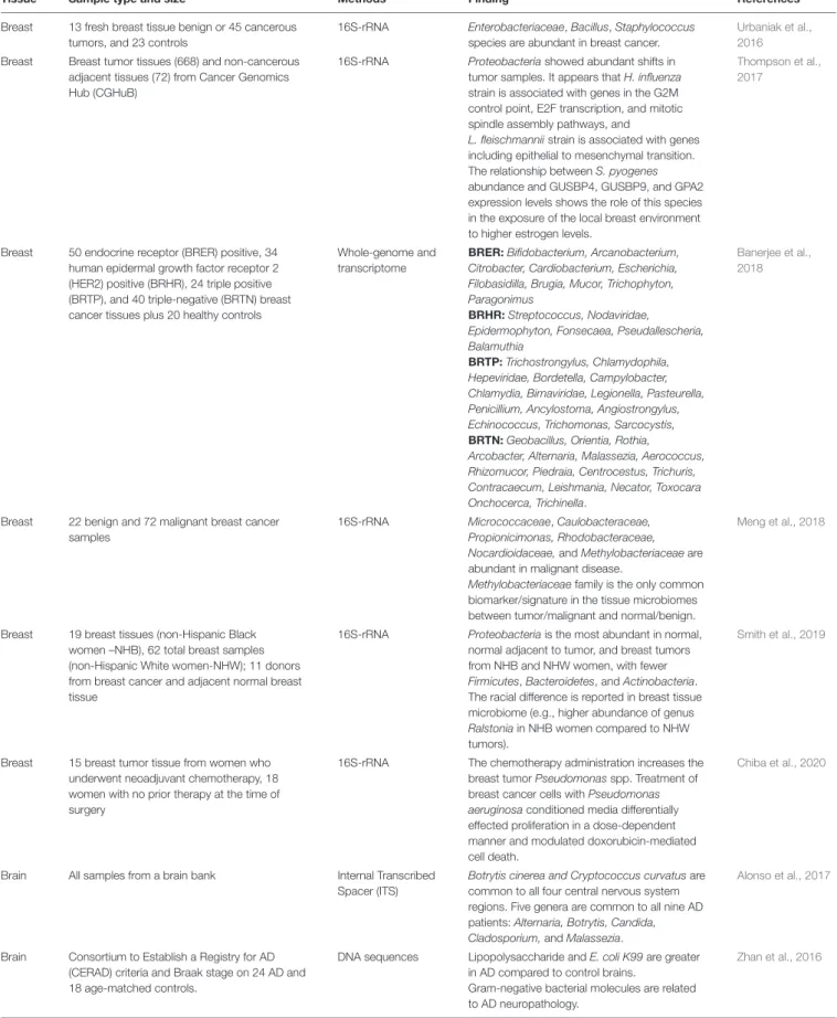
TABLE 1 | Summary of recent findings on the breast, brain and vagina tissue microbiome.
Tissue Sample type and size Methods Finding References
Breast 13 fresh breast tissue benign or 45 cancerous tumors, and 23 controls
16S-rRNA Enterobacteriaceae, Bacillus, Staphylococcus species are abundant in breast cancer.
Urbaniak et al., 2016
Breast Breast tumor tissues (668) and non-cancerous adjacent tissues (72) from Cancer Genomics Hub (CGHuB)
16S-rRNA Proteobacteria showed abundant shifts in tumor samples. It appears that H. influenza strain is associated with genes in the G2M control point, E2F transcription, and mitotic spindle assembly pathways, and
L. fleischmannii strain is associated with genes including epithelial to mesenchymal transition. The relationship between S. pyogenes abundance and GUSBP4, GUSBP9, and GPA2 expression levels shows the role of this species in the exposure of the local breast environment to higher estrogen levels.
Thompson et al., 2017
Breast 50 endocrine receptor (BRER) positive, 34 human epidermal growth factor receptor 2 (HER2) positive (BRHR), 24 triple positive (BRTP), and 40 triple-negative (BRTN) breast cancer tissues plus 20 healthy controls
Whole-genome and transcriptome
BRER: Bifidobacterium, Arcanobacterium, Citrobacter, Cardiobacterium, Escherichia, Filobasidilla, Brugia, Mucor, Trichophyton, Paragonimus
BRHR: Streptococcus, Nodaviridae, Epidermophyton, Fonsecaea, Pseudallescheria, Balamuthia
BRTP: Trichostrongylus, Chlamydophila, Hepeviridae, Bordetella, Campylobacter, Chlamydia, Birnaviridae, Legionella, Pasteurella, Penicillium, Ancylostoma, Angiostrongylus, Echinococcus, Trichomonas, Sarcocystis, BRTN: Geobacillus, Orientia, Rothia, Arcobacter, Alternaria, Malassezia, Aerococcus, Rhizomucor, Piedraia, Centrocestus, Trichuris, Contracaecum, Leishmania, Necator, Toxocara Onchocerca, Trichinella.
Banerjee et al., 2018
Breast 22 benign and 72 malignant breast cancer samples
16S-rRNA Micrococcaceae, Caulobacteraceae, Propionicimonas, Rhodobacteraceae, Nocardioidaceae, and Methylobacteriaceae are abundant in malignant disease.
Methylobacteriaceae family is the only common biomarker/signature in the tissue microbiomes between tumor/malignant and normal/benign.
Meng et al., 2018
Breast 19 breast tissues (non-Hispanic Black women –NHB), 62 total breast samples (non-Hispanic White women-NHW); 11 donors from breast cancer and adjacent normal breast tissue
16S-rRNA Proteobacteria is the most abundant in normal, normal adjacent to tumor, and breast tumors from NHB and NHW women, with fewer Firmicutes, Bacteroidetes, and Actinobacteria. The racial difference is reported in breast tissue microbiome (e.g., higher abundance of genus Ralstonia in NHB women compared to NHW tumors).
Smith et al., 2019
Breast 15 breast tumor tissue from women who underwent neoadjuvant chemotherapy, 18 women with no prior therapy at the time of surgery
16S-rRNA The chemotherapy administration increases the breast tumor Pseudomonas spp. Treatment of breast cancer cells with Pseudomonas aeruginosa conditioned media differentially effected proliferation in a dose-dependent manner and modulated doxorubicin-mediated cell death.
Chiba et al., 2020
Brain All samples from a brain bank Internal Transcribed Spacer (ITS)
Botrytis cinerea and Cryptococcus curvatus are common to all four central nervous system regions. Five genera are common to all nine AD patients: Alternaria, Botrytis, Candida, Cladosporium, and Malassezia.
Alonso et al., 2017
Brain Consortium to Establish a Registry for AD (CERAD) criteria and Braak stage on 24 AD and 18 age-matched controls.
DNA sequences Lipopolysaccharide and E. coli K99 are greater in AD compared to control brains.
Gram-negative bacterial molecules are related to AD neuropathology.
Zhan et al., 2016
TABLE 1 | Continued
Tissue Sample type and size Methods Finding References
Brain Control = 12, AD = 14 16S-rRNA The control brain has similar bacterial profiles
with the blood, but the AD brain displays a larger proportion of Actinobacteria.
Emery et al., 2017
Brain Accelerating Medicines Partnership AD; Knowledge Portal
16S-rRNA The increase in the abundance of HHV-6A, HHV-7, and herpes simplex virus (HSV-1) genomes in banked postmortem brains from subjects with AD compared to controls.
Readhead et al., 2019
Brain Postmortem hippocampal formation specimens from 10 neurological control and 10 AD cases, 22 AD specimens, and 19 neurological controls from the hippocampus; 12 control and 20 AD cerebellum samples
16S-rRNA The most abundant phyla in both cancer and normal cases are Proteobacteria, Firmicutes, Actinobacteria, and Bacteroidetes.
There is a significant role of brain region on the presence of microbial DNA revealed by the variations in beta diversity in hippocampal and cerebellum samples.
Westfall et al., 2020
Brain Examination of the olfactory bulbs from autopsy of two AD cases
Bacterial DNA The HSV1 is a strong risk factor when it is present in the brain of carriers of the type 4 allele of the gene for APOE-ε4. Another important risk factor is the bacterium, Chlamydia pneumoniae identified and localized in the AD brain.
Itzhaki et al., 2004
Brain Samples of tissue from brain donors, AD, and control individuals
16S-rRNA No evidence for expression of early (ICP0) or late (ICP5) proteins of the HSV-1 in brain. A polyclonal antibody against Borrelia detected structures that appeared not related to spirochetes, but rather to fungi, revealing the presence of several bacteria in brain tissue of AD patients.
Pisa et al., 2017
Brain 118 human viruses, and PCR from 711 AD and non-AD control brains
Whole transcriptome
There is no clear relation between HHV-6 and AD, whereas the Epstein–Barr virus (EBV) and cytomegalovirus (CMV) are comparable.
Allnutt et al., 2020
Vagina 151 healthy women (65 HPV+ and 86 HPV−) 16S rDNA Bacteroides plebeius, Acinetobacter lwoffii, and Prevotella buccae are significantly seen in HPV-positive women.
Chao et al., 2019
Vagina Women with the age of 18–29 who have applied to the sexually transmitted infections (STIs) clinic have symptoms
Whole transcriptome
The samples from women with STI infection only contain pathogen-specific sequences (3–38% transcriptome coverage).
O’Connell et al., 2019
claimed that the repeated activation of herpes simplex virus 1
(HSV-1) promotes the neurodegeneration aspect of AD (
Deatly
et al., 1990
;
Mori et al., 2004
;
Mancuso et al., 2014
). Microbial
colonization is also considered as a possible risk factor for
Huntington’s disease (HD) (
Alonso et al., 2019
). Alzheimer’s
disease (AD) is a neurodegenerative disorder whose pathogenesis
is not only limited to the neuronal compartment but also
accompanied by a significant interaction with the immunological
mechanisms in the brain (
Heneka et al., 2015
). Even though the
etiopathogenesis of AD has not been well documented, some
investigations point to the role of the microbiome (Figure 1)
(gut,
Vogt et al., 2017
; oral,
Shoemark and Allen, 2015
), and the
etiology of AD has been considered as microbial (
Bhattacharjee
and Lukiw, 2013
;
Pisa et al., 2017
).
The role of human herpesvirus (HHV) in the etiology of AD
has been evident recently, yet studies on the different viruses
of the herpes family [e.g., HHV-6, -7, cytomegalovirus (CMV),
Epstein–Barr virus (EBV)] are limited (
Carbone et al., 2014
).
Nevertheless, there are some recent additional attempts to reveal
the relation between HHV and AD (
Eimer et al., 2018
;
Readhead
et al., 2018, 2019
). For instance, HHV-6A and 7 have been
detected in AD patients with a higher viral abundance of amyloid
precursor protein (APP) (
O’Brien and Wong, 2011
) metabolism
(i.e., induction of APBB2, APPBP2, BIN1, BACE1, and CLU)
(
Readhead et al., 2018
). The oligomers of Amyloid-
β (Aβ) peptide
bind HHV surface glycoproteins and accelerate the deposition
of
β-amyloid (
Eimer et al., 2018
). This leads to protective
viral entrapment activity against neurotropic HSV-1 and also
HHV6A-B that are linked to AD (
Eimer et al., 2018
). Besides,
the increase of the KIR2DL2/C1 genotype in AD patients and
the lower anti-herpetic activity of KIR2DL2 positive natural killer
(NK) cells support the role of HHV infection in AD development
and also increase the susceptibility to HHV-6A infection (
Rizzo
et al., 2019
). Most studies also suggest the prevalence of HHV-6A
in the AD brain compared to others (
Eimer et al., 2018
;
Readhead
et al., 2019
;
Rizzo et al., 2019
). Besides, the effect of HSV-1 in the
development of AD among people with the genetic susceptibility
factor of the apolipoprotein E (APOE4) allele has been evident
(
Lindman et al., 2019
;
Linard et al., 2020
).
The contribution of infectious microbial components and
also virulence factors rhamnolipids (RLs) to the pathophysiology
of the human central nervous system (CNS), including AD,
has been potentially important (
Andreadou et al., 2017
). The
presence of RLs is attributed to chronic bacterial infections
forming bacterial virulence factors secreted by a wide variety of
pathogens (
Andreadou et al., 2017
). The fungi in the samples
of the frontal cortex in AD brains are distinct, indicating the
varying microbial compositions among brain regions (
Alonso
et al., 2018
;
Westfall et al., 2020
) (Table 1). A polyclonal antibody
against
Borrelia has been identified in structures associated
with fungi (
Pisa et al., 2017
). Two independent
Chlamydophila
antibodies have revealed several structures similar to fungal cells
and hyphae and prokaryotic cells, but are most likely unrelated
to
Chlamydophila spp. Several bacteria in the AD patients
suggested that the polymicrobial infections are comprised
of fungi and bacteria occurred in their brain tissues (
Pisa
et al., 2017
) (Table 1). The fungal species found in the CNS
of AD patients have been investigated by next-generation
sequencing (NGS) that revealed the most common species,
namely,
Botrytis cinerea and Cryptococcus curvatus (
Alonso
et al., 2017
).
Burkholderiaceae and Staphylococcaceae are more
abundant in AD brains compared to normal brains (
Alonso
et al., 2018
). Studies on that subject aiming at investigating the
link between fungal species with the AD are rather essential
for providing antifungal therapy and also for the evolution and
severity stages of clinical symptoms in AD patients (
Alonso et al.,
2017
) (Table 1). On the other hand, microbiological attack or
change is thought to be one of the factors causing CNS disorders,
also evident for the AD that shows an increase in bacterial
populations (e.g., large amount of
Actinobacteria;
Emery et al.,
2017
) (Table 1).
Huntington’s disease is caused by a triplet expansion in the
Huntingtin (HTT) gene (
Vonsattel and Difiglia, 1998
).
Alonso
et al. (2019)
first identified the role of some prevalent bacterial
genera (Pseudomonas, Acinetobacter, and Burkholderia) in the
brain microbiome of HD patients. RNA-seq analysis of human
neurodegenerative disease tissues (except for AD) reported
no significant difference compared to cytotoxic T-lymphocytes
(CTL) tissues, indicating that the sub-clinical infections do
not result in the inflammation related to the tissue of many
neurodegenerative diseases such as amyotrophic lateral sclerosis
(ALS) and Parkinson’s disease (PD) (
Bennett et al., 2019
).
VAGINAL MICROBIOME
The relation between the vaginal microbiome (Figure 1) and
the high-risk HPV infection has been propounded by several
studies (
Chao et al., 2019, 2020
;
Keller et al., 2019
;
Liu et al.,
2019
;
Nené et al., 2019
;
Zhou et al., 2019
;
Abudula et al.,
2020
;
Jiang et al., 2020
). The sequencing of 16S rRNA genes
reveals that some anaerobic bacteria (e.g.,
Bacteroides plebeius
and
Acinetobacter lwoffii) are significantly more common in HPV
positive women, suggesting a specific microbiome as a biomarker
to detect changes in the cervical microenvironment indicating
HPV infection (
Chao et al., 2019
). The genus
Prevotella,
Porphyromonas, and Enterococcus are the highest in the cervical
permanent HPV infection, whereas the
Bacteroides genus is the
lowest (
Chao et al., 2020
).
The expression studies on cervical lesions to explore the
possible relation of HPV with cervical cancer are also crucial.
Toll-like receptor 4 (TLR4) expression supports the claim
of a distinct relation between tumor formation and
HPV-positive cervical cancer (
Jiang et al., 2020
), suggesting that TLR4
somehow enables the formation of a local immunosuppressive
microenvironment.
Human papillomavirus, human immunodeficiency virus
(HIV), and HSV have been associated with the growth of
genital-related cancers.
Keller et al. (2019)
proclaim that the increase in
microbial diversity and cervicovaginal inflammation in women
with HIV+ and HSV+ significantly perturbs genital health. It is
thought that neither the inception of antiretroviral therapy (ART)
nor the restructuring of the immune system affects the vagina
microbiome of HIV-infected women (
Liu et al., 2019
). Several 16S
rRNA sequencing studies (
Nené et al., 2019
;
Zhou et al., 2019
)
demonstrate a decrease in bacterial diversity in ovarian cancer
tissues compared to normal ones. Some bacterial implications are
suggested as biomarkers for the early detection of ovarian tumors,
such as higher ratios of two phyla for
Proteobacteria/Firmicutes,
and the increase of genus
Acinetobacter and decrease of genus
Lactococcus (
Zhou et al., 2019
).
DISCUSSION AND CONCLUSION
In this mini-review, we mainly discuss the possible effects
of human tissue microbiome in the development of some
common cancers and neurodegenerative diseases. There might
be two important implications of our comprehensive literature
compilation: the first implication being the presentation of
the limited number of studies that deal with the microbiome
differences in healthy and unhealthy/cancer tissues, for example,
AD is more commonly studied for its relationship with the
oral and gastrointestinal microbiome. The second implication
of our literature compilation is to list the shared bacteria (e.g.,
Proteobacteria, Bacteroides, and Firmicutes) that display either
positive or negative anomalies common to all cancer tissues
discussed above (Figure 1).
Many of the recent findings summarized in Table 1 are now
possible due to the ability of the whole transcriptome analysis
to (1) provide abundance information along with the taxonomic
diversity and (2) provide a vaster picture of the expression profile
of the microbiome comprising of fungal, viral, and bacterial along
with host expression profile. In some of the cases summarized in
Table 1
, observed differences in each patient are due to fungal
species which could not be discovered if a whole transcriptomic
approach is not taken (
Alonso et al., 2017, 2019
).
Variations in the microbiome of cancer patients with different
cancer stages have already been known in the literature. For
instance, the species of
Firmicutes and Bacteroides are dominant
in the invasive and benign BC, whereas some of the species such
as
Fusobacterium, Atopobium, and Lactobacillus are enriched in
malignant breast tissues (e.g.,
Hieken et al., 2016
;
Chadha et al.,
2020
). The recent studies about the role of the microbiome in
different cancers and neurodegenerative diseases reveal that there
are some common species observed in all cancer tissues. For
instance, some of these species (e.g.,
Bacteroides) are enriched
in the AD tissues, while they are not abundant in cervical
and BC tissues. Additionally,
Proteobacteria and Firmicutes are
enriched in all types of cancer tissues. Such similarities and/or
differences may be attributed to differences in tissue structure.
Our mini-review will attract more attention to the reprocessing
of the publicly available RNA-seq data to distinguish microbial
contamination from tissue microbiome via
in silico techniques
to clarify the relative composition, abundance, and impact of
microbiome in cancer tissues.
Most of the studies on the possible link between microbiome
and diseases associating the presence of many potential microbial
biomarkers and their pathways with the advanced stage of
the diseases offer new insights into the diagnostic staging
(e.g.,
Wozniak et al., 2005
;
Alonso et al., 2017
;
Meng et al.,
2018
;
Rizzo et al., 2019
). There is also some evidence that
the profiles of microbiome in healthy and unhealthy tissues
(e.g., for BC;
Urbaniak et al., 2016
) are identical, and
hence the role of tissue microbiome for the development of
diseases/cancers should be further investigated by increasing
the number of available studies. As the Next-Generation
Sequence (NGS) technology becomes more precise and novel
in silico techniques are getting developed to discard the studies
with bacterial contamination and non-standardized analysis
pipelines, the re-analysis of the existing transcriptome data
offers a huge potential for the future
in silico data-mining
microbiome studies from the vast amount of publicly available
transcriptomic data.
AUTHOR CONTRIBUTIONS
RS wrote the manuscript and TÖS supervised the project.
Both authors discussed the results and contributed to the
final manuscript.
FUNDING
RS was funded by YOK 100/2000 program and TÖS was funded
by NIH Intramural program. The open-access publication cost
was funded by the corresponding author.
ACKNOWLEDGMENTS
We thank the NIH Fellows Editorial Board (FEB) for
editorial assistance.
REFERENCES
Abudula, A., Rouzi, N., Xu, L., Yang, Y., and Hasimu, A. (2020). Tissue-based metabolomics reveals potential biomarkers for cervical carcinoma and HPV infection.Bosn. J. Basic Med. Sci. 20, 78–87. doi: 10.17305/bjbms.2019.4359 Al-Badry, B. J., Al-Muswie, R. T., and Jameel, S. (2019). Histopathological study of
Human Papillomavirus (HPV) in breast cancer patients.J. Phys. 1294:062080. doi: 10.1088/1742-6596/1294/6/062080
Allnutt, M. A., Johnson, K., Bennett, D. A., Connor, S. M., Troncoso, J. C., Pletnikova, O., et al. (2020). Human herpesvirus 6 detection in Alzheimer’s disease cases and controls across multiple cohorts. Neuron 105, D1027– D1035.e2. doi: 10.1016/j.neuron.2019.12.031
Alonso, R., Pisa, D., Aguado, B., and Carrasco, L. (2017). Identification of fungal species in brain tissue from Alzheimer’s disease by next-generation sequencing. J. Alzheimer Dis. 58, 55–67. doi: 10.3233/JAD-170058
Alonso, R., Pisa, D., and Carrasco, L. (2019). Brain microbiota in Huntington’s disease patients.Front. Microbiol. 10:2622. doi: 10.3389/fmicb.2019.02622 Alonso, R., Pisa, D., Fernández-Fernández, A. M., and Carrasco, L. (2018).
Infection of fungi and bacteria in brain tissue from elderly persons and patients with Alzheimer’s disease.Front. Aging Neurosci. 10:159. doi: 10.3389/fnagi.2018. 00159
Andreadou, E., Pantazaki, A. A., Daniilidou, M., and Tsolaki, M. (2017). Rhamnolipids, microbial virulence factors, in Alzheimer’s disease.J. Alzheimer Dis. 59, 209–222. doi: 10.3233/JAD-161020
Balcı, F. L., Uras, C., and Feldman, S. M. (2019). Is human papillomavirus associated with breast cancer or papilloma presenting with pathologic nipple discharge?Cancer Treat. Res. Commun. 19:100122. doi: 10.1016/j.ctarc.2019. 100122
Banerjee, S., Tian, T., Wei, Z., Shih, N., Feldman, M. D., Peck, K. N., et al. (2018). Distinct microbial signatures associated with different breast cancer types.Front. Microbiol. 9:951. doi: 10.3389/fmicb.2018.00951
Bennett, J. P. Jr., Keeney, P. M., and Brohawn, D. G. (2019). RNA sequencing reveals small and variable contributions of infectious agents to transcriptomes of postmortem nervous tissues from amyotrophic lateral sclerosis, Alzheimer’s disease and Parkinson’s disease subjects, and increased expression of genes from disease-activated microglia.Front. Neurosci. 13:235. doi: 10.3389/fnins.2019. 00235
Bhattacharjee, S., and Lukiw, W. J. (2013). Alzheimer’s disease and the microbiome. Front. Cell. Neurosci. 7:153. doi: 10.3389/fncel.2013.00153
Carbone, I., Lazzarotto, T., Ianni, M., Porcellini, E., Forti, P., Masliah, E., et al. (2014). Herpes virus in Alzheimer’s disease: relation to progression of the disease.Neurobiol. Aging 35, 122–129. doi: 10.1016/j.neurobiolaging.2013.06. 024
Chadha, J., Nandi, D., Atri, Y., and Nag, A. (2020). Significance of human microbiome in breast cancer: tale of an invisible and an invincible.Semin. Cancer Biol. (in press). doi: 10.1016/j.semcancer.2020.07.010
Champer, M., Wong, A. M., Champer, J., Brito, I. L., Messer, P. W., Hou, J. Y., et al. (2018). The role of the vaginal microbiome in gynaecological cancer.Intern. J. Obstet. Gynaecol. 125, 309–315. doi: 10.1111/1471-0528.14631
Chao, X., Sun, T., Wang, S., Tan, X., Fan, Q., Shi, H., et al. (2020). Research of the potential biomarkers in vaginal microbiome for persistent high-risk human papillomavirus infection.Ann. Transl. Med. 8:100. doi: 10.21037/atm.2019.12. 115
Chao, X. P., Sun, T. T., Wang, S., Fan, Q. B., Shi, H. H., Zhu, L., et al. (2019). Correlation between the diversity of vaginal microbiota and the risk of high-risk human papillomavirus infection.Intern. J. Gynecol. Cancer 29:32. doi: 10.1136/ijgc-2018-000032
Chiba, A., Bawaneh, A., Velazquez, C., Clear, K. Y., Wilson, A. S., Howard-McNatt, M., et al. (2020). Neoadjuvant chemotherapy shifts breast tumor microbiota populations to regulate drug responsiveness and the development of metastasis. Mol. Cancer Res. 18, 130–139. doi: 10.1158/1541-7786.MCR-19-0451 Costantini, L., Magno, S., Albanese, D., Donati, C., Molinari, R., Filippone, A., et al.
(2018). Characterization of human breast tissue microbiota from core needle biopsies through the analysis of multi hypervariable 16S-rRNA gene regions. Sci. Rep. 8:16893. doi: 10.1038/s41598-018-35329-z
De Carolis, S., Storci, G., Ceccarelli, C., Savini, C., Gallucci, L., Sansone, P., et al. (2019). HPV DNA associates with breast cancer malignancy and it is transferred to breast cancer stromal cells by extracellular vesicles.Front. Oncol. 9:860. doi: 10.3389/fonc.2019.00860
Deatly, A. M., Haase, A. T., Fewster, P. H., Lewis, E., and Ball, M. J. (1990). Human herpes virus infections and Alzheimer’s disease.Neuropathol. Appl. Neurobiol. 16, 213–223. doi: 10.1111/j.1365-2990.1990.tb01158.x
Di Lonardo, A., Venuti, A., and Marcante, M. L. (1992). Human papillomavirus in breast cancer.Breast Cancer Res. Treat. 21, 95–100. doi: 10.1007/BF01836955
Eimer, W. A., Kumar, D. K. V., Shanmugam, N. K. N., Rodriguez, A. S., Mitchell, T., Washicosky, K. J., et al. (2018). Alzheimer’s disease-associatedβ-amyloid is rapidly seeded by herpesviridae to protect against brain infection.Neuron 99, 56–63. doi: 10.1016/j.neuron.2018.06.030
Emery, D. C., Shoemark, D. K., Batstone, T. E., Waterfall, C. M., Coghill, J. A., Cerajewska, T. L., et al. (2017). 16S rRNA next generation sequencing analysis shows bacteria in Alzheimer’s post-mortem brain.Front. Aging Neurosci. 9:195. doi: 10.3389/fnagi.2017.00195
Harris, S. A., and Harris, E. A. (2018). Molecular mechanisms for herpes simplex virus type 1 pathogenesis in Alzheimer’s disease.Front. Aging Neurosci. 10:48. doi: 10.3389/fnagi.2018.00048
Heneka, M. T., Carson, M. J., El Khoury, J., Landreth, G. E., Brosseron, F., Feinstein, D. L., et al. (2015). Neuroinflammation in Alzheimer’s disease.Lancet Neurol. 14, 388–405. doi: 10.1016/S1474-4422(15)70016-5
Heng, B., Glenn, W. K., Ye, Y., Tran, B., Delprado, W., Lutze-Mann, L., et al. (2009). Human papilloma virus is associated with breast cancer.Br. J. Cancer 101, 1345–1350. doi: 10.1038/sj.bjc.6605282
Hieken, T. J., Chen, J., Hoskin, T. L., Walther-Antonio, M., Johnson, S., Ramaker, S., et al. (2016). The microbiome of aseptically collected human breast tissue in benign and malignant disease.Sci. Rep. 6:30751. doi: 10.1038/srep30751 Itzhaki, R. F., Wozniak, M. A., Appelt, D. M., and Balin, B. J. (2004). Infiltration
of the brain by pathogens causes Alzheimer’s disease. Neurobiol. Aging 25, 619–627. doi: 10.1016/j.neurobiolaging.2003.12.021
Jiang, N., Xie, F., Chen, L., Chen, F., and Sui, L. (2020). The effect of TLR4 on the growth and local inflammatory microenvironment of HPV-related cervical cancer in vivo.Infect. Agents Cancer 15, 1–10. doi: 10.1186/s13027-020-0279-9 Jiang, Z., Zhou, X., Li, R., Michal, J. J., Zhang, S., Dodson, M. V., et al. (2015). Whole transcriptome analysis with sequencing: methods, challenges and potential solutions.Cell. Mol. Life Sci. 72, 3425–3439. doi: 10.1007/s00018-015-1934-y Kan, C. Y., Iacopetta, B. J., Lawson, J. S., and Whitaker, N. J. (2005). Identification
of human papillomavirus DNA gene sequences in human breast cancer.Br. J. Cancer 93, 946–948. doi: 10.1038/sj.bjc.6602778
Keller, M. J., Huber, A., Espinoza, L., Serrano, M. G., Parikh, H. I., Buck, G. A., et al. (2019). Impact of herpes simplex virus type 2 and human immunodeficiency virus dual infection on female genital tract mucosal immunity and the vaginal microbiome.J. Infect. Dis. 220, 852–861. doi: 10.1093/infdis/jiz203
Khodabandehlou, N., Mostafaei, S., Etemadi, A., Ghasemi, A., Payandeh, M., Hadifar, S., et al. (2019). Human papilloma virus and breast cancer: the role of inflammation and viral expressed proteins.BMC Cancer 19:61. doi: 10.1186/ s12885-019-5286-0
Komorowski, A. S., and Pezo, R. C. (2020). Untapped “-omics”: the microbial metagenome, estrobolome, and their influence on the development of breast cancer and response to treatment. Breast Cancer Res. Treat. 179, 287–300. doi: 10.1007/s10549-019-05472-w
Lawson, J. S., Glenn, W. K., and Whitaker, N. J. (2016). Human papilloma viruses and breast cancer-assessment of causality.Front. Oncol. 6:207. doi: 10.3389/ fonc.2016.00207
Linard, M., Letenneur, L., Garrigue, I., Doize, A., Dartigues, J. F., and Helmer, C. (2020). Interaction between APOE4 and herpes simplex virus type 1 in Alzheimer’s disease.Alzheimer Dement. 16, 200–208. doi: 10.1002/alz.12008 Lindman, K. L., Weidung, B., Olsson, J., Josefsson, M., Kok, E., and Johansson, A.
(2019). A genetic signature including apolipoprotein Eε4 potentiates the risk of herpes simplex-associated Alzheimer’s disease.Alzheimer Dement. 5, 697–704. doi: 10.1016/j.trci.2019.09.014
Liu, C. M., Packman, Z. R., Abraham, A. G., Serwadda, D. M., Nalugoda, F., Aziz, M., et al. (2019). The effect of antiretroviral therapy initiation on the vaginal microbiome in HIV-infected women.Open Forum Infect. Dis. 6:ofz328. doi: 10.1093/ofid/ofz328
Ma, B., Forney, L. J., and Ravel, J. (2012). Vaginal microbiome: rethinking health and disease.Ann. Rev. Microbiol. 66, 371–389. doi: 10.1146/annurev-micro-092611-150157
Mancuso, R., Baglio, F., Agostini, S., Cabinio, M. C., Laganà, M. M., Hernis, A., et al. (2014). Relationship between herpes simplex virus-1-specific antibody titers and cortical brain damage in Alzheimer’s disease and amnestic mild cognitive impairment.Front. Aging Neurosci. 6:285. doi: 10.3389/fnagi.2014.00285 Marchesi, J. R., and Ravel, J. (2015). The vocabulary of microbiome research: a
proposal.Marchesi Ravel Microb. 3:31. doi: 10.1186/s40168-015-0094-5
Marwaha, A. K., Morris, J. A., and Rigby, R. J. (2020). Hypothesis: bacterial induced inflammation disrupts the orderly progression of the stem cell hierarchy and has a role in the pathogenesis of breast cancer.Med. Hypothes. 136:109530. doi: 10.1016/j.mehy.2019.109530
Meng, S., Chen, B., Yang, J., Wang, J., Zhu, D., Meng, Q., et al. (2018). Study of microbiomes in aseptically collected samples of human breast tissue using needle biopsy and the potential role of in situ tissue microbiomes for promoting malignancy.Front. Oncol. 8:318. doi: 10.3389/fonc.2018.00318
Morgan, X. C., and Huttenhower, C. (2012). Human microbiome analysis.PLoS Comput. Biol. 8:e1002808. doi: 10.1371/journal.pcbi.1002808
Mori, I., Kimura, Y., Naiki, H., Matsubara, R., Takeuchi, T., Yokochi, T., et al. (2004). Reactivation of HSV-1 in the brain of patients with familial Alzheimer’s disease.J. Med. Virol. 73, 605–611. doi: 10.1002/jmv.20133
Muls, A., Andreyev, J., Lalondrelle, S., Taylor, A., Norton, C., and Hart, A. (2017). Systematic review: the impact of cancer treatment on the gut and vaginal microbiome in women with a gynecological malignancy.Intern. J. Gynecol. Cancer 27, 1550–1559.
Nejman, D., Livyatan, I., Fuks, G., Gavert, N., Zwang, Y., Geller, L. T., et al. (2020). The human tumor microbiome is composed of tumor type-specific intracellular bacteria.Science 368, 973–980. doi: 10.1126/science.aay9189
Nené, N. R., Reisel, D., Leimbach, A., Franchi, D., Jones, A., Evans, I., et al. (2019). Association between the cervicovaginal microbiome, BRCA1 mutation status, and risk of ovarian cancer: a case-control study.Lancet Oncol. 20, 1171–1182. doi: 10.1016/S1470-2045(19)30340-7
O’Brien, R. J., and Wong, P. C. (2011). Amyloid precursor protein processing and Alzheimer’s disease.Annu. Rev. Neurosci. 34, 185–204. doi: 10.1146/annurev-neuro-061010-113613
O’Connell, C. M., Brochu, H., Girardi, J., Harrell, E., Jones, A., Darville, T., et al. (2019). Simultaneous profiling of sexually transmitted bacterial pathogens, microbiome, and concordant host response in cervical samples using whole transcriptome sequencing analysis.Microb. Cell 6:177. doi: 10.15698/mic2019. 03.672
Ozsolak, F., and Milos, P. M. (2011). RNA sequencing: advances, challenges and opportunities.Nat. Rev. Genet. 12, 87–98. doi: 10.1038/nrg2934
Philley, J. V., Kannan, A., Olusola, P., McGaha, P., Singh, K. P., Samten, B., et al. (2019). Microbiome diversity in sputum of Nontuberculous Mycobacteria infected women with a history of breast cancer.Cell. Physiol. Biochem. 52, 263–279. doi: 10.33594/00000002
Piacentini, R., De Chiara, G., Li Puma, D. D., Ripoli, C., Marcocci, M. E., Garaci, E., et al. (2014). HSV-1 and Alzheimer’s disease: more than a hypothesis.Front. Pharmacol. 5:97. doi: 10.3389/fphar.2014.00097
Pisa, D., Alonso, R., Fernández-Fernández, A. M., Rábano, A., and Carrasco, L. (2017). Polymicrobial infections in brain tissue from Alzheimer’s disease patients.Sci. Rep. 7, 1–14. doi: 10.1038/s41598-017-05903-y
Pisa, D., Alonso, R., Rábano, A., Rodal, I., and Carrasco, L. (2015). Different brain regions are infected with fungi in Alzheimer’s disease.Sci. Rep. 5:15015. doi: 10.1038/srep15015
Razzauti, M., Galan, M., Bernard, M., Maman, S., Klopp, C., Charbonnel, N., et al. (2015). A Comparison between transcriptome sequencing and 16S metagenomics for detection of bacterial pathogens in wildlife.PLoS Negl. Trop. Dis. 9:e0003929. doi: 10.1371/journal.pntd.0003929
Readhead, B., Haure-Mirande, J. V., Ehrlich, M. E., Gandy, S., and Dudley, J. T. (2019). Further evidence of increased human Herpesvirus in Alzheimer’s disease.bioRxiv [Preprint], doi: 10.1101/858050
Readhead, B., Haure-Mirande, J. V., Funk, C. C., Richards, M. A., Shannon, P., Haroutunian, V., et al. (2018). Multiscale analysis of independent Alzheimer’s cohorts finds disruption of molecular, genetic, and clinical networks by human herpesvirus.Neuron 99, 64–82. doi: 10.1016/j.neuron.2018.05.023
Rizzo, R., Bortolotti, D., Gentili, V., Rotola, A., Bolzani, S., Caselli, E., et al. (2019). KIR2DS2/KIR2DL2/HLA-C1 haplotype is associated with Alzheimer’s disease: implication for the role of herpesvirus infections.J. Alzheimer Dis. 67, 1379–1389. doi: 10.3233/JAD-180777
Rosean, C. B., Bostic, R. R., Ferey, J. C., Feng, T. Y., Azar, F. N., Tung, K. S., et al. (2019). Preexisting Commensal Dysbiosis is a host-intrinsic regulator of tissue inflammation and tumor cell dissemination in hormone receptor-positive breast cancer.Cancer Res. 79, 3662–3675. doi: 10.1158/0008-5472.CAN-18-3464
Sancesario, G. M., and Bernardini, S. (2018). Alzheimer’s disease in the omics era. Clin. Biochem. 59, 9–16. doi: 10.1016/j.clinbiochem.2018.06.011
Shakya, M., Lo, C. C., and Chain, P. S. (2019). Advances and challenges in metatranscriptomic analysis. Front. Genet. 10:904. doi: 10.3389/fgene.2019. 00904
Sher, G., Salman, N. A., Kulinski, M., Fadel, R. A., Gupta, V. K., Anand, A., et al. (2020). Prevalence and type distribution of high-risk human Papillomavirus (HPV) in breast cancer: a qatar based study.Cancers 12:1528. doi: 10.3390/ cancers12061528
Shoemark, D. K., and Allen, S. J. (2015). The microbiome and disease: reviewing the links between the oral microbiome, aging, and Alzheimer’s disease.J. Alzheimer Dis. 43, 725–738. doi: 10.3233/JAD-141170
Smith, A., Pierre, J. F., Makowski, L., Tolley, E., Lyn-Cook, B., Lu, L., et al. (2019). Distinct microbial communities that differ by race, stage, or breast-tumor subtype in breast tissues of non-Hispanic Black and non-Hispanic White women.Sci. Rep. 9, 1–10. doi: 10.1038/s41598-019-48348-1
Thompson, K. J., Ingle, J. N., Tang, X., Chia, N., Jeraldo, P. R., Walther-Antonio, M. R., et al. (2017). A comprehensive analysis of breast cancer microbiota and host gene expression.PLoS One 12:e0188873. doi: 10.1371/journal.pone. 0188873
Urbaniak, C., Gloor, G. B., Brackstone, M., Scott, L., Tangney, M., and Reid, G. (2016). The microbiota of breast tissue and its association with breast cancer. Appl. Environ. Microbiol. 82, 5039–5048. doi: 10.1128/AEM.01235-16 Vogt, N. M., Kerby, R. L., Dill-McFarland, K. A., Harding, S. J., Merluzzi, A. P.,
Johnson, S. C., et al. (2017). Gut microbiome alterations in Alzheimer’s disease. Sci. Rep. 7, 1–11. doi: 10.1038/s41598-017-13601-y
Vonsattel, J. P. G., and Difiglia, M. (1998). Huntington disease.J. Neuropathol. Exper. Neurol. 57:369.
Westfall, S., Dinh, D. M., and Pasinetti, G. M. (2020). Investigation of potential brain microbiome in Alzheimer’s Disease: implications of study bias. J. Alzheimer Dis. 75, 559–570. doi: 10.3233/JAD-191328
Widschwendter, A., Brunhuber, T., Wiedemair, A., Mueller-Holzner, E., and Marth, C. (2004). Detection of human papillomavirus DNA in breast cancer of patients with cervical cancer history.J. Clin. Virol. 31, 292–297. doi: 10.1016/j.jcv.2004. 06.009
Wozniak, M. A., Shipley, S. J., Combrinck, M., Wilcock, G. K., and Itzhaki, R. F. (2005). Productive herpes simplex virus in brain of elderly normal subjects and Alzheimer’s disease patients.J. Med. Virol. 75, 300–306. doi: 10.1002/jmv. 20271
Xu, J., Peng, J. J., Yang, W., Fu, K., and Zhang, Y. (2020). Vaginal microbiomes and ovarian cancer: a review.Am. J. Cancer Res. 10:743.
Zhan, X., Stamova, B., Jin, L. W., DeCarli, C., Phinney, B., and Sharp, F. R. (2016). Gram-negative bacterial molecules associate with Alzheimer disease pathology. Neurology 87, 2324–2332. doi: 10.1212/WNL.0000000000003391
Zhou, B., Sun, C., Huang, J., Xia, M., Guo, E., Li, N., et al. (2019). The biodiversity composition of microbiome in ovarian carcinoma patients.Sci. Rep. 9, 1–11. doi: 10.1038/s41598-018-38031-2
Zumwalde, N. A., Haag, J. D., Gould, M. N., and Gumperz, J. E. (2018). Mucosal associated invariant T cells from human breast ducts mediate a Th17-skewed response to bacterially exposed breast carcinoma cells. Breast Cancer Res. 20:111. doi: 10.1186/s13058-018-1036-5
Conflict of Interest:The authors declare that the research was conducted in the absence of any commercial or financial relationships that could be construed as a potential conflict of interest.
Copyright © 2021 Saliho˘glu and Önal-Süzek. This is an open-access article distributed under the terms of the Creative Commons Attribution License (CC BY). The use, distribution or reproduction in other forums is permitted, provided the original author(s) and the copyright owner(s) are credited and that the original publication in this journal is cited, in accordance with accepted academic practice. No use, distribution or reproduction is permitted which does not comply with these terms.