Journal of Istanbul Aydın University Faculty of Dentistry
Yıl 3 Sayı 1 Nisan 2017 ISSN 2149-5572
ORIGINAL ARTICLES
Er,Cr:YSGG Laser as a Surface Detoxification Method in Enhancement of Osseointegration
Cihan UYSAL, Esra ERCAN, Levent KARA, Tuna ARIN
CASE REPORTS
Focal Epithelial Hyperplasia (Heck’s Disease) Treated with Using a Diode Laser
Murat ÖZLE, Sercan KÜÇÜKKURT, Gizem DİMİLİLER, Burcu SENGUVEN, Sedat ÇETİNER
A Multidisciplinary Orthodontic Case: Orthodontics, Periodontics, Implant and Prosthodontics
Fatma YILDIRIM, Orhan AKSOY, Utku Gaye DİKME GÜVELİ, Erol AKIN
Double Inverted Mesiodenses Diagnosed Using CBCT: An Exceptional Entity
Mehmet Ali ELÇİN, Gizem ÇOLAKOĞLU, Sercan KÜÇÜKKURT
Needle Breakage During Dental Anesthesia in The Maxilla: Report of a Case and Literature Review
Gökhan GÜRLER, Çağrı DELİLBAŞI, İpek KAÇAR
Endodontik Sodyum Hipoklorit Komplikasyonlarinin Değerlendirilmesi ve Olgu Bildirisi
Celalettin TOPBAŞ, Işıl KAYA BÜYÜKBAYRAM, Tarık TOKER, Nilay BUDAK, Rüstem Kemal SÜBAY
REVIEWS
Maksillofasiyal Cerrahide CAD/CAM Sistemlerinin Kullanımı
Onur ADEMHAN, Can TÜKEL, Sercan KÜÇÜKKURT
Probiyotikler: Periodontolojide Antibiyotiklere Alternatif Olabilir Mi?
Gülbahar USTAOĞLU, Elif BILGIN, Esra ERCAN, Ali Osman KILIÇ
Ağız Sağlığı ile İlişkili Yaşam Kalitesi ve Kullanılan Ölçekler
Gülhan Yıldırım, Funda Erol, Melahat Güven Çelik
AYDIN DENTAL
Year 3 Issue 1 - April 2017
ISTANBUL AYDIN UNIVERSITY
JOURNAL OF FACULTY OF DENTISTRY
ISTANBUL AYDIN UNIVERSITY JOURNAL OF FACULTY OF DENTISTRY
AYDIN DENTAL
Ahu URAZ Gazi University, Turkey Ali ZAIMOGLU Istanbul Aydın University, Turkey Arzu ATAY Gulhane Military Medical Academy, Turkey Aylin BAYSAN The London School of Medicine and Dentistry, London, U.K.
Ayşen NEKORA AZAK Istanbul Aydın University, Turkey Behçet EROL Istanbul Aydın University, Turkey Bilgin GİRAY Istanbul Aydın University, Turkey Bora ÖZDEN Ondokuz Mayıs University, Turkey
Can DÖRTER Istanbul University, Turkey Cansu ALPASLAN Gazi University, Turkey
Cem TANYEL Istanbul University, Turkey Cemal ERONAT Ege University, Izmir, Turkey Didem ÖNER ÖZDAŞ Istanbul Aydın University, Turkey Elif KALYONCUOĞLU Ondokuz Mayıs University, Turkey Engin Fırat CAKAN, Istanbul Aydın University, Turkey Erman BULENT TUNCER Istanbul Aydın University, Turkey Ersin YILDIRIM Gulhane Military Medical Academy, Turkey
Esra SOMTÜRK Istanbul Aydın University, Turkey Feyza OTAN ÖZDEN Ondokuz Mayıs University, Turkey Fulya TOKSOY TOPÇU Gulhane Military Medical Academy, Turkey
Gülce ALP Istanbul Aydın University, Turkey
Günseli GÜVEN POLAT Gülhane Military Medical Academy, Turkey
Hakan ÖZBAŞ Istanbul University, Turkey Handan ERSEV Istanbul University, Turkey Kadriye DEMİRKAYA Gulhane Military Medical Academy, Turkey
Kemal SÜBAY Istanbul Aydın University, Turkey Korkud DEMİREL Istanbul University, Turkey Leyla KURU Marmara University, Istanbul, Turkey Mehmet CUDİ BALKAYA Istanbul Aydın University, Turkey
Raif ERİŞEN Istanbul University, Turkey Rezzan ÖZER Dicle University, Turkey Rüdiger JUNKER Danube Private University, Austria
Sedat ÇETİNER Gazi University, Turkey Sema BELLİ Selçuk University, Turkey Sema ÇELENK Dicle University, Turkey Semih BERKSUN Ankara University, Turkey Serap KARAKIŞ Istanbul Aydın University, Turkey
Serdar CİNTAN Istanbul University, Turkey Simel AYYILDIZ Gulhane Military Medical Academy, Turkey Şeniz KARAÇAY Gulhane Military Medical Academy, Turkey
Şölen GÜNAL İstanbul Aydın University, Turkey Ümit KARAÇAYLI Gulhane Military Medical Academy, Turkey
Vesela STEFANOVA Medical University of Plovdiv, Bulgaria Proprietor - Sahibi
Mustafa Aydın
Editor-in-Chief - Yazı İşleri Müdürü Zeynep AKYAR
Editor - Editör
Jülide Özen Assistant Editor - Yardımcı Editör Sercan Küçükkurt
Editorial Board - Yayın Kurulu Jülide Özen
Sercan Küçükkurt
Academic Studies Coordination Office (ASCO) Akademik Çalışmalar Koordinasyon Ofisi İdari Koordinatör/Administrative Coordinator Gamze AYDIN
Technical Editor - Teknik Editör Merve KELEŞ
Language - Dili English - Türkçe
Publication Period - Yayın Periyodu Published twice a year - Yılda iki kere yayınlanır April and October - Nisan ve Ekim
Correspondence Address - Yazışma Adresi Beşyol Mahallesi, İnönü Caddesi, No: 38 Sefaköy, 34295 Küçükçekmece/İstanbul Tel: 0212 4441428 - Fax: 0212 425 57 97 web: www.aydin.edu.tr - e-mail: [email protected] Printed by - Baskı
Özgün Ofset Ticaret LTD. ŞTİ.
Yeşilce Mah. Oto Sanayi Sitesi Aytekin Sk. No: 21 Kağıthane / İSTANBUL
Tel: 0212 280 00 09 Fax: 0212 264 74 33 E-mail: [email protected]
ISSN: 2149-5572
Scientific Board
İstanbul Aydın Üniversitesi, Diş Hekimliği Fakültesi, Aydın Dental Dergisi özgün bilimsel araştırmalar ile uygulama çalışmalarına yer veren ve bu niteliği ile hem araştırmacılara hem de uygulamadaki akademisyenlere seslenmeyi amaçlayan hakem sistemini kullanan bir dergidir.
İÇİNDEKİLER - CONTENTS
ARAŞTIRMA / RESEARCH
Er,Cr:YSGG Laser as a Surface Detoxification Method in Enhancement of Osseointegration
Osseointegrasyonun Iyileştirilmesinde Yüzey Detoksifikasyon Yöntemi Olarak Er,Cr:YSGG Lazer Kullanımı
Cihan UYSAL, Esra ERCAN, Levent KARA, Tuna ARIN... 1 OLGU SUNUMU / CASE REPORTS
Focal Epithelial Hyperplasia (Heck’s Disease) Treated with Using a Diode Laser
Fokal Epitelyal Hiperplazinin (Heck Hastalığı) Diode Lazer Kullanılarak Tedavisi
Murat ÖZLE, Sercan KÜÇÜKKURT, Gizem DİMİLİLER, Burcu SENGUVEN, Sedat ÇETİNER... 11
A Multidisciplinary Orthodontic Case: Orthodontics, Periodontics, Implant and Prosthodontics
Multidisipliner Bir Ortodontik Tedavi Vakası: Ortodonti, Periodontoloji, İmplant ve Protez
Fatma YILDIRIM, Orhan AKSOY, Utku Gaye DİKME GÜVELİ, Erol AKIN... 17
Double Inverted Mesiodenses Diagnosed Using CBCT: An Exceptional Entity
CBCT Kullanılarak Teşhis Edilen Çift Ters Mesiodens:Bir İstisnai Vaka
Mehmet Ali ELÇİN, Gizem ÇOLAKOĞLU, Sercan KÜÇÜKKURT... 21
Needle Breakage During Dental Anesthesia in The Maxilla: Report of a Case and Literature Review
Maxilla’da Dental Anestezi Sırasında İğne Kırılması: Olgu Sunumu ve Literatür İncelemesi
Gökhan GÜRLER, Çağrı DELİLBAŞI, İpek KAÇAR...27
Endodontik Sodyum Hipoklorit Komplikasyonlarinin Değerlendirilmesi ve Olgu Bildirisi
Evaluation of Sodium Hypoclorite Complications in Endodontic Treatment and a Case Report
Celalettin TOPBAŞ, Işıl KAYA BÜYÜKBAYRAM, Tarık TOKER, Nilay BUDAK, Rüstem Kemal SÜBAY...35
DERLEME / REVIEWS
Maksillofasiyal Cerrahide CAD/CAM Sistemlerinin Kullanımı
Use Of CAD/CAM Systems in Maxillofacial Surgery
Onur ADEMHAN, Can TÜKEL, Sercan KÜÇÜKKURT... 43
Probiyotikler: Periodontolojide Antibiyotiklere Alternatif Olabilir Mi?
Probiotics: Can Be Alternative to Antibiotics in Periodontology?
Gülbahar USTAOĞLU, Elif BILGIN, Esra ERCAN, Ali Osman KILIÇ... 53
Ağız Sağlığı ile İlişkili Yaşam Kalitesi ve Kullanılan Ölçekler
1
© 2017 Published by Istanbul Aydın University, Faculty of Dentistry. All rights reserved Aydın Dental - Year 3 Issue 1 - April 2017 (1-10)
ER,CR:YSGG LASER AS A SURFACE
DETOXIFICATION METHOD IN
ENHANCEMENT OF OSSEOINTEGRATION
Cihan UYSAL1, Esra ERCAN2, Levent KARA3, Tuna ARIN11 Private Practice, İstanbul, Turkey
2 Karadeniz Technical University, Faculty of Dentistry, Department of Periodontology, Trabzon, Turkey 3 Erzincan University Faculty of Engineering, Department of Mechanical Engineering, Erzincan, Turkey
ABSTRACT
Purpose: The aim of the current study was to establish protocols for Erbium Chromium-doped Yttrium-Scandium-Gallium-Garnet (Er,Cr:YSGG) laser application for detoxification of implant surface, preservation of surface biocompatibility and enhancement of osseointegration. In this purpose, four different variables including power (W), frequency (Hz), distance (mm) and duration (sn) were investigated at 3 different levels.
Material and Methods: Grade 5 titanium discs infected by S.aureus were detoxified with Er, Cr: YSGG laser according to various protocols. After laser application, surface morphology and surface roughness of titanium discs as well as cellular morphology and proliferation of osteoblasts-like cells at the end of 24 and 48 hours (SaOs-2 cell culture) were examined.
Results: The most remarkable changes on the surface of titanium discs were observed in group Test 8 (3 W-25 Hz-2 mm-45 sn) which was exposed to the highest power density (W/cm2).. In this protocol, melting and flattening on the surface was observed most prominently and surface roughness (Ra) was lowest. Proliferation indicators in groups Test 1 and Test 7 were found to be statistically significantly increased compared to the control group at the end of 48 hours. Furthermore, Ra values of these 2 groups (Test 1 and Test 7) were similar to that of control group.
Discussion: To conclude, our results have shown that power intensity, which is linked with distance, was the leading parameter for alteration of surface morphology. We suggest that cellular proliferation during reosseointegration is facilitated by conditions that maintain surface roughness in its initial form and amplify surface biocompatibility.
Keywords: Periimplantitis, Detoxification, Er,Cr:YSGG, Osteoblast, Osseointegration, Titanium Disc.
ÖZET
Amaç: Bu çalışmanın amacı; enfekte implant yüzeyini en uygun şekilde detoksifiye edecek ve aynı zamanda yüzey biyouyumluluğunu koruyarak, iyileşme sürecinde osteoblastların yeniden osseointegrasyonunu kolaylaştıracak Er,Cr:YSGG lazer uygulama protokollerini ortaya koymaktır. Bu amaçla lazer ile ilgili dört farklı değişken (güç-W, frekans-Hz, mesafe-mm ve süre-sn.) üç farklı düzeyde incelendi. Materyal ve Metot: S.aureus ile enfecte edilen Grade 5 titanium diskler Erbium Chromium-doped Yttrium-Scandium-Gallium-Garnet (Er,Cr:YSGG) lazer ile farklı protokollerde detoksifiye edildi. Lazer uygulamasından sonra, titanyum disklerin yüzey morfolojileri, yüzey pürüzlülükleri, 24 saat ve 48 saat sonundaki osteoblast hücre proliferasyonları (SaOs-2 hücre kültürü) ve osteoblast hücre morfolojileri incelendi.
Bulgular: Çalışma sonucunda; titanyum disk yüzeyinde en fazla morfolojik değişikliğe neden olan protokolün güç yoğunluğunun (W/cm2) en fazla olduğu test 8 grubu (3 W- 25 Hz-2 mm-45 sn) olduğu görüldü. Bu protokolde yüzeydeki ergime ve düzleşmenin en fazla, yüzey pürüzlülük değerinin (Ra) ise en düşük olduğu belirlendi. Hücresel proliferasyon değerleri incelendiğinde, 48 saat sonundaki proliferasyon değerlerine göre test 1 ve test 7 gruplarındaki proliferasyon değerlerinin kontrol grubuna göre istatistiksel olarak anlamlı derecede arttığı gösterildi. Ayrıca, bu iki test grubunun (test 1 ve test 7) Ra değerleri incelendiğinde, kontrol grubuna oldukça benzer Ra değerlerine sahip oldukları belirlendi. Sonuç: Sonuçta, yüzey morfolojisinin değişiminde en etkili parametrenin güç yoğunluğu olduğu bununda doğrudan uygulama mesafesi ile ilgili olduğu görüldü. Ayrıca yüzey pürüzlülüğünü neredeyse değiştirmeden, ilk haline yakın olacak şekilde koruyarak, yüzey biyouyumluluğunu arttıran uygulama koşullarının reosseointegrayon sürecinde hücresel proliferasyona olumlu katkı sağladığı görüldü.
Anahtar kelimeler: Periimplantitis, Detoksification,
Er,Cr:YSGG, Osteoblast, Osseointegrasyon, Titanium Disk
Er,Cr:YSGG Laser as a Surface Detoxification Method in Enhancement of Osseointegration
INTRODUCTION
Peri-implant diseases are infectious diseases that affect the peri-implant tissues. A lot of bacterial species such as Staphyloccoccus spp., gram-negative anaerobic periopathogens and opportunistic microorganisms mentioned to play a role in peri-implantitis1. In fact, S. aereus can be detected on titanium based dental implants, within an hour following surgical insertion2. Cleansing of remnants on the surface and detoxification of implant surface are crucial in the treatment of periimplantitis. Preservation of titanium surface and maintenance of biocompatibility are important points during detoxification of implant surface. Various techniques including mechanical cleansing with plastic and titanium surface curettes, ultrasonic devices, citric acid application and laser treatment are currently used for surface detoxification3. Contemporarily, a popular treatment in periimplantitis is laser application 4, 5.
In dentistry practice, laser application has been used as an effective therapeutic modality for a long time. This method possesses both advantages and disadvantages and the most notable advantages offered by laser in periodontal surgery is coagulation, vaporization, sterilization and its selective effect on tissues. On the other hand, long duration of procedure and increased cost are disadvantages6.
The current practice of treatment in periimplantitis includes many types of laser such as CO2, Er:YAG, Nd:YAG and Er,Cr:YSGG. Maintenance of biocompatibility of the implant surface during detoxification by laser is critical for the reosseointegration process in the healing period. However, laser treatment protocols that provide a safe and
effective detoxification without damage on the implant surface have not been established yet 7-9.
The objective of the present study was to determine the principles for safe and effective management of perimplantitis using application of Er,Cr:YSGG laser on titanium surface using a model that simulates of infected peri-implant surface by using S. aereus - one of the early detected bacteria at dental implant surface-.
MATERIALS AND METHODS
In this study, a total of 40 grade 5 titanium discs (Implance, AGS Medikal, Turkey) having a diameter of 10 mm and a thickness of 1 mm with Resorbable Blast Media (RBM) surface were utilized. These discs were packed separately before sterilization in the autoclave. Ten of these discs were used for examination with scanning electron microscopy (SEM) for evaluation of surface morphology and surface roughness after laser application and the remaining 30 discs were used for cell culture study.
1. Experimental infection of discs with S. Aureus
Staphylococcus aureus ATCC 25923-standart strain-, has been growth on blood agar 37°C, then one colony inoculated into brain-heart infusion liquid medium, and incubated for 18 hours at 37°C. Bacterial working suspension in Phosphate Buffered Saline (PBS) was prepared at concentration of McFarland 0.5 turbidity (1.5x10⁸bacteria/ml) using the broth culture, next the suspension was washed twice with PBS. Ten microliters liquid from S.aureus suspansion (1.5x10⁸ bacteria/ml) that reconstituted using PBS was spreaded on titanium disc surfaces and waited for 30
Cihan UYSAL, Esra ERCAN, Levent KARA, Tuna ARIN
3
Aydın Dental - Year 3 Issue 1 - April 2017 (1-10)
minutes at Class II working cabin for getting dry.
2. Laser procedure
In the current study, we used Er,Cr:YSGG laser (Biolase, California). Laser energy was applied using G4 type tips applied at an angle of 90° to the titanium disc under cooling with 15% water and 30% air and H mode. After placement of titanium discs on sterile glass, laser was applied after adjustment of appropriate angle, position, distance and dose (Fig. 1).
Figure 1. The laser treatment of Ti dics In this experimental design, we would have to repeat the tests in 81 (34) different settings in order to compare 4 factors (power, frequency, distance and duration) with variables at 3 different levels (1-2-3W, 20-25-30 Hz, 2-4-6mm and 15-30-45 sn). To reduce this number, we alternatively used Taguschi method, which is a combination of mathematical and statistical techniques 10. Accordingly, 10 groups including 9 experimental groups and 1 control group were constituted using ANOVA program. Protocols used in test groups and laser energy values used for these protocols are presented in Table 1 and Table 2, respectively.
Table 1. Laser Protocols
Groups Protocols Test group 1 1 W-20 Hz-2 mm-15 sn Test group 2 1 W-25 Hz-4 mm-30 sn Test group 3 1 W-30 Hz-6 mm-45 sn Test group 4 2 W-20 Hz-4 mm-45 sn Test group 5 2 W-25 Hz-6 mm-15 sn Test group 6 2 W-30 Hz-2 mm-30 sn Test group 7 3 W-20 Hz-6 mm-30 sn Test group 8 3 W-25 Hz-2 mm-45 sn Test group 9 3 W-30 Hz-4 mm-15 sn
Control No laser application
Table 2. Laser Energy Values
Spot Size (2r) Spot Area (mm2) Power Density (W/ mm2) Energy Density (W*sn/ mm2) Pulse energy (W/Hz) Pulse Duration (μs) Test 1 0.80 0.50 1.99 29.85 50 140 Test 2 0.90 0.63 1.57 47.18 40 140 Test 3 1.05 0.86 1.15 51.99 33 140 Test 4 1.30 1.32 1.50 67.84 100 140 Test 5 1.40 1.53 1.29 19.49 80 140 Test 6 0.90 0.63 3.14 94.36 67 140 Test 7 2.10 3.46 0.86 25.99 150 140 Test 8 1.00 0.78 3.82 171.97 120 140 Test 9 1.40 1.53 1.94 29.24 100 140
3. Examination of surface roughness
Ten out of 40 titanium discs were used for assessment of surface roughness under examination with SEM. Mean surface roughness (Ra) was measured with a profilometer (MarSurf PS1, Germany) at the center of the disc, laser treated area. For each group, Ra values were calculated as the average of 3 consecutive measurements.
4. Scanning electron microscopic examination
Any morphological changes such as melting, scratch and crack occurring after laser application were examined and documented
Er,Cr:YSGG Laser as a Surface Detoxification Method in Enhancement of Osseointegration with SEM (EVO LS10; Zeiss, Cambridge,
UK) under X2000 magnification.
5. Osteoblast cell culture
Human osteoblast-like cells SaOs-2 (ATCC85-HTB) purchased from an American tissue bank were incubated in 10% fetal bovine serum (FBS, Lonza, USA), and Minimum Essential Medium Alpha (α-MEM, Lonza, USA) containing 2 mm L-glutamine, penicillin/streptomycine and fungisone using T75 culture flasks in 5% CO2 incubator at 37 °C. When cellular proliferation covered 80%-90% of culture flask surface, culture medium was removed and cells were irrigated with phosphate buffered saline (PBS) solution devoid of calcium and magnesium. After preparation of a suspension using trypsin/EDTA (Lonza, USA), cell count was performed. Each disc was placed on the wells of a 24 well plate culture and put in incubator following 6.4X10⁴ of cell suspension. A glass slide of 1 cm diameter was used as a positive control. Since surface area of a disc is 0.785 cm2, cell count per disc is 3.2X10⁴.
After 24 and 48 hours, discs were removed using a sterile pincette and put into wells of 24 well plate culture containing 0.5 ml of 2% PBS added α-MEM culture medium. Into each well, 0.05 ml of 3-(4,5-dimethyl-thiazol-2)-2,5-diphenyl-SH-tetrazolium bromide (MTT) solution was added and mixed slowly. The culture plate was kept at 5% CO2 incubator at 37°C for 3 hours. At the end of this period, MTT solution was completely eliminated with a micropipet, 0.5 ml dimethyl sulfoxide was put and incubated for 30 minutes. To measure the absorbance of blue-violet color, samples of 0.1 ml was collected and placed in wells of 96-well culture area and read by microplate reader (Tecan Sunrise) at 570/630 nm.
6. Examination of cellular morphology of osteoblasts
After 48 hours, culture medium in wells was removed, and discs were gently washed with PBS and subsequently fixed with 3% formaldehyde in PBS (pH 7.4) for 30 min at room temperature. Discs were dried by dry air and sputtered with gold prior to SEM examination and microscopic evaluation was performed under 2000 magnification.
Statistical analysis
Data was analyzed using Statistical Package for the Social Science program version 13.0 (SPSS; Chicago, IL) for Windows. A p value ≤0.05 was considered as statistically significant.
Comparison of test groups and the control group was implemented with Mann-Whitney U test. Surface roughness (Ra) and cell proliferation values for every test group were compared with control group in terms of mean absolute percentage error.
RESULTS
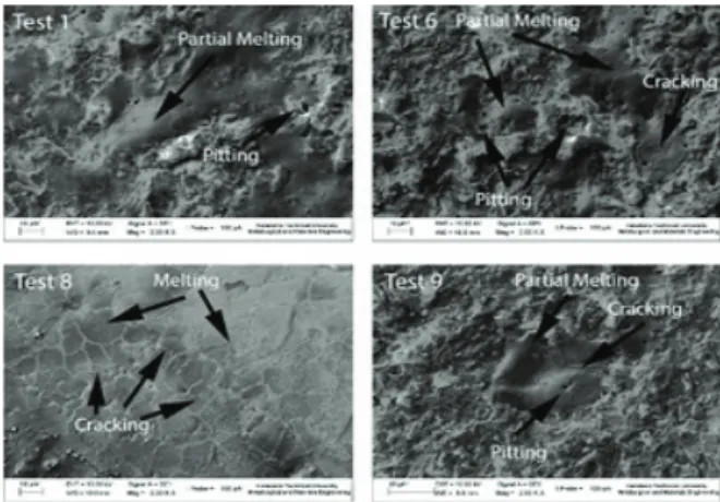
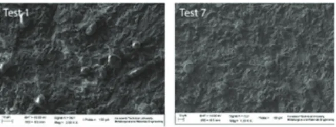
Images were taken for assessment of changes on the surface of titanium discs after laser application with Er,Cr:YSGG. Magnification of 2000 was used for viewing under SEM. Assessment of these images revealed that the most remarkable surface changes were evident in the group Test 8. Flattening of the surface was seen in conjunction with melting, scratch and pitting. Melting and pitting on the surface of discs were less noteworthy in the other test groups.
In the group Test 1, melting and scarce number of pits were detected compared to the control group. No cracks were seen on the surface. Test 2 exhibited melting and roughness on the
Cihan UYSAL, Esra ERCAN, Levent KARA, Tuna ARIN
5
Aydın Dental - Year 3 Issue 1 - April 2017 (1-10)
surface. In Test 3, only melting was evident on the surface. Test 4 demonstrated melting and pitting in addition to a few cracks.
Test 5 displayed mild degrees of melting on the surface compared to the control group. In Test 6, melting and pits were observed on the surface but there were no cracks. Test 7 demonstrated a few pits on the surface. Test 8 has shown remarkable melting and deep scratches on the surface. Test 9 exhibited only a few findings consistent with melting and other findings were similar to the control group. Consequently, experimental groups else than group Test 8 yielded similar findings with the control group in terms of macro changes in surface morphology (Fig. 2, 3) (Table 3).
Figure 2. SEM images of some of the test groups
Figure 3. SEM image of control group
Table 3. Morphological Changes on Ti dics surfaces of Test Groups
Test Groups Morphological Changes Test 1 Surface melting and a few pits Test 2 Surface melting and roughness Test 3 Melting
Test 4 Melting, pitting, and a few cracks Test 5 Mild melting
Test 6 Melting, pitting , and a few cracks Test 7 A few pits
Test 8 Marked melting and deep scratches Test 9 Partial melting, cracking and pitting Table 4 presents surface roughness (Ra) after laser application. In groups Test 1 and Test 7, Ra values were similar to that of control group which has not been exposed to laser. No statistically significant difference was noted between control group and test groups with respect to Ra values.
Table 4. Mean Ra Values
Ra (nm) Test 1 1.320±0.48 Test 2 1.129±0.52 Test 3 1.279±0.47 Test 4 1.252±0.96 Test 5 1.230±0.33 Test 6 1.189±0.84 Test 7 1.302±0.27 Test 8 1.120±0.37 Test 9 1.237±0.22 Control 1.360±0.96
Evaluation of cellular morphology after an incubation of 48 hours, SaOs-2 cells were attached on the surface in a similar fashion with the control group. In this aspect, no
Er,Cr:YSGG Laser as a Surface Detoxification Method in Enhancement of Osseointegration differences were detected between test groups
and the control group.
Moreover, cells having spindle-like structures were encountered in test and control groups (Fig. 4, 5).
Figure 4. Cellular morphology of some of the test groups
Figure 5. Cellular morphology of control group
Proliferation values at the end of 24 hours is demonstrated in Table 5. There was no statistically significant difference between test groups and control in terms of proliferation indicators (p≥0.05).
Proliferation values at the end of 48 hours is presented in Table 6. In this aspect, Test 1 and Test 7 displayed remarkably higher proliferation values compared to the control group. There was no statistically significant difference between the control group and
other test groups. Comparison of proliferation in two different periods is established in Table 5 and 6.
Table 5. Proliferation Values at the end of 24 hours
Groups OD values Sig.(2-tailed)
Test 1 0.322±0.003 p≥0.05 Test 2 0.369±0.002 p≥0.05 Test 3 0.224±0.003 p≥0.05 Test 4 0.268±0.002 p≥0.05 Test 5 0.348±0.003 p≥0.05 Test 6 0.376±0.006 p≥0.05 Test 7 0.359±0.001 p≥0.05 Test 8 0.368±0.001 p≥0.05 Test 9 0.373±0.003 p≥0.05 Control 0.345±0.002 *p˂0.05 is statistically difference Table 6. Proliferation Values at the end of 48
hours
Groups OD values Sig.(2-tailed)
Test 1 0.536±0.013 p=0.029* Test 2 0.538±0.034 p≥0.05 Test 3 0.519±0.011 p≥0.05 Test 4 0.467±0.008 p≥0.05 Test 5 0.466±0.006 p≥0.05 Test 6 0.506±0.015 p≥0.05 Test 7 0.564±0.011 p=0.021* Test 8 0.514±0.016 p≥0.05 Test 9 0.508±0.017 p≥0.05 Control 0.480±0.014 *p˂0.05 is statistically difference
Cihan UYSAL, Esra ERCAN, Levent KARA, Tuna ARIN
7
Aydın Dental - Year 3 Issue 1 - April 2017 (1-10)
DISCUSSION
Effective cleansing of the implant surface, avoidance of production of heat on the surface and maintenance of surface biocompatibility are key points for a successful osseointegration process in the healing period during periimplantitis treatment8, 11-13.
Few reports have been published on the use of Er,Cr:YSGG in the treatment of periodontitis14-20. No consensus exists on the standardization of the measures such as power, frequency and distance of application and only 2 studies have focused on all of these variables19, 20.
Miller et al. suggested that decontamination of titanium surface with Er,Cr:YSGG laser was a reliable and effective method 12. Interestingly, they reported no change was observed in the surface morphology despite the high power (6 W) and long duration (3 minutes) of application. This difference may be attributed to the variability in type of tip used and duration of the procedure. In addition, no data is available on the distance of laser application in that trial. On the same topic, Ercan et al. proposed that the most important determinant affecting the changes on surface morphology was intensity of power (W/mm2) and power is directly influenced by the distance19,20. They concluded that optimization of laser protocols was crucial for establishment of safe and effective treatment regimen besides preservation of the surface titanium discs.
Kreisler et al. have investigated the impacts of Er:YAG laser on implant surfaces21. Consequently, not only decontamination was satisfactory for 3 types of titanium discs at 2 energy levels, but also no excessive heat
formation was observed nor there were any remarkable changes on surface morphology. In harmony with their study design, we contaminated sterile implant surfaces with bacteria (S.aureus) to simulate the clinical situation. However, we did not perform any biofilm only put bacteria on the surface and wait approximately for an hour. We only want to benefit the presence of bacteria on surface because we know that any cells found on the surface will absorb the some of the laser energy. We did not perform any microbiological analysis and this is one of the limitations of this study. The microbiological analysis of the discs after laser treatment will be another important study.
One of the most remarkable results of our study is that; the groups test 1 and test 7 had similar Ra values with control group and the proliferations were statistically significantly increased in comparison to the control group at the end of 48 hours. Surface roughness is important osseointegration and many studies have been conducted to investigate the effects of the impacts of surface roughness on proliferation and morphology of osteoblasts 22-25. Wenneberg et al. have classified Ra values and they determined Ra<0.5 μm as smooth, Ra between 0.5-1 μm as minimally rough, Ra between 1-2 μm as moderately rough and Ra>2 μm as rough22. They claimed that the most appropriate Ra value for reosseointegration was detected in moderately rough (1-2 μm) surfaces. In agreement with this data, Shalabi et al. reported that contact of bone implant was improved on moderately rough surfaces23. The Ra value during detoxification procedure, may has a critical role in cellular proliferation. Alternatively, this increase in proliferation was attributed to the settlement of osteoblasts into the pits formed after laser application on the surface and surface energy may be
Er,Cr:YSGG Laser as a Surface Detoxification Method in Enhancement of Osseointegration changed. Another reason may be dampening
due to flattening of sharp and pointed edges allowing easier attachment of osteoblasts on larger surfaces. However, more studies with long time follow-up periods are needed to determine the factors that affect the osteoblast behavior.
Park et al. employed 3 different types of laser (Er,Cr:YSGG, Er:YAG, CO2) at various power levels (1, 2, 3, 4 and 5 W) to titanium discs and assessed the changes on surface 17. Their results demonstrated that Er,Cr:YSGG should be used maximally at a level of 3 W. Therefore, we have determined the maximal level of power in our study as 3 W.
In 2007, Huang et al. employed Er,Cr:YSGG laser at different energy levels (125 j/mm2 and 10 j/mm2) on titanium discs polished for improvement of initial biocompatibility14. The distance between the laser tip and titanium surface was set as 2 mm. Melting and surface roughness were found to be increased with higher levels of power applied on discs. In both experimental groups, CPI (cell proliferation index) was statistically significantly increased compared to the control group (p<0.001), while there was no difference between two experimental groups. Evaluation of cellular morphology at the end of first day demonstrated that spindle like cellular morphology of cell bodies in the group receiving 190 j/m2 was more evident compared to control group14. These findings may imply that Er,Cr:YSGG laser amplifies surface biocompatibility. In our study, since no difference was determined between experimental groups and control group at the end of first day with respect to cellular proliferation, laser application seems not to have any adverse effects on cellular proliferation.
In another study published by Ayobian et al. in 2015, impacts of Er:YAG laser on roughness, dampening and biocompatibility of titanium discs with SLA surface26. Evaluation of cellular proliferation in SaOs-2 culture at the end of 5 days revealed that cellular proliferation was more prominent in the group receiving laser treatment. Results of this study are in harmony with our data since we noted that Ra values were lower and cellular proliferation was significantly increased in groups Test 1 and Test 7 at the end of 48 hours.
Limitation of the present study is lack of evaluation of cellular activity. Long term trials focusing on proliferation, adhesion and cellular activity are warranted for understanding cellular biocompatibility and osteoblast cell behaviors in periimplantitis after detoxification.
Acknowledgments:
The authors report no conflict of interest. The authors thanks to Dr. Kurtuluş Buruk for his supports during microbiological part.
REFERENCES
[1] Mombelli A, Decaillet F. The characteristics of biofilms in peri-implant disease. J Clin Periodontol. 2011;38 Suppl 11:203-13. [2] Salvi GE, Furst MM, Lang NP, Persson
GR. One-year bacterial colonization patterns of Staphylococcus aureus and other bacteria at implants and adjacent teeth. Clin Oral Implants Res. 2008;19:242-8.
[3] Okayasu K, Wang HL. Decision tree for the management of periimplant diseases. Implant Dent. 2011;20:256-61.
[4] Persson LG, Mouhyi J, Berglundh T, Sennerby L, Lindhe J. Carbon dioxide laser and hydrogen peroxide conditioning in the treatment of periimplantitis: an experimental study in the dog. Clin
Cihan UYSAL, Esra ERCAN, Levent KARA, Tuna ARIN
9
Aydın Dental - Year 3 Issue 1 - April 2017 (1-10) 9
Implant Dent Relat Res. 2004;6:230-8. [5] Renvert S, Lindahl C, Roos Jansaker
AM, Persson GR. Treatment of peri-implantitis using an Er:YAG laser or an air-abrasive device: a randomized clinical trial. J Clin Periodontol. 2011;38:65-73.
[6] Romanos GE, Gupta B, Yunker M, Romanos EB, Malmstrom H. Lasers use in dental implantology. Implant Dent. 2013;22:282-8. [7] Javed F, Hussain HA, Romanos GE.
Re-stability of dental implants following treatment of peri-implantitis. Interv Med Appl Sci. 2013;5:116-21.
[8] Schwarz F, Rothamel D, Sculean A, Georg T, Scherbaum W, Becker J. Effects of an Er:YAG laser and the Vector
ultrasonic system on the biocompatibility of titanium implants in cultures of human osteoblast-like cells. Clin Oral Implants Res. 2003;14:784-92.
[9] Aoki A, Sasaki KM, Watanabe H, Ishikawa I. Lasers in nonsurgical periodontal therapy. Periodontol 2000. 2004;36:59-97.
[10] Asghar A, Abdul Raman AA, Daud WM. A comparison of central composite
design and Taguchi method for optimizing Fenton process. Scientific Worl Journal. 2014;2014:869120.
[11] Schwarz F, Sculean A, Romanos G, Herten M, Horn N, Scherbaum W, et al.
Influence of different treatment approaches on the removal of early plaque biofilms and the viability of SAOS2
osteoblasts grown on titanium implants. Clin Oral Investig. 2005;9:111-7.
[12] Miller RJ. Treatment of the contaminated implant surface using the Er,Cr:YSGG laser. Implant Dent. 2004;13:165-70. [13] Azzeh MM. Er,Cr:YSGG laser-
assisted surgical treatment of peri-implantitis with 1-year reentry and 18-month follow-up.
J Periodontol. 2008;79:2000-5. [14] Huang HH, Chuang YC, Chen ZH,
Lee TL, Chen CC. Improving the initial biocompatibility of a titanium surface using an Er,Cr:YSGG laser-powered hydrokinetic system. Dent Mater. 2007;23:410-4.
[15] Natto ZS, Aladmawy M, Levi PA, Jr., Wang HL. Comparison of the efficacy of
different types of lasers for the treatment of peri-implantitis: a systematic review. Int J Oral Maxillofac Implants. 2015;30:338-45.
[16] Romanos G, Crespi R, Barone A, Covani U. Osteoblast attachment on titanium disks after laser irradiation. Int J Oral Maxillofac Implants. 2006;21:232-6. [17] Park JH, Heo SJ, Koak JY, Kim SK,
Han CH, Lee JH. Effects of laser irradiation on machined and anodized titanium disks. Int J Oral Maxillofac Implants.
2012;27:265-72.
[18] Schwarz F, Nuesry E, Bieling K,
Herten M, Becker J. Influence of an erbium, chromium-doped yttrium, scandium, gallium, and garnet (Er,Cr:YSGG) laser on the reestablishment of the
biocompatibility of contaminated titanium implant surfaces.
J Periodontol. 2006;77:1820-7.
[19] Ercan E, Arin T, Kara L, Candirli C, Uysal C. Effects of Er,Cr:YSGG laser irradiation on the surface characteristics of titanium discs: an in vitro study. Lasers Med Sci. 2014;29:875-80.
[20] Ercan E, Candirli C, Arin T, Kara L, Uysal C. The effect of Er,Cr:YSGG laser irradiation on titanium
discs with microtextured surface morphology. Lasers Med Sci. 2015;30:11-5.
[21] Kreisler M, Kohnen W, Marinello C, Gotz H, Duschner H, Jansen B, et al. Bactericidal effect of the Er:YAG laser on dental implant surfaces: an in vitro study. J Periodontol.
Er,Cr:YSGG Laser as a Surface Detoxification Method in Enhancement of Osseointegration 2002;73:1292-8.
[22] Wennerberg A, Albrektsson T. Effects of titanium surface topography on bone integration: a systematic review. Clin Oral Implants
Res. 2009;20 Suppl 4:172-84.
[23] Shalabi MM, Gortemaker A, Van’t Hof MA, Jansen JA, Creugers NH. Implant surface roughness and bone healing: a systematic review. J Dent Res. 2006;85:496-500. [24] Shibli JA, Grassi S, de Figueiredo LC,
Feres M, Marcantonio E, Jr., Iezzi G, et al. Influence of implant surface topography on early osseointegration: a histological study in human jaws. J Biomed Mater Res B Appl Biomater. 2007;80:377-85.
[25] Sul YT. The significance of the surface properties of oxidized titanium to the bone response: special emphasis on potential biochemical bonding
of oxidized titanium implant. Biomaterials. 2003;24:3893-907.
[26] Ayobian-Markazi N, Karimi M, Safar-Hajhosseini A. Effects of Er: YAG laser irradiation on wettability, surface roughness, and biocompatibility of SLA titanium surfaces: an
11
© 2017 Published by Istanbul Aydın University, Faculty of Dentistry. All rights reserved Aydın Dental - Year 3 Issue 1 - April 2017 (11-16)
FOCAL EPITHELIAL HYPERPLASIA (HECK’S
DISEASE) TREATED WITH USING A DIODE
LASER
Murat ÖZLE1, Sercan KÜÇÜKKURT2, Gizem DİMİLİLER1, Burcu SENGUVEN3, Sedat ÇETİNER1
1 Gazi Üniversitesi Diş Hekimliği Fakültesi, Ağız, Diş ve Çene Cerrahisi, ABD 2 İstanbul Aydın Üniversitesi Diş Hekimliği Fakültesi, Ağız, Diş ve Çene Cerrahisi, ABD 3 Gazi Üniversitesi Diş Hekimliği Fakültesi, Oral Patoloji, ABD
ABSTRACT
Focal epithelial hyperplasia (FEH) or Heck’s disease is an uncommon asymptomatic proliferation of oral mucosa. It usually presents with multiple asymptomatic and occasionally painful exophytic papules or nodules on the buccal mucosa, gingiva, tongue or lips. Although the lesions may resolve after months or years with or without any specific treatment, they may also indefinitely persist or frequently recur. These lesions can be surgically removed if the lesions are exposed to occlusal trauma or the patient has aesthetic concerns. A 35-year-old female patient was admitted to our clinic with the complaint of multiple nodules on her lip and buccal mucosa. After an intraoral examination, we detected multiple, soft, pink, non-inflammatory, and exophytic 2 to 8 mm papules on the oral mucosa including buccal mucosa and lower lip. Histopathologic examination of the incisional biopsy of a papule confirmed the diagnosis of focal epithelial hyperplasia. Oral lesions were treated with serial partial excisions using diode laser. After 24 months of a follow-up period, there were no signs of recurrence. Although Heck’s disease is generally seen in childhood and adolescence, a rare case of FEH seen in a female adult patient who has got treatment with diode laser and 24 months of follow-up.
Keywords: Focal Epithelial Hyperplasia, Heck’s Disease, Human Papillomavirus (HPV), Diode Laser.
ÖZET
Fokal epitelyal hiperplazi (FEH) veya Heck hastalığı, oral mukozanın nadir görülen, asemptomatik bir proliferasyonudur. Genellikle bukkal mukoza, gingiva, dilde veya dudaklarda birden fazla sayıda, çoğu zaman asemptomatik ancak zaman zaman ağrılı ekzofitik papüller veya nodüller olarak görülür. Lezyonlar herhangi bir tedaviye bağlı olarak ya da olmadan aylar ya da yıllar içerisinde düzelebilse de, süresiz olarak devam edebilir ya da sık sık tekrarlanabilirler. Bu lezyonların, oklüzal travmaya maruz kalacağına karar verilirse veya hastada estetik endişelere neden olursa cerrahi olarak çıkarılabilirler. Kliniğimize 35 yaşında bir kadın hasta, dudağı ve bukkal mukozasında birden fazla nodül yakınması ile başvurdu. Ağız içi muayenesinde, sol bukkal mukoza ve alt dudak olmak üzere çoklu, yumuşak, pembe, inflamatuar, ekzofitik, 2-8 mm’lik papüller görüldü. Papüllerden birinin eksizyonel biyopsisinin histopatolojik incelemesi FEH tanısını teyit etti. Teşhis konulmasının ardından oral lezyonlar, diyot lazer kullanılarak seri eksizyonlarla tedavi edildi. 24 aylık takip sonrasında nüks bulgularına rastlanmamıştır. FEH büyük oranda çocukluk ve ergenlik döneminde görülen bir hastalık olmasına rağmen, bu vaka raporunda, erişkin bir kadın hastada teşhis edilen FEH, diode lazer ile tedavi edilip ve izleyen 24 aylık süreçte takibi yapılmıştır.
Anahtar Kelimeler: Fokal Epitelyal Hiperplazi, Heck Hastalığı, İnsan İapillomavirüs (HPV), Diode Lazer
Focal Epithelial Hyperplasia (Heck’s Disease) Treated With Using a Diode Laser
INTRODUCTION
Focal epithelial hyperplasia (FEH) or Heck’s disease is a rare asymptomatic proliferation of oral mucosa, which was first diagnosed by Archard and Heck in 1965 in the Native American population.1 The incidence of the disease ranges from 0.002% to 35% depending on the population and the investigated geographical area.2 It affects mostly children and young adults regardless gender difference.3 Recently, FEH has been also described as multifocal papillomavirus epithelial hyperplasia.4
Focal papillomavirus epithelial hyperplasia is strongly associated with human papilloma virus (HPV) infection; DNA of HPV was detected in 80.3% of FEH lesions.5 The etiologic agent of FEH was first described in 1983, as HPV 13 related to HPV 6 and HPV 11.6 In the following years, another HPV strain was isolated from FEH and similarly named as HPV 32 in relation to HPV 6 and HPV 13.7 Although the diagnosis of FEH can sometimes be made by clinical examination, ethnicity and social anamnesis, the biopsy is still the gold standard for definitive diagnosis.8 In the diagnosis, histological evaluation is important and it allows the identification of the characteristics of lesion such as hyperparakeratosis, acanthosis, elongation and anastomosis of the rete ridges, classical koilocyte, perinuclear cytoplasmic halos and nuclear dysplasia.9-11 FEH is usually detected clinically as exophytic soft papules or nodules in the gingiva, tongue or lips in the same color as the neighboring healthy mucosa, and mostly asymptomatic but rarely painful, smoothly restricted on the cheek mucosa.9, 12-14 Lesions may recover within months or years
without requiring any treatment, but some cases may not improve and recur repeatedly. Nevertheless, FEH has a benign nature. For this reason, it may not always require treatment. However, exophytic lesions can be surgically removed because of an occlusal trauma or aesthetic concerns.3, 9
This study reports a case of focal epithelial hyperplasia in a middle-aged Turkish woman, which occurred on her lips and cheek mucosa, and its treatment with diode laser excision and her 24 months of follow-up.
CASE REPORT
A 35-year-old female patient was admitted to our department with the diagnosis of multiple nodules on her lips and buccal mucosa. The patient reported that painless and soft consistency lesions has been spontaneously developed about two years ago. According to the anamnesis, the patient had no systemic disease other than oral complaints, and no history of allergies or hereditary disease. Moreover, the patient stated that no one from her close family members have had similar oral lesions
Since such oral lesions may be oral symptoms of HIV infection, additional laboratory tests have been required in order to detect and eliminate the HIV infection and other sexually transmitted diseases. According to test results, patient’s routine blood tests were within the normal limits. Anti-HIV, VDRL and FT-ABS tests were negative, however only Anti-HBc IgG and Anti-HBs tests were positive.
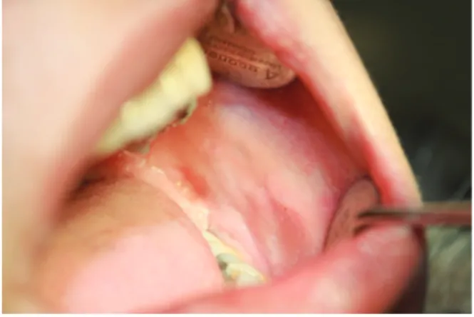
Intraoral examination revealed multiple, soft, pink, inflammatory, exophytic, 2-8 mm papules on the left buccal mucosa (Fig. 1). Radiographic examination revealed that there
Murat ÖZLE, Sercan KÜÇÜKKURT, Gizem DİMİLİLER, Burcu SENGUVEN, Sedat ÇETİNER
13
Aydın Dental - Year 3 Issue 1 - April 2017 (11-16)
was no hard tissue involved in the lesion area.
Figure 1. Pre-operative intraoral image of the patient
Under local anesthesia, a few of the lesions were removed by excisional biopsy with the help of scalpel in order to distinguish the lesions from other similar pathological lesions. In order to protect the wounded areas from infection, the patient was prescribed a mouthwash containing 0.12% chlorhexidine that should be used 3 three times a day. After the excisional biopsy, postoperative proposals were made.
Biopsy specimen with dimensions between 1 and 3 mm was sent to Gazi University, Faculty of Dentistry, Department of Oral Pathology. Specimens were embedded in paraffin after the routine procedures and sections had been taken with a microtome and with a thickness of 0,5μ and then, stained with Hematoxylin-Eosin (H&E) and examined under light microscope. In the histological examination, we detected oral epithelium and acanthosis in the confusion. Furthermore, areas with coilocitas cells and cells with mitotic-like nuclear degeneration (mitosoid cells) were observed. The basal layer was not damaged and chronic inflammatory infiltration has been found in the connective tissue. Based on the
clinical and histopathologic features, a clinical diagnosis of FEH was made.
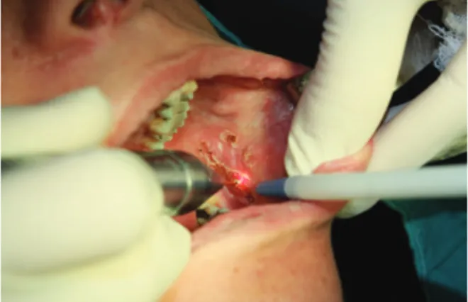
Oral lesions were treated with serial partial excision using diode laser. (Fig. 2) The diode laser was applied with excitation wavelengths of 810 nm, 4.0 W power, 0.5 MS continuous wave and 1000 Hz frequency power.
Figure 2. Excision of the lesion with diode laser
No suture was applied after the excision and the area was left for secondary healing (Fig. 3)
Focal Epithelial Hyperplasia (Heck’s Disease) Treated With Using a Diode Laser No complications were observed during a
routine follow up period following the heal-ing period. No recurrence occurred after 24 months of follow-up (Fig. 4).
Figure 4. Intraoral image after 24 months
DISCUSSION
HPV is a type of virus that infects skin and mucous membranes and can induce the formation of both benign and malignant tumors. Infection begins when the virus enters the host from micro-breaks.10 FEH is a benign infectious disease caused by HPV and its symptoms can be only seen in the mouth. FEH is most commonly found in Eskimos, North and Central American Indians.9,12 In addition, various cases have been reported among some ethnic groups in the countries such as Spain, Netherlands, Brazil, South Africa, and in Turkey.2,3,15
The correct diagnosis is achieved by screening other benign oral and oropharyngeal lesions associated with HPV, such as squamous cell papilloma, condyloma acuminata, common warts and malignant oral lesions, especially squamous cell carcinoma.10 Diagnosis can be made based on the clinical, histological and cytological features of the lesions and can be supported by immunohistochemical assays
which detect the presence of HPV types 13 and 32, molecular biological tests such as in situ hybridization, Southern blot and PCR (Polymerase chain reaction).16 Although the etiology of FEH is associated with a virus, it is considered to be a genetic predisposition because it is a well-known trend in some ethnic groups.11,13,15 In our case, the possibility of genetic transmission was not taken into account because no other case could be identified in the family and the ethnic origin of the family was not one of the trending origins. Major histologic features of FEH include epidermal acanthosis and parakeratosis, thickening and extension of rete ridges, and marked koilocytosis with characteristic mitosoid numbers.17 These lesions may persist for a few years, but they do not become malignant and most tend to regress spontaneously.17 FEH is seldom associated with immunodeficiency or suppression of the immune system.18 In our case, the patient’s immune status was normal. However, FEH is an oral manifestation of HIV infection, so it is the primary duty of clinicians to make an early diagnosis of the disease and to bear the probability of HIV infection in mind.19 In our case, the possibility of such infectious blood diseases has been eliminated with the blood tests.
FEH diagnosis is based on clinical, histopathological and HPV genotypes. It is important to make an early diagnosis of FEH, because there are more serious diseases which should be considered as differential diagnosis such as inflammatory fibrous hyperplasia, inflammatory papillary hyperplasia, verruciform xanthoma, verrucous carcinoma, Cowden’s disease, condyloma acuminatum and focal dermal hypoplasia syndrome.4,14 The first three types of lesions mentioned above
Murat ÖZLE, Sercan KÜÇÜKKURT, Gizem DİMİLİLER, Burcu SENGUVEN, Sedat ÇETİNER
15
Aydın Dental - Year 3 Issue 1 - April 2017 (11-16)
are reactive lesions. Therefore, an irritant agent must be detected as an indication of the presence of lesions. In our case, no irritant factor has been identified like removable prosthesis or malocclusion which can cause occlusal trauma. Verrucous carcinoma is a neoplasia of a different age group with epidemiological features typically found in oral carcinomas. Cowden’s disease fibroepithelial polyps, which indicate characteristics of an older age group, have more consistent, less mobile and different intraoral topography.20,21
Condyloma acuminatum is important for differential diagnosis because the clinical appearance of isolated lesions is similar for both diseases, as they are both HPV-derived.22 Nevertheless, the medical history of the patient is very useful for differential diagnosis. In this case, FEH lesions were more flat and numerous than HPV lesions. In addition, the location of FEH lesions (lip, tongue, and buccal mucosa) is very characteristic.4 Considering the age of the patient, medical history, blood tests, cytological examinations, and the clinical appearance of the lesions, FEH was considered as a preliminary diagnosis. The lesions are removed with a scalpel only if they are exposed to occlusal trauma during mouth function or if the patient has requested the removal upon aesthetic concerns.5,15 Other treatment options include cryotherapy, electrocoagulation, laser, chemical agents such as retinoic acid, and immunostimulants such as interferon.15,23 In our case, the excision was performed with the help of a diode laser, because primary closure of the lesion would not be possible to after excision with a scalpel and also using diode laser could provide an easier hemorrhage control after the excision. Recurrence is not predictable, therefore,
follow-up of patients is essential. When the symptoms of the disease recur, it is uncertain whether there is a new infection or it is caused by a viral delay or the weak immune system of the patient.10 The patient in this case was followed up for 24 months and no recurrence occurred during this period. Yet, the patient should be monitored further.
REFERENCES
[1] Archard HO, Heck JW, Stanley HR. Focal Epithelial Hyperplasia: An Unusual Oral Mucosal Lesion Found in Indian Children. Oral Surg Oral Med Oral Pathol. 1965;20:201-12.
[2] Liu N, Li Y, Zhou Y, Zeng X. Focal epithelial hyperplasia (Heck’s disease) in two
Chinese females. Int J Oral Maxillofac Surg. 2012;41:1001-4.
[3] Ozden B, Gunduz K, Gunhan O,
Ozden FO. A Case Report of Focal Epithelial Hyperplasia (Heck’s disease) with
PCR Detection of Human Papillomavirus. J Maxillofac Oral Surg. 2011;10:357-60. [4] Borborema-Santos CM, Castro MM,
Santos PJ, Talhari S, Astolfi-Filho S. Oral focal epithelial hyperplasia: report of five cases. Braz Dent J. 2006;17:79-82. [5] Flaitz CM. Focal epithelial hyperplasia:
a multifocal oral human papillomavirus infection. Pediatr Dent. 2000;22:153-4. [6] Pfister H, Hettich I, Runne U,
Gissmann L, Chilf GN. Characterization of human papillomavirus type 13 from focal epithelial hyperplasia Heck lesions. J Virol. 1983;47:363-6.
[7] Beaudenon S, Praetorius F, Kremsdorf D, Lutzner M, Worsaae N, Pehau-Arnaudet G, et al. A new type of human papillomavirus associated with oral focal epithelial
hyperplasia. J Invest Dermatol. 1987;88:130-5.
Focal Epithelial Hyperplasia (Heck’s Disease) Treated With Using a Diode Laser [8] Said AK, Leao JC, Fedele S, Porter SR.
Focal epithelial hyperplasia - an update. J Oral Pathol Med. 2013;42:435-42. [9] Dos Santos-Pinto L, Giro EM, Pansani
CA, Ferrari J, Massucato EM, Spolidorio LC. An uncommon focal epithelial hyperplasia manifestation. J Dent Child (Chic). 2009;76:233-6. [10] Castro TP, Bussoloti Filho
I. Prevalence of human papillomavirus (HPV) in oral cavity and oropharynx. Braz J Otorhinolaryngol. 2006;72:272-82. [11] Gonzalez LV, Gaviria AM, Sanclemente G, Rady P, Tyring SK, Carlos R, et al. Clinical, histopathological and virological findings in patients with focal epithelial hyperplasia from Colombia. Int J Dermatol. 2005;44:274-9.
[12] Morrow DJ, Sandhu HS, Daley TD. Focal epithelial hyperplasia (Heck’s disease) with generalized lesions of the gingiva. A case report. J Periodontol. 1993;64:63-5.
[13] Ledesma-Montes C, Vega-Memije E, Garces- Ortiz M, Cardiel-Nieves M, Juarez-
Luna C. Multifocal epithelial
hyperplasia. Report of nine cases. Med Oral Patol Oral Cir Bucal. 2005;10:394-401. [14] Terezhalmy GT, Riley CK, Moore WS. Focal
epithelial hyperplasia (Heck’s disease). Quintessence Int. 2001;32:664-5.
[15] Martins WD, de Lima AA, Vieira S. Focal epithelial hyperplasia (Heck’s disease): report of a case in a girl of Brazilian Indian descent. Int J Paediatr Dent. 2006;16:65-8.
[16] Jayasooriya PR, Abeyratne S, Ranasinghe AW, Tilakaratne WM. Focal epithelial q hyperplasia (Heck’s disease): report
of two cases with PCR detection of human papillomavirus DNA. Oral Dis. 2004;10:240-3. [17] Cuberos V, Perez J, Lopez CJ, Castro
F, Gonzalez LV, Correa LA, et
al. Molecular and serological evidence of the epidemiological association of HPV 13
with focal epithelial
hyperplasia: a case-control study. J Clin Virol. 2006;37:21-6.
[18] Steinhoff M, Metze D, Stockfleth E, Luger TA. Successful topical treatment of focal epithelial hyperplasia (Heck’s disease) with interferon-beta. Br J Dermatol. 2001;144:1067-9.
[19] Puriene A, Rimkevicius A, Gaigalas M. Focal zepithelial hyperplasia: Case report. Stomatologija. 2011;13:102-4.
[20] Reddy KV, Anusha A, Maloth KN, Sunitha K, Thakur M. Mucocutaneous manifestations of Cowden’s syndrome. Indian Dermatol Online J. 2016;7:512-5.
[21] Wright DD, Whitney J. Multiple hamartoma syndrome (Cowden’s syndrome): case report and literature review. Gen Dent.
2006;54:417-9.
[22] Pringle GA. The role of human papillomavirus in oral disease. Dent Clin North Am. 2014;58:385-99. [23] Durso BC, Pinto JM, Jorge J, Jr., de
Almeida OP. Extensive focal
epithelial hyperplasia: case report. J Can Dent Assoc. 2005;71:769-71.
17
© 2017 Published by Istanbul Aydın University, Faculty of Dentistry. All rights reserved Aydın Dental - Year 3 Issue 1 - April 2017 (17-20)
A MULTIDISCIPLINARY ORTHODONTIC
CASE: ORTHODONTICS, PERIODONTICS,
IMPLANT AND PROSTHODONTICS
Fatma YILDIRIM1, Orhan AKSOY1, Utku Gaye DİKME GÜVELİ2, Erol AKIN3
1 Istanbul Aydın University, Faculty of Dentistry, Department of Orthodontics 2 Istanbul Aydın University, Faculty of Dentistry, Department of Periodontics 3 Private Practice, Orthodontist
ABSTRACT
Diastemas are frequent orthodontic problems caused by multiple etiologies which can be physiologic and dentoalveolar like missing tooth, peg lateral, supernumerary teeth, prominent frenulum, proclination of the labial dental segment; or self-inflicted pathologies. The continuing presence of an anterior diastema in adults, has often been considered as an aesthetic problem. In this case, a multidisciplinary treatment of an adult patient who had a missing tooth and a prominent upper labial frenulum, which includes orthodontics, periodontics, implants and prosthetics will be presented.
Keywords: Diastema, Labial Frenulum, Multidisciplinary Treatment
ÖZET
Diastemalar; diş kaybı, kama lateral, süpernümerer diş, belirgin frenulum ve labial dental segmentin proklinasyonu gibi fizyolojik nedenlerle veya patolojik sebeplere bağlı olarak sık görülen ortodontik problemlerdir. Yetişkinlerde anterior diastemanın varlığı, sıklıkla estetik bir sorun olarak kabul edilir. Bu vakada, yetişkin bir hastada, diş kaybına ve belirgin labial frenuluma bağlı ortaya çıkan diastemanın; ortodontik, periodontal, implant ve protetik işlemlerini içeren multidisipliner bir tedavisi sunulacaktır.
Anahtar Kelimeler: Diastema, labial frenulum, multidisipliner tedavi
A Multidisciplinary Orthodontic Case: Orthodontics, Periodontics, Implant and Prosthodontics
INTRODUCTION
A space between adjacent teeth is called a diastema. Diastemas are frequent orthodontic problems caused by multiple etiologies which can be physiologic and dentoalveolar like missing tooth, peg lateral, supernumerary teeth, prominent frenulum, proclination of the labial dental segment; or self-inflicted pathologies.1 Midline diastema refers to anterior midline spacing between the two central incisors. Midline diastema occurs in approximately 98% of 6 year olds, 49% of 11 year olds and 7% of 12–18 year olds.2 It is a part of normal dental development during mixed dentition which is called ugly duckling stage by Broadbent.4 It is a self-correcting anomaly after the eruption of the permanent canines. The continuing presence of a midline diastema is not physiologic and considered as an aesthetic problem in adults. Numerous etiologies have been proposed for maxillary midline diastema including tooth size or jaw size discrepancies, parafunctional habits, tooth loss, periodontal disease, deep bites, aberrant labial frenulum attachments and maxillary midline pathologies. 3, 5-12
The frenulum is a mucous membrane fold that attaches the lip and the cheek to the alveolar mucosa, the gingiva, and the underlying periosteum3. The aberrant labial frenulum is a problem which creates midline diastema and it can be treated by frenectomy or by frenotomy procedures. These procedures are technically different from each other. Frenectomy is the complete removal of the frenulum, including its attachment to the underlying bone, while frenotomy is the incision and the relocation of the frenal attachment 3.
A healthy and attractive smile is very important in today’s society. Many patients
seek closure of a diastema for aesthetic reasons. Many treatment options have been used, varying from restorative procedures to surgery (frenectomy) and orthodontics. A carefully developed differential diagnosis allows the practitioner to choose the most effective treatment objectives. In this case, a multidisciplinary treatment of an adult patient who had a missing tooth and a prominent upper labial frenulum, which includes orthodontics, periodontics, implants and prosthetics will be presented.
CASE REPORT
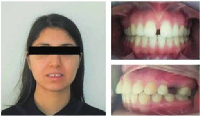
A 26 year old female reported to the orthodontic clinic with the chief complaint of upper jaw midline diastema. She has Angle Class I malocclusion and diastemas due to missing upper left second premolar teeth and prominent upper labial frenulum attachment. In lateral cephalometric examination, we determined skeletal Class I relationship and normal vertical pattern. The patient’s past medical and dental history were not contributory; but in family history, she noted that her sister also had upper prominent labial frenulum and this information led us to think that genetic factors can be contributing factor in diastema etiology. (Fig. 1)
Fatma YILDIRIM, Orhan AKSOY, Utku Gaye DİKME GÜVELİ, Erol AKIN
19
Aydın Dental - Year 3 Issue 1 - April 2017 (17-20)
Multidisciplinary treatment which includes orthodontics, periodontics, implants, and prosthetics was planned for the case. Orthodontic treatment was started at first and the midline diastema was closed with fixed bonded braces and mechanics. The space caused by tooth extraction was maintained for dental implant procedures. Before the finishing phase, when the upper midline diastema closed, patient was directed to the periodontology department. Because of the width and thickness of the frenulum attachment frenecomy was chosen instead of frenotomy. Frenectomy was carried out by periodontist under local anesthesia with incision using No. 15 Bard Parker blade. Sutures were placed and periodontal pack was placed for a week. Healing was successful and pleasant. (Fig. 2, 3)
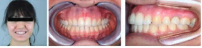
Figure 2. The photographic records before upper labial frenectomy and after healing.
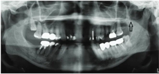
Figure 3. The panoramic radiographs after orthodontic debonding and implant surgery. After these procedures, braces were removed and fixed lingual retainers were bonded
canine to canine on lower ach and between first premolars on upper arch. Implant surgery was performed and a week later sutures were removed. A removable Hawley appliance used for retention until the osseointegration process was complete. After osseointegration, the patient was rehabilitated with crown prosthesis. At the end of the treatment, the upper third molars were extracted. (Fig. 4)
Figure 4. The photographic records after 1 year retention period.
DISCUSSION
In the recent times, due to increasing expectations of the patients for an esthetically and functionally stable treatment result, an interdisciplinary treatment will yield the best result for the patient and the clinicians. Evaluating the patient history prior to treatment is vital for the treatment planning. In this case, we determine the upper labial frenulum as an etiological cause of upper midline diastema. Patient also stated that her sister has a similar midline diastema hence it suggests that genetic factors may be contributing factor in this etiology. Gaas et al.13 reported that, heritability of maxillary midline diastemas was estimated to be %32 in white population and % 0,4 in black population. According to their study, the pedigree data suggest an autosomal dominant mode of inheritance for maxillary midline diastema and a possible genetic influence in the expression of maxillary midline diastema in both the black and the white sample populations. In this case, the diastema was closed orthodontically and then frenectomy
A Multidisciplinary Orthodontic Case: Orthodontics, Periodontics, Implant and Prosthodontics performed. We should not do frenectomy
prior to orthodontic treatment because the scar tissue can inhibit the tooth movement. In these types of cases, the fixed retainers are recommended to curb the high possibility of relapse.14
In the treatment of midline diastema and other diastemas due to missing tooth, a multidisciplinary approach is necessary to achieve a successful outcome. All specialists should set realistic treatment objectives in order to meet the needs of the patient.
CONCLUSION
Today, many adults demand orthodontic treatment to improve their appearance and dental health. Multidisciplinary treatment approaches should be offered to these patients. Orthodontic procedures are routinely used to close diastemas; however more stable and aesthetic results can be achieved with interdisciplinary treatments which includes orthodontics, periodontics, implants, and prosthetics.
REFERENCES
[1] Hussain, U., Ayub, A., & Farhan, M. (2013). Etiology and treatment of midline diastema: A review of literature. Pakistan Orthodontic Journal, 5(1), 27-33.
[2] Foster TD, Grundy MC. Occlusal changes from primary to permanent dentitions. J Ortho. 1986; 13: 187–93.
[3] Devishree, Gujjari SK, Shubhashini PV. Frenectomy: A Review with the Reports of Surgical Techniques. Journal of Clinical and Diagnostic Research :
JCDR. 2012; 6(9):1587-1592.
[4] Broadbent BH: The face of the normal child (diagnosis, development).
Angle Orthod 1937; 7:183-208.
[5] Angle EH. Treatment of malocclusion of the teeth. 7th ed. Philadelphia: S.S. White Dental Manufacturing Co; 1907. [6] Sicher H. Oral anatomy. 2nd ed. St. Louis:
C. V. Mosby; 1952.
[7] Gardiner JH. Midline spaces. Dent Pract Dent Rec 1967; 17:287-97.
[8] Edwards JG. The diastema, the frenum, the frenectomy: a clinical study. Am J Orthod 1977; 71:489-508.
[9] Sullivan TC, Turpin DL, Artun J.
A postretention study of patients presenting with a maxillary median diastema.
Angle Orthod 1996; 66:131-8.
[10] Tait CH. The median frenum of the upper lip and its influence on the spacing of the upper central incisor teeth. Dent Cosmos
1934; 76:991-2.
[11] James GA. Clinical implications of a follow-up study after frenectomy. Dent Pract Dent Rec 1967; 17:299-305.
[12] Oesterle LJ, Shellhart WC. Maxillary midline diastemas: a look at the causes. J Am Dent Assoc 1999; 130:85-94.
[13] Gass, J. R., Valiathan, M., Tiwari, H. K., Hans, M. G., & Elston, R. C. (2003). Familial correlations and heritability of maxillary midline diastema. American journal of orthodontics and dentofacial orthopedics, 123(1), 35-39.
[14] De Morais, J. F., de Freitas, M. R., de Freitas, K. M. S., Janson, G., & Castello Branco, N. (2014). Postretention stability after
orthodontic closure of maxillary interincisor diastemas. Journal of Applied Oral Science, 22(5), 409–415.