See discussions, stats, and author profiles for this publication at: https://www.researchgate.net/publication/332207442
The Frequency Of Laryngeal Involvement In Lipoid Proteinosis Patients
Article · April 2019 DOI: 10.5455/annalsmedres.2019.01.059 CITATIONS 0 READS 23 7 authors, including:Some of the authors of this publication are also working on these related projects:
Working on thesisView project
A cese of erythema multiforme developing after levetiracetam therapyView project Ahmet Yükkaldiran
Şanlıurfa eğitim araştırma hastanesi 7 PUBLICATIONS 20 CITATIONS
SEE PROFILE
Yavuz Yeşilova
Special Lokman Physician Hospital, Van, TurkeySpecial Lokman Physician Hospital, … 86 PUBLICATIONS 368 CITATIONS SEE PROFILE Ismail Iynen Harran University 22 PUBLICATIONS 138 CITATIONS SEE PROFILE İsa an 47 PUBLICATIONS 17 CITATIONS SEE PROFILE
DOI: 10.5455/annalsmedres.2019.01.059
The frequency of laryngeal involvement in lipoid
proteinosis patients
Ahmet Yukkaldiran1, Mustafa Aksoy2, Yavuz Yesilova3, Mahmut Demir4, Ismail Iynen5, Isa An6, Osman Tanrikulu7
1Sanliurfa Training and Research Hospital, Department of Otorhinolaryngology, Sanliurfa Turkey 2Harran University, Faculty of Medicine Department of Dermatology, Sanliurfa, Turkey
3Special Lokman Physican Hospital, Department of Dermatology, Van, Turkey 4Harran University, Faculty of Medicine Department of Pediatrics, Sanliurfa, Turkey
5Harran University, Faculty of Medicine Department of Otorhinolaryngology, Sanliurfa, Turkey 6Sanliurfa Training and Research Hospital, Department of Dermatology, Sanliurfa, Turkey 7Special Batman Life Hospital, Department of Dermatology, Batman, Turkey
Copyright © 2019 by authors and Annals of Medical Research Publishing Inc.
Abstract
Aim: Lipoid proteinosis (LP) is a rare autosomal recessive disorder that occurs with the accumulation of hyaline substance in oral
cavity, laryngeal mucosa and skin. The aim of this study was to investigate the incidence of laryngeal involvement in patients with LP which have hoarseness and skin lesions, and the areas of hyaline substance accumulation in the larynx.
Material and Methods: In this study, 21 patients who were admitted to our Dermatology outpatient clinic between January 2013 and
December 2017 and diagnosed as LP with punch biopsy were evaluated. Laryngeal imaging was performed in otorhinolaryngology clinic, and the frequency of larynx involvement and the areas of hyaline accumulation in the larynx were evaluated.
Results: Twenty-one patients with hoarseness and diagnosed by skin biopsy as LP were included in the study. Thickening of
the vocal cords in 21 patients (100%); thickening and hypertrophic changes of arytenoids in 13 patients (61.9%); thickening and hypertrophic changes of the interarytenoid area in 10 patients (47.6%); thickeningand hypertrophic changes of epiglottis in 3 (14.3%) patients; hypertrophic changes and thickening of the aryepiglottic fold in 3 patients (14.3%); thickening and hypertrophic changes of ventricular folds in 2 patients (9.5%) were observed.
Conclusion: In our study, similar to the data in the literature, the pathological changes were most frequently seen in the vocal cords
and the least in the ventricle band, aryepiglottic fold and epiglottis. In order to better understand the laryngeal involvement and the areas of involvement in LP patients, studies with more patients are needed.
Keywords: Arytenoid; Larynx; Lipoid Proteinosis; Vocal Cord.
Received: 30.01.2019 Accepted: 27.03.2019 Available online: 03.04.2019
Corresponding Author: Isa An, Sanliurfa Training and Research Hospital, Department of Dermatology, Sanliurfa, Turkey E-mail: [email protected]
1
INTRODUCTION
Lipoid proteinosis (LP), also known as Urbach-Wiethe disease or hyalinosis cutis et mucosa, is a rare, autosomal recessive genetic disease characterized by deposition of amorphous hyaline substance in the skin, oral mucosa, larynx, eyelids and internal organs (1,2). The incidence and prevalence of LP are not fully known. Up to 300 patients have been described in the literature so far (1,3,4,5). LP cases are increasing gradually in our country due to consanguineous marriages which are a social problem in rural areas of Turkey (5-8).
Skin lesions usually occur in the first two years of life. Laryngeal involvement is recognized immediately after birth as hoarseness or scrawniness in the crying voice
and is usually the first symptom of the disease. Therefore, it is very important to recognize the main features of this disease as otorhinolaryngologists (9-11).
The aim of this study was to investigate the incidence of laryngeal involvement in patients with LP which have hoarseness and skin lesions, and the areas of hyaline substance accumulation in the larynx.
MATERIAL and METHODS
In this study, 21 patients who were admitted to our Dermatology outpatient clinic between January 2013 and December 2017 and were diagnosed as LP with punch biopsy were evaluated. Laryngeal imaging was performed in otorhinolaryngology clinic, and the frequency of larynx involvement and the areas of hyaline accumulation in
Ann Med Res 2019;
the larynx were evaluated. In the larynx imaging of the patients, a 3ccd full hd camera system with a 1920 *(X) 1080 pixel image capture and a lateral-sighted, wide angle 70 degree rigid telescope with a diameter of 4 mm and a length of 18 mm were used. For the study, ethics committee numbered 4300 and dated 14.05.2018 was approved by the ethics committee of Harran University Medical Faculty Hospital. Written informed consent was obtained from the parents of children and adult patients themselves. Statistical analyzes were performed using the SPSS 21.0 for Windows (SPSS Inc., Chicago, IL, USA) package program. Demographic data were expressed as frequency, mean and percentage.
RESULTS
The study included 21 patients with hoarseness and skin lesions who were diagnosed as LP by skin biopsy (Figure 1). Thirteen (61.9%) of the patients were female and 8 (38.1%) were male. The mean age of the women was 13.9 ± 8.04 and the mean age of men was 17.3 ± 8.38.
Figure 1A. Hyalinized material (arrows) is observed around the
vessels in the papillary dermis at the sections (H & Ex200). B) Cross sections show hyalinized material (arrows) positively stained with PAS around the vessels in the papillary dermis (PASx400)
Dermatologic examination revealed common acneiform scars in 21 (100%) patients, pearly papules in 1 (4.7%) and palmoplantar hyperkeratosis in 1 (4.7%) patient. In the laryngoscopic examination of 21 patients, vocal cord thickening was present in all patients and only 7 (33.3%) patients had isolated vocal cord thickening. In addition, thickening and hypertrophic changes of arytenoids and interarytenoid area in 5 (23.8%) patients; thickening and hypertrophic changes of epiglottis, arytenoids, aryepiglottic fold, interarytenoid area and ventricular folds in 2 (9.5%) patients; thickening and hypertrophic changes of arytenoids in 4 (19%) patients; thickening and hypertrophic changes of epiglottis, arytenoid and interarytenoid area in 1 (4.8%) patient; thickening and hypertrophic changes of arytenoids, aryepiglottic fold, interarytenoid area in 1 (4.8%) patient; thickening and hypertrophic changes of the interarytenoid area in 1 (4.8%) patient were observed.
When all patients were evaluated, thickening of the vocal cords in 21 (100%) patients; thickening and hypertrophic changes of arytenoids in 13 (61.9%) patients; thickening and hypertrophic changes of the interarytenoid area in 10 (47.6%) patients; thickening and hypertrophic changes of the epiglottis in 3 (14.3%) patients; thickening and hypertrophic changes of the aryepiglottic folds in 3
(14.3%) patients; thickening and hypertrophic changes of the ventricular folds in 2 (9.5%) patients were observed. (Table 1), (Figure 2-4).
Table 1. Demographic findings and larynx examination findings of lipoid proteinosis patients
Age(mean±Sd) Female 13.9±8.04
Male 17.3±8.38
Sex FemaleMale 13 (61.9%)8 (38.1%)
Larynx examination findings
Thickening-hypertrophic changes
of epiglottis 3 (14.3%) Thickening of vocal cords 21 (100%) Thickening-hypertrophic changes of arytenoids 13 (61.9%) Thickening-hypertrophic changes of interarytenoid area 10 (47.6%) Thickening-hypertrophic changes of aryepiglottic folds 3 (14.3%) Thickening-hypertrophic changes of ventricular folds 2 (9.5%)
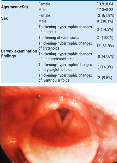
Figure 2. Thickening of the vocal cords and hypertrophic changes
in the interarytenoid area are seen
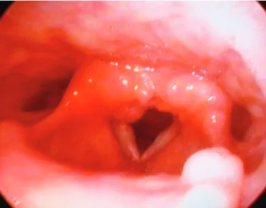
Figure 3. Thickening of the vocal cords, epiglottis, aryepiglottic
fold, interarytenoid area, arytenoid, hypertrophic changes and thickening of the ventricular folds
Figure 4. Thickening of the vocal cords, thickening and
hypertrophic changes in interarytenoid area and arytenoid
DISCUSSION
LP is a rare, disease characterized by accumulation of amorphous hyaline substance in the skin, oral mucosa and larynx. This disease, which affects both sexes equally, is caused by a decrease in the expression of the extracellular matrix protein 1 (ECM1) gene in the first chromosome. To date, more than 50 mutations have been reported on the 1q21 chromosome in the ECM1 gene. The ECM1 gene plays an important role in wound healing by affecting angiogenesis, epidermal differentiation, dermal collagen and proteoglycans. The loss of normal function of ECM1 in LP leads to infiltration with hyaline-like material in the skin, mucosa and internal organs (4,12).
The main symptom of LP is usually hoarseness, which is recognized at birth but may occur in later years. This symptom is often confused by clinicians with more common diseases such as chronic laryngitis. The cause of hoarseness is the deterioration of wave formation due to the accumulation of subepithelial hyaline material and incomplete closure of the vocal cords with air leakage during vocalization (13-16).
While infiltration in the tongue limits the movement of it, infiltration in the salivary glands may cause recurrent parotitis attacks. Dental anomalies are in the form of hyperplasia or aplasia of upper cutters, premolar and molars. Patients usually lose their teeth at an early age. Neurological findings occur as seizures and less frequently as behavior disorders. Calcifications can be seen in bilateral temporal lobes (3,6,8,9).
Skin lesions occur in two periods. In the first period, it is in the form of vesicles and hemorrhagic bullae in areas such as face, lip and limb which are open to trauma. In the second period, diffuse thickening, papules, plaques and nodules are caused by hyaline infiltrations in dermis. Face, axilla and scrotum are the most common sites. Verrucous lesions can be observed in the elbows (6,8,9).
The most prominent finding in histopathology is the accumulation of hyaline material around the dermis and vessels. Hyperkeratosis is seen in the epidermis and basal membrane thickening is seen in the dermoepidermal junction. Concentric layers of basement membrane-like material include type 2 and type 4 collagen and laminin. In addition, hyaline accumulations are PAS positive, indicating the presence of neutral mucopolysaccharides (1,3,5).
The treatment of LP is symptomatic. Despite the presence of various treatment alternatives, including dimethyl sulfoxide, oral corticosteroids, D-penicillamine, acitretin, carbon dioxide laser and surgical intervention, there is no single effective treatment for LP (3,17-20). Although LP is rarely a life-threatening condition, death due to respiratory obstruction has been reported in the literature. These complications can be prevented by early diagnosis of the disease and proper management of respiratory diseases (21). It has been shown that removal of lesions using microlaryngoscopy instruments improves airway and sound quality. (22,23).
The cases of LP in the literature are usually published in dermatology journals and the dermatological features of the disease have been emphasized (24-26). Savage et al. reviewed the literature on otolaryngologic findings of LP patients and reported that hoarseness occurred in congenital or early infancy in about two-thirds of cases. It has been reported that hoarseness is a direct result of hyaline accumulation in the vocal cords and most skin lesions occur a few years after the onset of sound changes (21).
Hofer et al. found hoarseness at birth in 26 of 27 patients diagnosed with LP. Changes in vocal cords were described in 75% of reported cases and epiglottic changes were defined in 45% of cases. Laryngeal lesions ranged from minor irregularities at the edges of the vocal cords to vegetative lesions in the larynx requiring tracheotomy (28). In the study conducted by Xu et al. with 22 patients with LP, hoarseness was reported as the first symptom in all patients. In 14 patients, the onset of hoarseness was seen in the neonatal period, and videolaryngoscopic examination revealed yellowish papules scattered along the edges of the vocal cord (29). Dertlioglu et al. evaluated 10 LP patients with hoarseness, and in the indirect laryngoscopic examination of 9 patients, the vocal cords that were thickened and had decreased movement were detected(5). In the video laryngoscopic examination of four siblings with LP, Acar et al. found two siblings with thickened epiglottis, edematous arythenoid and aryepiglotic folds, while they found a general thickened mucosal and bilateral edematous vocal cords in the larynx of other two siblings(9). Oz et al. found irregular vocal cords due to yellowish papules in two cases with LP but did not detect any pathology in epiglottis (16).
In our study, all of the LP patients had hoarseness. Thickening of the vocal cords in 21 patients (100%); thickening and hypertrophic changes of arytenoids in 13 3
Ann Med Res 2019;
patients (61.9%); thickening and hypertrophic changes of the interarytenoid area in 10 patients (47.6%); thickening and hypertrophic changes of epiglottis in 3 (14.3%) patients; hypertrophic changes and thickening of the aryepiglottic fold in 3 patients (14.3%); thickening and hypertrophic changes of ventricular folds in 2 patients (9.5%) were observed.
CONCLUSION
In our study, similar to the data in the literature, the pathological changes were most frequently seen in the vocal cords and the least in the ventricle, aryepiglottic fold and epiglottis. In order to better understand the laryngeal involvement and the areas of involvement in LP patients, studies with more patients are needed.
Acknowledgements
We would like to thank Dr.Muhammet Emin Güldür from Harran Unıversity Department of Pathology for his help
Ahmet Yukkaldiran ORCID: 0000-0002-1335-4110 Mustafa Aksoy ORCID:0000-0002-4966-1737 Yavuz Yesilova ORCID: 0000-0002-4177-1501 Mahmut Demir ORCID: 0000-0002-0983-9457 Ismail Iynen ORCID: 0000-0002-5937-3720 Isa An ORCID: 0000-0003-3366-4551
Osman Tanrikulu ORCID: 0000-0002-5755-0594
Competing interests: The authors declare that they have no competing interest.
Financial Disclosure: There are no financial supports
Ethical approval: For the study, ethics committee numbered 4300 and dated 14.05.2018 was approved by the ethics committee of Harran University Medical Faculty Hospital.
REFERENCES
1. Mcgrath JA. Lipoid proteinosis. Handb Clin Neurol 2015;132:317-22.
2. Urbach E, Wiethe C. Lipoidosis cutis et mucosae. Virchows Arch A Pathol (Pathol Anat) 1929;273:285-319.
3. Desmet S, Devos SA, Chan I, et al. Clinical and molecular abnormalities in lipoid proteinosis. Eur J Dermatol 2005;15:344-6.
4. Gao D, Ma X, Lian P, et al. Pathogenetic mechanism of lipoid proteinosis caused by mutation of the extracellular matrix protein 1 gene. Mol Med Rep 2018;17:8087-90.
5. Dertlioğlu SB, Çalık M, Çiçek D. Demographic, clinical, and radiologic signs and treatment responses of lipoid proteinosis patients: a 10-case series from Şanlıurfa. Int J Dermatol 2014;53:516-23.
6. Baykal C, Topkarci Z, Yazganoglu KD, et al. Lipoid proteinosis: a case series from Istanbul. Int J Dermatol 2007;46:1011-6. 7. Güvener G, Kocabay C, Huq GE, et al. Lipoid proteinosis in
the mouth and laryngeal structures: a case report. Kulak Burun Bogaz Ihtis Derg 2007;17:105-7.
8. Kartal D, Çınar SL, Kartal L, et al. Lipoid proteinosis. Acta
Dermatovenerol Alp Pannonica Adriat 2016;25:19-21. 9. Acar A, Eryilmaz A, Gocer C, et al. Lipoid proteinosis of
larynx: review of four cases. Int J Pediatr Otorhinolaryngol 2004;68:1557-61.
10. Sriwastava S, Desai A, Yuliati A, et al. Child with a hoarse cry and intracranial calcification. Pediatr Neurol 2018;87:78-9. 11. Koganti D. Urbach-wiethe disease: a rare cause of
hoarseness of voice. Int J Phonosurg Laryngol 2013;3:61-4. 12. Bai X, Liu JW, Ma DL. Novel mutations in extracellular matrix
protein 1 gene in a chinese patient with lipoid proteinosis. Chin Med J (Engl) 2016;129:2765-6.
13. Ally M, Kinshuck AJ, Sandison A, et al. The management of laryngeal lipoid proteinosis. J Laryngol Otol 2018;132:936-9.
14. Loos E, Kerkhofs L, Laureyns G. Lipoid proteinosis: a rare cause of hoarseness. J Voice 2019;33:155-8.
15. Karaman H, Soyuer I, Külahcı İ, et al. Lipoid proteinosis (Urbach-Wiethe’s Disease) Kulak Burun Bogaz Ihtis Derg 2015;25:357-60.
16. Oz F, Kalekoğlu N, Karakullukçu B, Lipoid proteinosis of the larynx. J Laryngol Otol 2002;116:736-9.
17. Kaya TI, Tursen U, Kokturk A, et al. The early erosive vesicular stage of lipoid proteinosis: clinical and histopathological features. Br J Dermatol 2003;148:380-2.
18. Kaya TI, Kokturk A, Tursen U, et al. D-penicillamine treatment for lipoid proteinosis. Pediatr Dermatol 2002;19:359-62. 19. Gruber F, Manestar D, Stasic A, et al. Treatment of
lipoid proteinosis with etretinate. Acta Derm Venereol 1996;76:154-5.
20. Kroukamp G, Lehmann K. Treatment of laryngeal lipoid proteinosis using CO2 laser. S Afr Med J 2007;97:90-2. 21. Savage MM, Crockett DM, McCabe BF. Lipoid proteinosis
of larynx: a cause of voice change in the infant and young child. Int J Pediatr Otorhinolaryngol 1988;15:33-8.
22. Bozdağ KE, Gül Y, Karaman A. Lipoid proteinosis. Int J Dermatol 2000;39:203-4.
23. Staut CC, Naidich TP. Urbach-Wiethe disease (Lipoid proteinosis). Pediatr Neurosurg 1998;28:212-4.
24. Harper JI, Duguid KP, Staughton RC, Oropharyngeal and laryngeal lesions in lipoid proteinosis. J Laryngol Otol 1983;97:877-80.
25. Aziz MT, Mandour MA, el-Ghazzawi IF, et al. Urbach-wiethe disease in O.R.L. practice. (a clinical and histochemical study of the laryngeal lesions). J Laryngol Otol 1980;94:1309-19. 26. Dıckey RF, Davıs S 2nd. Lipoidd proteinosis (Urbach-wiethe);
report of a case and revıew. Ann Otol Rhinol Laryngol 1964;73:287-96.
27. Hofer PA, Ohman J. Laryngeal lesions in Urbach-Wiethe disease (lipoglycoproteinosis; lipoid proteinosis; hyalinosis cutis et mucosae). A histopathological and clinical study, including direct laryngoscopical examinations. Acta Pathol Microbiol Scand A 1974;82:547-58.
28. Xu W, Wang L, Zhang L, Otolaryngological manifestations and genetic characteristics of lipoid proteinosis. Ann Otol Rhinol Laryngol 2010;119:767-71.