Full Terms & Conditions of access and use can be found at
https://www.tandfonline.com/action/journalInformation?journalCode=tbcp21
Psychiatry and Clinical Psychopharmacology
ISSN: 2475-0573 (Print) 2475-0581 (Online) Journal homepage: https://www.tandfonline.com/loi/tbcp21
Effects of agmatine on cognitive functions during
vascular dementia in biological aging through
eNOS and BDNF expression
Bülent Bağcı, Tijen Utkan, Yusufhan Yazir, Feyza Aricioglu, Gökçe Sevim
Öztürk & Yusuf Sarioglu
To cite this article: Bülent Bağcı, Tijen Utkan, Yusufhan Yazir, Feyza Aricioglu, Gökçe Sevim Öztürk & Yusuf Sarioglu (2017) Effects of agmatine on cognitive functions during vascular dementia in biological aging through eNOS and BDNF expression, Psychiatry and Clinical Psychopharmacology, 27:2, 106-115, DOI: 10.1080/24750573.2017.1309090
To link to this article: https://doi.org/10.1080/24750573.2017.1309090
© 2017 The Author(s). Published by Informa UK Limited, trading as Taylor & Francis Group
Accepted author version posted online: 21 Mar 2017.
Published online: 19 Apr 2017. Submit your article to this journal Article views: 1699
View related articles View Crossmark data
Effects of agmatine on cognitive functions during vascular dementia in
biological aging through eNOS and BDNF expression
Bülent Bağcıa, Tijen Utkanb, Yusufhan Yazirc, Feyza Aricioglud, Gökçe Sevim Öztürkeand Yusuf Sariogluf
a
Gilead Sciences Ilaç Ltd.Şti, Istanbul, Turkey;bDepartment of Pharmacology and Experimental Medical Research and Application Unit, Kocaeli University Faculty of Medicine, Kocaeli, Turkey;cDepartment of Histology and Embryology and Stem Cell and Gene Therapy Research and Application Center, Kocaeli University Faculty of Medicine, Kocaeli, Turkey;dFaculty of Pharmacy, Department of
Pharmacology and Psychopharmacology Research Unit, Marmara University, Istanbul, Turkey;eDepartment of Medical Pharmacology, Gazi University, Medical School, Ankara, Turkey;fIstinye University Faculty of Medicine, Istanbul, Turkey
ABSTRACT
Objective: Biological aging has been recognized to cause impairment of memory and the development of vascular dementia. Based on our previous work, agmatine has been shown to have a beneficial effect and might have therapeutic potential on cognitive functions, including learning and memory. The aim of the present study was to examine the possible effect of agmatine on biological aging-induced vascular endothelial dysfunction and associated dementia in rats.
Methods: We used three different age groups (4-month-olds, 18-month-olds and 24-month-olds; n = 12 in each group) of control and agmatine-treated rats. Control animals received physiological saline for 8 weeks. Agmatine sulfate (40 mg/kg, twice daily) was given to the agmatine groups orally for 8 weeks. Herein, we investigated the effects of agmatine on systolic blood pressure (SBP), nitric oxide (NO)-mediated endothelium-dependent and -independent vasorelaxant responses in thoracic aorta, cognitive performance (passive avoidance test; PAT, and Morris water maze test; MWMT), endothelial nitric oxide synthase (eNOS) expression and both hippocampal and amygdaloid brain-derived neurotrophic factor (BDNF) expression in aged rats.
Results: We found cognitive decline, endothelial dysfunction and reduced eNOS and BDNF expression in aged rats. All these changes may result from aging-induced vascular dementia. We also found that chronic treatment with agmatine may improve amygdala-dependent emotional and spatial learning and memorial performance, and endothelial function, and may regulate eNOS and BDNF protein expression in aged rats.
Conclusion: Results of the current study point out that chronic agmatine treatment may prevent endothelial dysfunction associated with vascular dementia through eNOS and BDNF expression in aged rats.
ARTICLE HISTORY
Received 2 February 2017 Accepted 15 March 2017
KEYWORDS
Aging; agmatine; passive avoidance; Morris water maze; vascular dementia
Introduction
Cognitive decline in biological aging is well known; how-ever, the exact cause of it is unclear. Several lines of evi-dence suggest that vascular causes of dementia are more common in older patients [1,2]. Vascular dementia, in which cerebrovascular pathologies are correlated with cognitive decline, is the most widely recognized type of dementia as well as Alzheimer’s disease [3]. Aging of the cerebrovascular circulation and the effects of vascu-lar changes on the brain are responsible for biological aging of the brain [4,5]. In addition, there is growing evi-dence that damage to the vascular system is related to an increased risk of cognitive decline in aging. Hemody-namic flow has been shown to be disturbed by cardiovas-cular risk factors, such as hypertension, and thus to result in cerebral hypoperfusion [6]. It is well known that chronic brain hypoperfusion can expedite the onset of
cognitive symptoms [7,8]. Previous clinical and preclini-cal researches demonstrate that aging is connected with systemic inflammation, vascular endothelial dysfunction [9], increased production of reactive oxygen species and decreased bioavailability of nitric oxide [10]. Taken together, we can introduce that drugs that improve endothelial dysfunction will have the ability to amelio-rate vascular cognitive deficits in aged individuals. This hypothesis was recently proposed by our study in dia-betic vascular dementia [11].
Agmatine, a putative neurotransmitter, was discov-ered in mammalian brains in 1994 [12], which interacts with several receptor subtypes, such as N-methyl-D-aspartate (NMDA) receptors, and inhibits neuronal and inducible nitric oxide synthase (NOS) (nNOS, iNOS) [13]; however, it stimulates endothelial nitric oxide synthase (eNOS) in the rat brain after cerebral ischemia [14]. Agmatine has a variety of pharmacological
© 2017 The Author(s). Published by Informa UK Limited, trading as Taylor & Francis Group
This is an Open Access article distributed under the terms of the Creative Commons Attribution License (http://creativecommons.org/licenses/by/4.0/), which permits unrestricted use, distribution, and reproduction in any medium, provided the original work is properly cited.
CONTACT Tijen Utkan [email protected] Department of Pharmacology and Experimental Medical Research and Application Unit, Kocaeli University Faculty of Medicine, Kocaeli, Turkey
PSYCHIATRY AND CLINICAL PSYCHOPHARMACOLOGY, 2017 VOL. 27, NO. 2, 106–115
effects in the central nervous system (CNS) such as antic-onvulsant [15], neuroprotective [16–18], anti-stress, anxiolytic and antidepressant activity [19,20] and pre-vents tolerance and attenuates withdrawal signs in mor-phine dependence [21,22], providing analgesia and reducing thermal and mechanic hyperalgesia in a neuro-pathic pain model [23,24]. Recently, endogenous agma-tine has been documented to have a crucial role as a neurotransmitter in the cognitive functions [25–27]. Aging-induced changes in the agmatine level were also found in memory-related structures of brain [28] and age-related cognitive decline and agmatine might be ben-efical to streptozotocin- [29] and scopolamine-induced dementia in rats [30] as well as aging [31,32]. Latest find-ings suggest that agmatine has a crucial role in the pro-cesses of cognition under normal and/or pathological situations. Therefore, herein our study, we examined the possible effect of agmatine on emotional and spatial memory, blood pressure and vascular response in young, middle-aged and aged male rats.
Materials and methods
Experimental animals
In the present study, male Wistar Albino rats aged 4 months old (220–300 g), 18 months old (300–350 g) and 24 months old (400–500 g) have been used. The rats were obtained from Kocaeli University Experimental Medical Research and Application Cen-ter (DETAB, Kocaeli, Turkey). Every behavioral trial was directed from 9:00 am to 12:00 pm under stan-dard conditions (22 ± 2°C room temperature; 12-h light/dark cycle with lights on at 7:00 am). The ani-mals were fed with tap water and food pellets ad libi-tum. Ethical approval was granted by the Kocaeli University Animal Research Ethics Committee (Pro-ject number: HADYEK-5/2-2010).
The rats were randomly divided into groups (n = 12 in each group) such as young (4 months old), middle-aged (18 months old), aged (24 months old) control groups and matching-aged agmatine groups. Control animals received physiological saline for 8 weeks, whereas agmatine sulfate (40 mg/kg, twice daily) was given to the agmatine groups (4 months, 18 months and 24 months) orally by gavage for 8 weeks.
Locomotor activity test
Locomotor movements were evaluated with an animal activity monitoring system (May AMS 9701, Commat Ltd., Ankara, Turkey) including a plexiglass box (40 cm × 40 cm × 30 cm), a computer and open-field activity software. Total locomotor activity was rep-resented as the sum of the vertical, ambulatory and stereotypic activities of the animals for 5 min.
Passive avoidance test
Passive avoidance test (PAT; model 7551, Ugo Basile, Italy) was utilized for the assessment of emotional mem-ory based on contextual fear conditioning, as previously described elsewhere [30]. Briefly, rats learn to avoid a specific place associated with an aversive event. The diminishment of latency to avoid was used as learning. A guillotine door separated two-compartment-contain-ing (light and dark chamber) apparatus was used. The rats were placed in the light chamber after 20 s, the guil-lotine door separating was opened and the initial latency to enter the dark chamber was recorded. When the rats entered the dark chamber, they were given a foot shock of 0.5 mA for 3 s through the grid floor of the dark com-partment. At that point, the rats came back to their home cage. Twenty-four hours later, the retention latency time was measured similarly as in the acqui-sition trial; however, foot shock was not conveyed. The cut-off time was limited to 300 s.
Morris water maze test
A water tank (150 cm in diameter) was used to measure spatial memory as previously described elsewhere [30]. The platform was put in the center of the southwest quadrant and submerged 1.5 cm below the surface of water, and small black plastic balls were put on the water surface. The platform was not changed during the first four days, and latency to discover the platform was determined. Every trial was ended when the rat had climbed onto the escape platform or at the end of 60 s. Each rat was permitted to remain on the platform for 20 s. The rats that could not discover the platform within 60 s were put on the platform and were permitted to stay there for 20 s. A probe trial was utilized to evalu-ate the rat’s spatial retention of the location of the hid-den platform on day 5. During this trial, the platform was removed from the maze and the rat was permitted to search the pool for 60 s in order to spend time in the quadrant that previously contained the hidden plat-form, the so-called target quadrant.
Systolic blood pressure measurement
An automatic blood pressure and heart rate recorder system (May BPHR 9610; Commat Ldt, Ankara, Tur-key) was used to assess indirect systolic blood pressure (SBP) and heart rate (HR). The average value of BP and HR in each rat was obtained from three consecutive inflation–deflation cycles of the cuff.
Organ bath studies
The rats were sacrificed by decapitation and the thor-acic aorta from the aortic arch to the diaphragm was extracted. Vessels were put in Krebs solution, dissected
cautiously and rings were prepared. The rings were placed in 20 ml organ baths containing Krebs solution that were maintained at 37°C by utilizing a thermo-regulated water circuit and aerated continuously with 95% O2 and 5% CO2. The pH of the fluid was 7.4. The rings were equilibrated for 60 min and during this period, the bath solution was changed each 15 min. Resting tension was set at 1 g by repeated adjustment and stayed unaltered throughout the inves-tigation. Each ring was connected to a force-displace-ment transducer (May FDT 10A; Commat Ltd., Ankara, Turkey) for the estimation of isometric force, which was displaced continuously and recorded online on a computer by utilizing a four-channel transducer data acquisition system (MP 30B-CE; Biopac Systems, Santa Barbara, CA, USA), using software (BSL Pro 3.7; Biopac Systems) that could analyze the data.
Agonist-induced contractions
Aortic rings were exposed to 80 mM KCl for 5 min. Tissues were then washed, and phenylephrine (10−9– 10−4M)-induced contractile responses were obtained cumulatively.
Agonist-induced relaxations
Each aortic ring was pre-contracted with a submaximal concentration of phenylephrine (10−6M). These con-centrations resulted in 85–87% of the maximal response to phenylephrine. After the phenylephrine-induced contraction had reached a plateau, the concen-tration–response relationships for carbachol (10−8 -10−5 M), sodium nitroprusside (SNP; 10−8–10−4 M) or papaverine (10−5–10−4 M) were obtained by the addition of one of these agents to the bath in a cumu-lative fashion. The agonist concentration in the bath was incremented approximately threefold at each step after the response to the previous dose had reached a plateau. Between successive concentration response curves, the tissues were rinsed with fresh buffer and they were allowed to recover for 30 min. During this period, tension returned to basal levels.
Immunohistochemical analyses for eNOS and brain-derived neurotrophic factor
Rats were fixed with 10% neutral buffered formalin. After routine histological tissue procedure, the tissues were cut into 3 µm thickness and embedded in paraffin. Routine immunohistological procedure was performed and sections were incubated with polyclonal primary antibody against eNOS (sc654, Santa Cruz, CA, USA) and with secondary biotinylated antibody, streptavidin peroxidase and diaminobenzidine (DAB) solution.
The primary rabbit polyclonal anti-brain-derived neurotrophic factor (BDNF) antibody (Santa Cruz Biotechnology, Santa Cruz, CA, USA) was applied overnight at a 1:100 dilution at room temperature.
Negative control samples were prepared by replacing the primary antibody with the antibody diluent sol-ution (Ab-diluent reagent solsol-ution, Invitrogen, Carls-bad, CA, USA) at the same concentration. After several washes, the slides were incubated with a bioti-nylated secondary antibody (Histostain-Plus Kit, Broad Spectrum, Invitrogen, Carlsbad, CA, USA) for 20 min at room temperature, and DAB (DAB Substrate Kit, Invitrogen, Carlsbad, CA, USA) was applied for visualization. The sections were briefly counterstained with Mayer’s hematoxylin (Invitrogen, Carlsbad, CA, USA) and mounted with ClearMount (Invitrogen, Carlsbad, CA, USA) on glass slides.
Both aorta and brain slides were examined under a light microscope (Olympus BX 50, Tokyo, Japan). The photomicrographs were taken with a Leica DM 100 system (Leica DFC 290HD, Wetzlar, Hessen, Germany). All samples were treated following the same protocol. One researcher who was blind to the current study graded the staining intensity based on a semi-quantitative scale ranging from no expression (0) to very weak (1+), moderate (2+), strong (3+) and very strong (4+) expression. The percentage of positive cells was defined as 0, < 5%; 1, 6–15%; 2, 16–50%; 3, 51–80%; and 4, > 80% positive cells.
Measurement of blood glucose
Blood glucose level was detected with a commercial glucose meter and glucose-sensitive dipsticks (Accu-trend-Alpha glucometer, Boehringer, Mannheim, Germany). In brief, blood was withdrawn from the tail vein. A drop of blood put on the glucometer strip was included in the glucometer for the determination of blood glucose level.
Drugs and treatments
Agmatine was dissolved in saline and given orally by gavage in a volume of 0.2 ml per 100 g body weight of the rat. Agmatine (40 mg/kg) was administered twice daily for 8 weeks. Behavioral testing commenced 24 h after the last drug treatment. The control group was received physiological saline (orally by gavage, 0.2 ml/ 100 g). Agmatine sulfate, phenylephrine hydrochloride, carbachol chloride, SNP and papaverine hydrochloride were used (obtained from Sigma Aldrich). Drugs were prepared fresh every day and kept in cold until injected.
Statistical analysis
Data are the mean ± standard error of the mean (SEM). Acquisition (1–4 day) latency scores in the Morris water maze test (MWMT) were measured by two-way analyses of variance (ANOVA). The follow-ing were measured by one-way ANOVA: scores of the time spent in the escape platform quadrant in the
MWMT; first day and retention latencies in PAT scores; total locomotor activity scores and foot shock sensitivity scores. Further statistical analyses for individual groups were carried out using the Bon-ferroni test. In isolated organ bath experiments, con-tractile force is expressed as milligrams of developed tension. The relaxant responses are expressed as the percentage of pre-contraction to phenylephrine. Con-centration–response curves were fitted by nonlinear regression with the simplex algorithm, and maximum responses (Em) and pD2(–log EC50) calculated using the software of the transducer data acquisition sys-tem. Briefly, the cumulative concentration–response curve data were fitted as described previously to a four-parameter logistic equation: E = Em/1 + (EC50/ [D]n), where E denotes the observed effect in grams of tension, [D] denotes the concentration of agonist, Em denotes the calculated maximal effect, EC50 denotes the [D] at 0.5 Em and n is the slope factor parameter. The significances were conducted with one-way ANOVA followed by a post hoc Tukey–Kra-mer test. The immunoreactivity scores were com-pared by the Kruskal–Wallis test following Dunn’s multiple comparison test; P < .05 was considered significant.
Results
Body weight was significantly different between groups (F(5,71)= 199, P < .0001), whereas agmatine treatment has no effect (Table 1).
Behavioral assessment Locomotor activity test
Agmatine administration of 40 mg/kg, twice daily for 8 weeks had no effect on locomotor activity in young and aged groups compared to controls (F(5,71)= 1.572, P = .183) (Table 1). There was no statistical difference in locomotor activity among the different age groups and agmatine-treated matching groups (Table 1).
Passive avoidance test
Retention times were evaluated in order to examine possible changes both by aging and by agmatine treat-ment by using PAT. There were no significant differ-ences between the groups (F(5,71)= 0.1667, P = .9739) on day 1 (training session) (Table 1).
On the second day, however, there was a significant reduction in retention time in 18-month-old and 24-month-old groups compared to controls. There was no significant change between 4-month-old rat responses with or without agmatine treatment, whereas agmatine treatment significantly reversed the reduction in retention time in both 18-month- and 24-month-old groups (F(5,71)=7.377,P < .0001) (Figure 1).
Morris water maze test
All rats exhibited lower latency time during the first 4 days of the test to get to the platform (F(3,264) = 16.90, P < .0001). There was a further statistical difference between groups by aging and by agmatine treatment
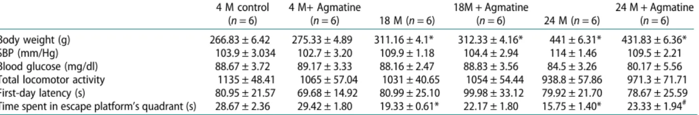
Table 1.Body weights (g), blood glucose (mg/dl) and systolic BP (mm/Hg), locomotor activity, passive avoidance acquisition test phases and MWMT of the time spent in the target quadrant on day 5 of rats in 4- (4 M), 18-(18 M), 24-month-old (24 M) and agmatine-treated age-matching groups.
4 M control (n = 6) 4 M+ Agmatine (n = 6) 18 M (n = 6) 18M + Agmatine (n = 6) 24 M (n = 6) 24 M + Agmatine (n = 6) Body weight (g) 266.83 ± 6.42 275.33 ± 4.89 311.16 ± 4.1* 312.33 ± 4.16* 441 ± 6.31* 431.83 ± 6.36* SBP (mm/Hg) 103.9 ± 3.034 102.7 ± 3.20 109.9 ± 1.18 104.4 ± 2.94 114 ± 1.46 109.5 ± 2.21 Blood glucose (mg/dl) 88.67 ± 3.72 89.17 ± 3.33 88.16 ± 2.47 88.83 ± 3.56 84.5 ± 3.26 80.17 ± 5.56 Total locomotor activity 1135 ± 48.41 1065 ± 57.04 1031 ± 40.65 1054 ± 54.44 938.8 ± 57.86 971.3 ± 71.71 First-day latency (s) 80.95 ± 21.57 69.68 ± 14.92 80.99 ± 25.10 99.98 ± 33.12 79.92 ± 21.70 78.67 ± 25.59 Time spent in escape platform’s quadrant (s) 28.67 ± 2.36 29.42 ± 1.80 19.33 ± 0.61* 22.17 ± 1.80 15.75 ± 1.40* 23.33 ± 1.94#
Note: Values are arithmetic means ± SEM,n = number of animals used.
*P < .05, statistically different from 4-month-old control rats and#P < .05, statistically different from 24-month-old rats.
Figure 1.Passive avoidance retention test phases of young (4-month-old), middle-aged (18-month-old), aged (24-month-old) and agmatine (40 mg/kg)-treated matching groups (n = 12 rats in each group). Data were presented as mean ± SEM and *P < .05 and ***P < .001 compared to the young control group (4-month-old).
(F(5,264 )= 24.82, P < .0001). There was no significant difference between days related to age and treatment (F(15 264)= 0.52,P = .9312) (Figure 2).
There was a significant difference between 18-month and 24-month groups on the 5th day of the experiment in terms of time spent in the escape platform quadrant during the probe trial of the MWMT. Also, agmatine treatment reversed the reduction in the time spent in escape platform’s quadrant back to control values (P < .001) (F(5,71)= 9.293,P < .001) (Table 1).
Blood glucose level estimation
Blood glucose levels were detected in all groups. There was no significant difference in blood glucose levels among the groups (Table 1).
Effect of aging and systemic administration of agmatine on the expression of BDNF protein
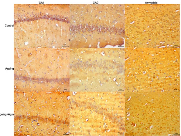
There was a significant decrease in BDNF protein expression in the hippocampal CA1 and CA3 regions in the aged rats (P < .05,Figure 3), whereas treatment with 40 mg/kg agmatine significantly increased the levels of BDNF protein in the hippocampal CA1 and CA3 regions in 24-month-old rats (P < .05,Figure 3). In 24-month-old rats receiving 40 mg/kg agmatine, BDNF protein expressions were similar to those in the control group. Similarly, in the amygdala, BDNF protein levels were significantly lower in the aging rats compared to the control rats (P < .05, Figure 3). In the 40 mg/kg agmatine group, levels of BDNF protein were significantly increased compared with those in the aging group (P < .05,Figure 3andTable 2).
Effects of aging and systemic administration of agmatine on SBP and vascular reactivity
Any significant change was not detected in the SBP of rats (old control = 103.9 ± 3.034 mmHg;
4-month-old control + agmatine = 102.7 ± 3.20; 18-month-4-month-old = 109.9 ± 1.18 mmHg; 18-month-old + agmatine = 104.4 ± 2.94 mmHg; month-old = 114 ± 1.46 mmHg; 24-month-old + agmatine = 109.5 ± 2.21 mmHg; n = 6 for each group;P > .05) (Table 1).
Contractile responses induced with 80 mM KCl did not differ in all groups. No significant difference was observed between the maximum responses of rings of the 4-, 18- and 24-month-old groups, or agmatine-treated rats (P > .05) (Table 3).
Carbachol and SNP induced relaxations in thoracic aortic rings pre-contracted with a submaximal concen-tration of phenylephrine (10−6M). There was no sig-nificant difference in the pre-contractile tone among the groups (Table 3). In rings pre-contracted with phenylephrine at submaximal concentrations, carba-chol (10−8–10−5M) evoked concentration-dependent relaxations. The carbachol-induced endothelium-mediated relaxation was decreased significantly in both the 18-month-old and 24-month-old groups compared with that in the 4-month-old control, agma-tine-treated 4-month-old control and agmaagma-tine-treated 18- and 24-month-old groups (P > .05, Figure 4). The cumulative concentration–response curve for carba-chol was shifted to the right with significantly lower values of Em and pD2 in the 18- and 24-month-old groups than in 4-month controls (P < .05, Table 3 and Figure 4). However, the impairment in the two agmatine treatment groups was returned to that seen in 4-month-old controls. No significant difference was found in the values of pD2 and Em between 4-month-old control and agmatine-treated rats (P > .05, Figure 4andTable 3).
In pre-contracted rings, SNP, an NO donor (10−8– 10−4M), induced concentration-dependent relaxation (Figure 5), but no significant differences were found in the values of Em and pD2 among the groups (Table 3). In addition, the papaverine-induced vasorelaxations were similar for all groups (Table 3).
Effects of aging and systemic administration of agmatine on the expression of eNOS protein
In the 4-month-old rats, eNOS immunopositivity was established in the cytoplasm of the endothelial cells of the aorta (Figure 6(a)). eNOS immunopositivity decreased in the 24-month-old group compared with that in 4-month-old controls (P < .01,Figure 6(b)). In the agmatine-treated aging rats, eNOS immunopositiv-ity was similar to that of the 4-month-old control group (Figure 6(c) andTable 2).
Discussion
This study revealed the behavioral function, endo-thelium-dependent vasorelaxant responses and the
Figure 2.Escape latency (s) in MWMT results of 1–4 days by rats in young (4-month-old), middle-aged (18-month-old), aged (24-month-old) and agmatine (40 mg/kg)-treated match-ing groups (n = 12 rats in each group). Data were presented as mean ± SEM and **P < .01 and ***P < .001 compared to the young control group (4-month-old).
age-induced changes in BDNF and eNOS expressions. Chronic agmatine administration increased the emotional (amygdala-dependent) and spatial learn-ing-memorial (hippocampus-dependent) performance of the aged rats, and maintained the endothelium-dependent vasorelaxant responses. Furthermore, the long-term agmatine treatment significantly prevented the age-related reduction in BDNF immunoreactivity in the hippocampus CA1 and CA3 zones and amyg-dala, and eNOS immunoreactivity in the thoracic
aorta, which may to some extent contribute to correct the behavioral functions in the aged rats.
Aging is an unavoidable process and it is well known that it is one of the causes of cognitive dysfunctions. Learning and memory deficits in aging are mostly related to hippocampal CA1, CA3 and dentate gyrus, (DG) entorhinal, perirhinal, parahippocampal and pre-frontal cortex dysfunctions. Besides, it has been shown in several studies that NO may play a major role in the aging process. It regulates cerebellar blood flow
Figure 3.Representative image illustrating BDNF expression (arrows) in hippocampal formation. Control (a), month (b) and 24-month + agmatine (c) groups in the CA1 region, and control (d), 24-24-month (e) and 24-24-month + agmatine (f) groups in the CA3 region. control (g), 24 month (h) and 24-month + agmatine (i) groups in the amygdala. BDNF expression was decreased in 24 month in the CA1 and CA3 regions of the hippocampus, and amygdala, whereas in 24-month + agmatine rats BDNF expression was similar to those in the control group in the all regions. Scale bars: 50μm.
Table 2.Semi-quantitative distribution of BDNF-immunoreactive neurons of the CA1 and CA3 regions of the hippocampus and amygdala and eNOSimmunoreactivity of aorta in rats.
Hippocampal CA1 Animal 1 Animal 2 Animal 3 Animal 4 Animal 5 Animal 6
4 M 2+ 1+ 2+ 3+ 2+ 3+ 24 M 1+ 1+ 2+ 1+ 2+ 0 24 M + agmatine 2+ 1+ 1+ 3+ 2+ 2+ Hippocampal CA3 4 M 2+ 2+ 3+ 2+ 2+ 2+ 24 M 1+ 2+ 1+ 0 0 2+ 24 M + agmatine 1+ 3+ 2+ 2+ 2+ 2+ Amygdala 4 M 3+ 2+ 3+ 2+ 2+ 3+ 24 M 1+ 1+ 1+ 2+ 2+ 1+ 24 M + agmatine 1+ 3+ 2+ 2+ 3+ 2+ eNOS 4 M 3+ 3+ 2+ 2+ 2+ 3+ 24 M 0 1+ 1+ 2+ 1+ 1+ 24 M + agmatine 2+ 3+ 2+ 2+ 3+ 1+
Note: The staining intensity was classified as no expression (0), very (1+), moderate (2+), strong (3+) or very strong (4+) expression. The immunopositivity was decreased in 24-month-old rats group (24M) compared to the 4-month-old control group (4M) (P < .01, Kruskal–Wallis test). In the agmatine-treated group, both BDNF and eNOS immunoreactivity were similar to that of the control group.
and several other functions at physiological levels. Though it functions as a neuronal messenger and regu-lates the cerebral blood flow at a physiological level, NO is a strong oxidizing agent that is excessively excreted through iNOS, and may cause inflammation, nitro-oxi-dative stress, apoptosis and mitochondrial dysfunction [33,34]. It has been demonstrated that an increase in iNOS expression and nNOS activity or protein expression might result in a number of cognitive dis-orders in aged rats [35–37]. Nevertheless, some other authors reported that the number of NOS-containing neurons reduced in the aged animals, accompanied with a reduction in the NOS activity as well as nNOS and eNOS protein expressions, and no iNOS activity was observed in the memory-related structures [38–43]. It was recently reported that the agmatine levels in the memory-associated cerebral structures demonstrated some changes specific to the age-related zone [39,40,44]. The recent empiric studies evoke that exogen-ously administered agmatine recovers the learning and memory functions, and has a therapeutic potential [45,46]. Both the behavioral and neurochemical useful
effects of agmatine in the aged rats were first reported by Rushaidhi et al. [31]. Agmatine (40 mg/kg once a day, i.p) corrected the spatial working memory and object recognition memory in the aged male rat after 10–16 injections, while the spatial reference memory and exploratory behavior demonstrated no change after a single injection of agmatine [31].
In this study, it was detected that the chronic agma-tine treatment corrected the emotional and spatial mem-ory broken down in the age-related PAT and water maze test, and these results are in compliance with the findings obtained by the previous researchers. For instance, sys-temically or centrally administered agmatine increases various learning and memory performances in naive animals, and ensures protection against the learning and memorial deficits caused by the administered amy-loid beta or scopolamine [41,47–50]. In addition to the behavioral studies, it was reported that chronically admi-nistered agmatine significantly repressed the age-related increase in total NOS activity in dentate gyrus and pre-frontal cortex [31,32].
In this study, chronically administered agmatine did not affect the locomotor activity of the aged rats. The animals were trained to find the hidden platform to determine spatial learning and memory. The retention time to find the platform was adversely affected in the
Figure 4.Carbachol concentration–response curves in isolated thoracic aortic rings pre-contracted with phenylephrine (10−6M). The concentration–response curve for carbachol was shifted to the right with significantly lower values ofEm
and pD2in the 24-month-old group than in 4-month-old
con-trols (*P < .05). Impairment of relaxation in the agmatine treat-ment group was returned to that seen in the controls. Each point is expressed as a percentage of the contraction induced by phenylephrine and is represented as the mean ± SEM. The values in parentheses indicate the number of preparations used; *P < .05, different from the response of tissue rings from 4-month-old young control rats.
Figure 5.Sodium nitroprusside concentration–response curves in isolated thoracic aortic rings pre-contracted with phenyl-ephrine (10−6M). The relaxation response to SNP was similar among vascular tissues from all groups. Each point is expressed as a percentage of the contraction induced by phenylephrine and is expressed as the mean ± SEM. The values in parentheses indicate the number of preparations used.
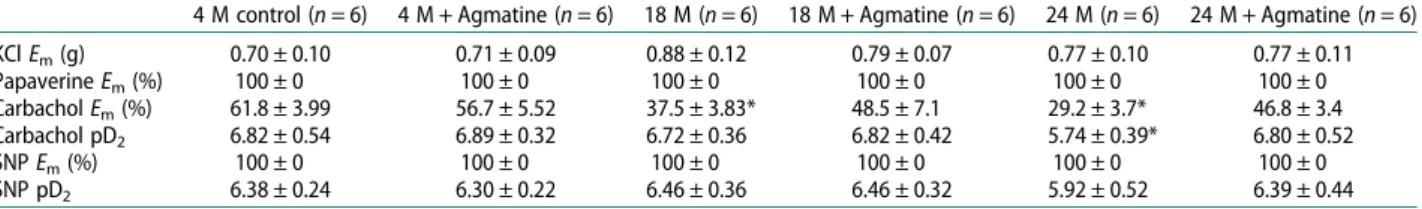
Table 3.Em(% of 10−6M phenylephrine) values for carbachol, sodium nitroprusside and papaverine,Emvalues (g) for 80 mM KCl
and pD2values (–log EC50) for carbachol, and sodium nitroprusside in rings of thoracic aorta obtained from 4- (4 M), 18-(18 M),
24-month-old (24 M) and agmatine-treated age-matching group rats.
4 M control (n = 6) 4 M + Agmatine (n = 6) 18 M (n = 6) 18 M + Agmatine (n = 6) 24 M (n = 6) 24 M + Agmatine (n = 6) KClEm(g) 0.70 ± 0.10 0.71 ± 0.09 0.88 ± 0.12 0.79 ± 0.07 0.77 ± 0.10 0.77 ± 0.11 PapaverineEm(%) 100 ± 0 100 ± 0 100 ± 0 100 ± 0 100 ± 0 100 ± 0 CarbacholEm(%) 61.8 ± 3.99 56.7 ± 5.52 37.5 ± 3.83* 48.5 ± 7.1 29.2 ± 3.7* 46.8 ± 3.4 Carbachol pD2 6.82 ± 0.54 6.89 ± 0.32 6.72 ± 0.36 6.82 ± 0.42 5.74 ± 0.39* 6.80 ± 0.52 SNPEm(%) 100 ± 0 100 ± 0 100 ± 0 100 ± 0 100 ± 0 100 ± 0 SNP pD2 6.38 ± 0.24 6.30 ± 0.22 6.46 ± 0.36 6.46 ± 0.32 5.92 ± 0.52 6.39 ± 0.44
Note: Values are arithmetic means ± SEM.n = the number of thoracic aortic rings used. *Statistical significance wasP < .05.
course of the memoric direction finding, and the exploratory time was reduced and the spatial memory functions were distorted in the platform area, depend-ing on the age compared to the young controls even in the probe test. During the locomotor activity test, no sig-nificant difference was found among the groups. On the last day of the water tank trial, a probe test was carried out to evaluate the performance of the animals in learn-ing the place of the platform. The animals in the two different aged age groups performed the explorative behavior in the quadrant of the secret platform in a shorter time, compared to the young controls, while the agmatine-treated aged animals demonstrated a similar performance to that of the young groups. In the PAT, the first-day performance was identical in all the groups, while the performances evaluated 24 hours later (on the second day) were found to be lowered depending on age. The agmatine-treated aged animals had a latency identical to the young controls on the second day. Considering all the behavioral data, the findings obtained in the present study are consistent with the previously published findings. In consequence, 40 mg/kg chronic agmatine treatment selectively cor-rects behavioral performance in the aged rats [31,32].
Agmatine has been reported to lower blood glucose levels in animal models of diabetes [51]. In our study, blood glucose levels were measured at the end of the treatment and no change in blood glucose levels have been found in any group. Hence, the effect of agmatine on cognitive dysfunction is independent from the glu-cose levels in aged rats.
NO and cGMP mediate physiological effects not only in the central nervous system, but also in the cardiovas-cular system. Reducing the NO-dependent signal trans-duction during biological brain aging may be an important factor in cognitive deficits. In addition to this decrease in the central nervous system, an age-related decrease in eNOS activity was reported, pointing out that the changes in vascular reactivity would likely result in age-related vascular dysfunction [52]. NO has a basic role in the regulation of vascular function [53]. It is reported that aging is closely related to vascular aging, and the vascular structural and functional changes
might be the most important cause of vascular aging [54]. In this study, the endothelium-mediated relaxing responses obtained by carbachol in the isolated aorta strips were significantly reduced depending on age, while no significant change was determined among the groups in terms of the NO-donor SNP relaxing responses. The agmatine-treated aged animals were determined to have such endothelium-dependent relax-ing responses as are comparable to those of the young control group. Likewise, the chronic agmatine treatment restored the reduced eNOS immunoreactivity detected in the aged animals to that of the young control groups. However, no age-related change was detected in the KCI and papaverine responses. In addition to such data, it was demonstrated that eNOS immunoreactivity in the thoracic aorta was reduced in the aged animals com-pared to the young animals, but a long-term agmatine application prevented this reduction. Moreover, in a stress model, down-regulated gene expressions of all stress-induced NLRP3 inflammasome components, such as NLRP3, IL-1β and IL-18, have been shown by exogenously administered agmatine in the prefrontal cortex and hippocampus. Agmatine decreased pro-inflammatory cytokine levels in these brain regions and in serum. In addition, stress decreased IL-4 and IL-10 levels and agmatine treatment restored these anti-inflammatory cytokines to normal in the prefrontal cortex. Therefore, it was claimed that the mechanisms underlying the neuroprotective role of agmatine would inhibit the TNF-α and IL-1β secretion, and clean the reactive oxygen types, or block the intrinsic apoptotic pathway, so that the NOS activity or protein expression might be affected [44,53,55–57].
Furthermore, it was reported that aging causes an increase in the iNOS activity, and a distortion in the eNOS/iNOS balance [58]. It is well known that agmatine plays an important role in regulating the NO production [59–61]. The treatment with agmatine by inhibiting iNOS has a neuroprotective effect, and may correct dis-torted balance. In addition to the age-related increase in iNOS activity, the levels of free oxygen radicals were found to be increased [62]. Agmatine is known to have a strong antioxidant effect, and it’s protective effect on
Figure 6.Representative light microscopy of control, 24-month-old and agmatine-treated 24-month-old groups in the endothelium of the aorta. Decreased eNOS (arrows) (b) immunoreactivity in the aorta in 24-month-old rats compared to 4-month-old controls (a) and increased eNOS (c) immunoreactivity in endothelial tissue in the agmatine-treated month-old group compared to 24-month-old groups (b). Scale bars: 20μm.
the age-related endothelial dysfunction might be explained also with the anti-oxidizing effect of agmatine [56].
BDNF, a member of the neurotrophine family of growth factors, has a crucial role in cognitive functions [63] and is shown to play a critical role in the aging and neurodegenerative process. The mechanism by which aging impairs memory involves decreased BDNF levels. Several lines of findings support this concept. Accord-ingly, our findings support that the significant decrease in BDNF immunoreactivity in the hippocampal CA1 and CA3 zones as well as amygdala of the aged rats in comparison with the young control group provides direct evidence for the role of BDNF in memory deficit. Furthermore, agmatine treatment caused a significant increase in the age-related BDNF reduction in the hip-pocampus and amygdala, thus showing a neuroprotec-tive effect.
Taken together, the findings of this study show that long-term agmatine administration increases the BDNF levels in both the hippocampus and amygdala, and also peripherally the NO synthesis and/or bioavail-ability, and corrects the age-related endothelial dys-function, and hence may help in recovering vascular aging and vascular dementia.
Disclosure statement
No potential conflict of interest was reported by the authors.
Funding
This study was supported by the Grant from the Gazi Üni-versitesi Research Fund [project number: 01/2010-80] and presented at the 27th European College of Neuropsycho-pharmacology Congress, 18–21 October 2014, Berlin, Germany. The funders had no role in the study design, data collection and analysis, decision to publish, or prep-aration of the manuscript.
References
[1] Rincon F, Wright CB. Vascular cognitive impairment. Curr Opin Neurol.2013;26:29–36.
[2] Ramos AR, Dib SI, Wright CB. Vascular dementia. Curr Transl Geriatr Exp Gerontol Rep.2013;2(3):188–195. [3] Sudduth TL, Powell DK, Smith CD, et al. Induction of
hyperhomocysteinemia models vascular dementia by induction of cerebral microhemorrhages and neuroin-flammation. J Cereb Blood Flow Metab.2013;33:708–715. [4] Kennedy KM, Raz N. Aging White matter and cogni-tion: differential effects of regional variations in diffu-sion properties on memory, executive functions, and speed. Neuropsychologia.2009;47:916–927.
[5] Akinyemi RO, Mukaetova-Ladinska EB, Attems J, et al. Vascular risk factors and neurodegeneration in ageing related dementias: Alzheimer’s disease and vascular dementia. Curr Alzheimer Res.2013;10(6):642–653. [6] de la Torre JC. Cardiovascular risk factors promote brain
hypoperfusion leading to cognitive decline and demen-tia. Cardiovasc Psychiatry Neurol.2012;2012:367516.
[7] Feldstein CA. Association between chronic blood pressure changes and development of Alzheimer’s dis-ease. J Alzheimers Dis.2012;32:753–763.
[8] Shah NS, Vidal JS, Masaki K, et al. Midlife blood pressure, plasma beta-amyloid, and the risk for Alzheimer’s disease: Honolulu Asia Aging Study. Hypertension.2012;59:780–789.
[9] Miralbell J, Soriano JJ, Spulber G, et al. Structural brain changes and cognition in relation to markers of vascular dysfunction. Neurobiol Aging. 2012;33 (5):1003.e9–17.
[10] Toth P, Tarantini S, Tucsek Z, et al. Resveratrol treat-ment rescues neurovascular coupling in aged mice: role of improved cerebromicrovascular endothelial func-tion and downregulafunc-tion of NADPH oxidase. Am J Physiol Heart Circ Physiol.2014;306:H299–H308. [11] Utkan T, Yazir Y, Karson A, et al. Etanercept improves
cognitive performance and increase eNOS and BDNF expression during experimental vascular dementia in streptozotocin-induced diabetes. Curr Neurovasc Res.
2015;12(2):135–146.
[12] Li G, Regunathan S, Barrow CJ, et al. Agmatine: an endogenous clonidine-displacing substance in the brain. Science.1994;263(5149):966–969.
[13] Auguet M, Viossat I, Marin JG, et al. Selective inhi-bition of inducible nitric oxide synthase by agmatine. Jpn J Pharmacol.1995;69:285–287.
[14] Mun CH, Lee WT, Park KA, et al. Regulation of endo-thelial nitric oxide synthase by agmatine after transient global cerebral ischemia in rat brain. Anat Cell Biol.
2010;43:230–240.
[15] Luszczki JJ, Czernecki R, Wojtal K, et al. Agmatine enhances the anticonvulsant action of phenobarbital and valproate in the mouse maximal electroshock sei-zure model. J Neural Transm.2008;115(1):1485–1494.
[16] Gilad GM, Salame K, Rabey JM, et al. Agmatine treat-ment is neuroprotective in rodent brain injury models. Life Sci.1995;58:PL41–PL46.
[17] Gilad GM, Gilad VH. Accelerated functional recovery and neuroprotection by agmatine after spinal cord ischemia. Neurosci Lett.2000;296(2–3):97–100. [18] Wang WP, Iyo AH, Miguel-Hidalgo J, et al. Agmatine
protects against cell damage induced by NMDA and glutamate in cultured hippocampal neurons. Brain Res.2006;1084(1):210–216.
[19] Aricioglu F, Altunbas H. Is agmatine an endogenous anxiolytic/antidepressant agent? Ann N Y Acad Sci.
2003;1009:136–140.
[20] Aricioglu F, Regunathan S. Agmatine attenuates stress-and lipopolysaccharide-induced fever in rats. Physiol Behav.2005;85(3):370–375.
[21] Aricioglu-Kartal F, Means A, Regunathan S. Effect of agmatine on the development of morphine depen-dence in rats: potential role of cAMP system. Eur J Pharmacol.2004;540:191–197.
[22] Regunathan S. Agmatine: biological role and thera-peutic potentials in morphine analgesia and depen-dence. AAPS J.2006;8:479–484.
[23] Aricioglu F, Korcegez E, Bozkurt A, et al. Effect of agmatine on acute and mononeuropathic pain. Ann N Y Acad Sci.2003;1009:106–115.
[24] Santos ARS, Gadotti VM, Oliveira GL, et al. Mechanisms involved in the antinociception caused by agmatine in mice. Neuropharmacol.2005;48:1021–1034.
[25] Liu P, Jing Y, Collie ND, et al. Memory-related changes in L-citrulline and agmatine in the rat brain. Hippocampus.2009;19:597–602.
[26] Leitch B, Shevtsova O, Reusch K, et al. Spatial learning induced increase in agmatine levels at hippocampal CA1 synapses. Synapse.2011;65:146–153.
[27] Seo S, Liu P, Leitch B. Spatial learning-induced accumu-lation of agmatine and glutamate at hippocampal CA1 synaptic terminals. Neuroscience.2011;192:28–36. [28] Liu P, Chary S, Devaraj R, et al. Effects of aging on
agmatine levels in memory-associated brain structures. Hippocampus.2008;18:853–856.
[29] Sirvanci-Yalabik M, Sehirli AO, Utkan T, et al. Agmatine, a metabolite of arginine, improves learning and memory in streptozotocin-induced Alzheimer’s disease model in rats. Klinik Psikofarmakoloji Bülteni-Bulletin of Clinical Psychopharmacology.2016;26(4):342–354. [30] Utkan T, Gocmez SS, Regunathan S, et al. Agmatine, a
metabolite L-arginine, reverses scopolamine-induced learning and memory impairment in rats. Pharmacol Biochem Behav.2012;102:578–584.
[31] Rushaidhi M, Collie ND, Zhang H, et al. Agmatine selec-tively improves behavioural function in aged Male Sprague-Dawley rats. Neuroscience.2012;218:206–215. [32] Rushaidhi M, Zhang H, Liu P. Effects of prolonged agmatine treatment in aged male Sprague-Dawley rats. Neuroscience.2013;234:116–124.
[33] Law A, Gauthier S, Quirion R. Say NO to Alzheimer’s disease: the putative links between nitric oxide and dementia of Alzheimer’s type. Brain Res Brain Res Rev.2001;35:73–96.
[34] Malinski T. Nitric oxide and nitroxidative stress in Alzheimer’s disease. J Alzheimers Dis.2007;11:207–218. [35] Sugaya K, Chouinard M, Greene R, et al. Molecular indices of neuronal and glial plasticity in the hippocam-pal formation in a rodent model of age-induced spatial learning impairment. J Neurosci.1996;16:3427–3443. [36] Law A, Dore S, Blackshaw S, et al. Alteration of
expression levels of neuronal nitric oxide synthase and haemoxygenase-2 messenger RNA in the hippo-campi and cortices of young adult and aged cognitively unimpaired and impaired Long-Evans rats. Neuroscience.2000;100:769–775.
[37] Law A, O’Donnell J, Gauthier S, et al. Neuronal and inducible nitric oxide synthase expressions and activi-ties in the hippocampi and cortices of young adult, aged cognitively unimpaired, and impaired Long-Evans rats. Neuroscience.2002;112:267–275.
[38] La Porta CA, Comolli R. Age-dependent modulation of PKC isoforms and NOS activity and expression in rat cortex, striatum, and hippocampus. Exp Gerontol.
1999;34:863–874.
[39] Liu P, Smith PF, Appleton I, et al. Nitric oxide synthase and arginase in the rat hippocampus and the entorhinal, perirhinal, postrhinal, and temporal cortices: regional variations and age-related changes. Hippocampus.
2003a;13:859–867.
[40] Liu P, Smith PF, Appleton I, et al. Regional variations and age-related changes in nitric oxide synthase and arginase in the sub-regions of the hippocampus. Neuroscience.2003b;119:679–687.
[41] Liu P, Smith PF, Appleton I, et al. Age related changes in nitric oxide synthase and arginase in the rat prefron-tal cortex. Neurobiol Aging.2004a;25:547–552. [42] Liu P, Smith PF, Appleton I, et al. Potential
involve-ment of NOS and arginase in age-related behavioural impairments. Exp Gerontol.2004b;39:1207–1222.
[43] Liu P, Smith PF, Appleton I, et al. Hippocampal nitric oxide synthase and arginase and age associated behav-ioral deficits. Hippocampus.2005;15:642–655.
[44] Feng Y, LeBlanc MH. Effect of agmatine on the time course of brain inflammatory cytokines after injury in rat pups. Ann N Y Acad Sci.2003;1009:152–156. [45] Molderings GJ, Haenisch B. Agmatine (decarboxylated
L-arginine): physiological role and therapeutic poten-tial. Pharmacol Ther.2012;133:351–365.
[46] Piletz JE, Aricioglu F, Cheng JT, et al. Agmatine: clini-cal applications after 100 years in translation. Drug Discov Today.2013;18(17–18):880–893.
[47] Liu P, Jing Y, Zhang H. Age-related changes in arginine and its metabolites in memory-associated brain struc-tures. Neuroscience.2009;164:611–628.
[48] Arteni NS, Lavinsky D, Rodrigues AL, et al. Agmatine facilitates memory of an inhibitory avoidance task in adult rats. Neurobiol Learn Mem.2002;78:465–469. [49] Bergin DH, Liu P. Agmatine protects against
β-amy-loid 25-35-induced memory impairments in the rat. Neuroscience.2010;169:794–811.
[50] Moosavi M, Khales GY, Abbasi L, et al. Agmatine protects against scopolamine-induced water maze performance impairment and hippocampal ERK and Akt inactivation. Neuropharmacology. 2012;62:2018– 2023.
[51] Jou SB, Liu IM, Cheng JT. Activation of imidazoline receptor by agmatine to lower plasma glucose in strep-tozotocin-induced diabetic rats. Neurosci Lett.
2004;368:111–111.
[52] Herrera MD, Mingorance C, Rodriguez-Rodriguez R, et al. Endothelial dysfunction and aging: an update. Ageing Res Rev.2010;9(2):142–152.
[53] Förstermann U, Sessa WC. Nitric oxide synthases: regu-lation and function. Eur Heart J.2012;33(7):829–837. [54] Lakatta EG. Arterial and cardiac aging: major share
holders in cardiovascular disease enterprises: part I: aging arteries: a “set up” for vascular disease. Circulation.2003;107(1):139–146.
[55] Regunathan S, Piletz JE. Regulation of inducible nitric oxide synthase and agmatine synthesis in macrophages and astrocytes. Ann N Y Acad Sci.
2003;1009:20–29.
[56] Arndt MA, Battaglia V, Parisi E, et al. The arginine metabolite agmatine protects mitochondrial function and confers resistance to cellular apoptosis. Am J Physiol Cell Physiol.2009;296:C1411–C1419.
[57] Hong S, Kim CY, Lee JE, et al. Agmatine protects cultured retinal ganglion cells from tumor necrosis factor-alpha induced apoptosis. Life Sci. 2009;84: 28–32.
[58] Satriano J. Agmatine: at the crossroads of the arginine pathways. Ann N Y Acad Sci.2003;1009:34–43. [59] Gong X, Ma Y, Ruan Y, et al. Long-term atorvastatin
improves age-related endothelial dysfunction by ameli-orating oxidative stress and normalizing eNOS/iNOS imbalance in rat aorta. Exper Ger.2014;52:9–17. [60] Halaris A, Piletz J. Agmatine: metabolic pathway and
spectrum of activity in brain. CNS Drugs. 2007;21: 885–900.
[61] Regunathan S, Feinstein DL, Reis DJ. Anti-proliferative and antiinflammatory actions of imidazoline agents. Are imidazoline receptors involved? Ann N Y Sci.
1999;881:410–419.
[62] van der Loo B, Labugger R, Skepper JN, et al. Enhanced peroxynitrite formation is associated with vascular aging. J Exp Med.2000;192:1731–1744.
[63] Yamada K, Nabeshima T. Brain-derived neurotrophic factor/TrkB signaling in memory processess. J Pharmacol Sci.2003;91:267–270.