Revie
w / D
erleme
Cite this article as: Şahin T, Balaban O. Lumbar Ultrasonography for Obstetric Neuraxial Blocks: Sonoanatomy and Literature Review. Turk J Anaesthesiol Reanim 2018; 46(4): 257-67.
Corresponding Author/Sorumlu Yazar: Onur Balaban E-mail: [email protected]
©Copyright 2018 by Turkish Anaesthesiology and Intensive Care Society - Available online at www.jtaics.org
©Telif Hakkı 2018 Türk Anesteziyoloji ve Reanimasyon Derneği - Makale metnine www.jtaics.org web sayfasından ulaşılabilir.
Received / Geliş Tarihi : 06.09.2017 Accepted / Kabul Tarihi : 02.04.2018
257
Lumbar Ultrasonography for Obstetric Neuraxial Blocks:
Sonoanatomy and Literature Review
Obstetrik Hastalarda Nöraksiyal Bloklar İçin Lomber Ultrasonografi: Sonoanatomi ve Literatür
Derlemesi
Tülay Şahin1 , Onur Balaban2
1Department of Anaesthesiology and Critical Care, Kocaeli University Hospital, Kocaeli, Turkey 2Department of Anaesthesiology and Pain Medicine, Dumlupınar University Hospital, Kütahya, Turkey
Ultrason anestezistler arasında son yıllarda büyük popülerlik ka-zanmıştır; periferik sinir blokları ve santral venöz kateterizasyonda yaygın olarak uygulanmaktadır. Nöraksiyal bloklarda ultrasonog-rafi uygulaması, özellikle iğne giriş yerinin doğru tespiti ve epidu-ral alanın ciltten derinliğinin saptanmasında faydalı bulunmuştur. Yapılan en son çalışmalarda, obstetrik hastalarda ultrason eşliğinde uygulanan epidural ve spinal bloklar değerlendirilmiş; işlem önce-sinde lomber ultrasonografi ve gerçek zamanlı ultrasonografi eşli-ğinde uygulanan nöraksiyal bloklar geniş olarak incelenmiştir. Bu derlemede, obstetrik hastalarda nöraksiyal anestezide lomber ultra-sonografisinin kullanımını irdelemeyi amaçladık. Lomber ultraso-nografi prensiplerini tanımlayarak, lomber sonoanatominin uygu-lamaya yönelik bir derlemesini sunduk. Daha önceki çalışmalara dayanarak, obstetrik nöraksiyal blokların geliştirilmesinde ultrason kılavuzluğunun klinik uygulamadaki potansiyel etki ve yararlarını ve klinik uygulamada kullanım sınırlamalarını gözden geçirdik.
Anahtar Kelimeler: Lomber ultrasonografi, nöraksiyel blok, obs-tetrik anestezi
Ultrasonography has gained popularity in recent years among an-aesthesiologists and being used widely for regional blocks and cen-tral venous catheterization. Ultrasonography for neuraxial blocks was found beneficial especially for determining the correct needle insertion site and estimating the needle insertion depth of epi-dural space. In many recent studies, ultrasound guided epiepi-dural and spinal blocks have been evaluated among obstetric patients. Pre-procedural lumbar ultrasonography and real time ultrasound guidance for neuraxial blocks was the subject of these studies. We aimed to evaluate lumbar ultrasonography for obstetric neuraxial blocks. We describe the principles of lumbar ultrasound scanning techniques and present a practical review of lumbar sonoanat-omy. We discuss the potential impact and benefits of the ultra-sound-guided technique in improving obstetric neuraxial blocks and limitations of its use in clinical practice, based on previous studies.
Keywords: Lumbar ultrasonography, neuraxial block, obstetric anesthesia
Abstract / Ö
z
ORCID IDs of the authors: T.Ş. 0000-0002-3921-8037; O.B. 0000-0003-0953-4191.
Introduction
U
ltrasonography (US) has brought a revolutionary improvement in anaesthesiology. Neuraxial US was first described in 1980 by Cork et al (1). Although multiple studies and evidence-based guidelines support US for safer and con-venient neuraxial blocks, ultrasound-guided neuraxial procedures were not common until recent years (2). In the conventional technique, the performance of neuraxial blocks relies primarily on the palpation of anatomical land-marks. This is a blind approach and the variability of patient anatomy may lead to complications occurring during block performance. The anatomic landmarks may be obscured in the case of obesity, oedema or anatomical variation. Regarding obstetric patients, the structure of the interspinous ligament becomes softer and inhomogeneous, which often causes a false sense of loss of resistance (3, 4). The distance from the skin to the epidural space increases, the potential gap is reduced and the interspinal space ascends at a steeper angle (4). These physiological changes during pregnancy contribute to a higher risk of procedure-related complications in obstetric patients. Such complications include multiple puncture attempts, difficult catheter placement, trauma to neuronal structures, unintentional dural puncture with an increased incidence of postdural puncture headache, paraesthesia and spinal haematoma (5, 6).Evidence was found to support US-guided neuraxial blockade for different patient populations, including an obese parturi-ent. An ultrasound examination prior to neuraxial blocks (pre-puncture US) increases the success rate on the first attempt,
reduces the number of attempts and improves technical and clinical outcomes (7, 8). Ultrasound can identify the midline of the spine, the intervertebral level, the proper needle inser-tion point, the optimal angle for needle inserinser-tion and can measure the depth of the epidural space (3, 8, 9). When used for obstetric epidural analgesia, it also improves the quality of analgesia, reduces procedure-related complications and im-proves patient satisfaction (3, 9).
The objective of this review was to introduce a practical as-sessment of lumbar US for neuraxial blocks in obstetric pa-tients. Clinical sonoanatomy of the lumbar spine regarding obstetric anaesthesia is described. A number of lumbar US images obtained in our institution from daily practice are pre-sented. The utilisation of lumbar US in different modalities is demonstrated. Finally, the literature was reviewed and the advantages and limitations of lumbar ultrasound in obstetric anaesthesia are discussed.
Ultrasonography Scanning of the Spine
Ultrasonography visualises various components of the lumbar spine from the thoracolumbar fascia superficially to the spinal canal (10-12). An US scan can be performed prior to epidural catheterisation or spinal puncture. Alternatively, puncture can be performed under a real-time US-guidance (10, 11). The spine and neuraxial structures are located at a depth of approx-imately 5-7 cm in adults. Considering the deep location of the anatomic structures, a low-frequency transducer that penetrates deeper into the body is preferred (10). Usually, a convex (curved array) transducer is used for lumbar US. The transducer fre-quencies range between 2 and 9 MHz in previously published studies (9). Convex transducers increase the field of view and improve the image quality of deeper structures but may worsen the assessment of superficial structures. A 5-MHz curved array transducer is used for lumbar US in our institute.
The patient can be both seated and bent forward or lay in the lateral decubitus position. The hips and knees of the patient may also be flexed. The seated position with the patient bent forward enlarges the interspinous spaces and provides better imaging of structures behind the bones. A proper ultrasound gel should be applied over the skin for acoustic coupling. For real-time ultrasound-guided neuraxial blocks, the US trans-ducer should be prepared by applying a thin layer of US gel on its footprint. Then, it should be covered with a sterile dressing, ensuring that there is no air trapped between the footprint and the sterile dressing (10). Sterile saline is recom-mended to be applied between the covered transducer and the skin as a substitute coupling agent.
A spinal ultrasound is particularly challenging because the neuraxial structures are not only deep, but bones also shield them. The bone impedes the passage of ultrasound waves be-cause of its high acoustic impedance and casts an acoustic shad-ow (10). It is necessary to find an acoustic windshad-ow, which is a gap between bony areas through which the ultrasound waves can be transmitted to visualise the deeper structures (5).
In addition, obesity is common in obstetric patients, which may lead to a decrease in image quality during spinal US. Also, excess fat increases the overall depth of scan, attenuates the transmission of ultrasound and causes scattering of the ultrasound beam (10, 13).
Lumbar Sonoanatomy
Lumbar US scanning can be performed in three different technical approaches: transverse median approach, para-median sagittal oblique approach and longitudinal para-median approach. Bony structures (spinous process, laminae, artic-ular processes, transverse processes and vertebral body) are visualised as hyperechoic (bright, white) structures with a hy-poechoic (dark) shadow underneath. The ligamentous struc-tures (interspinous ligament, posterior longitudinal ligament and ligamentum flavum) and membranes (dura mater) are less hyperechoic. Fat and muscles are relatively hypoechoic and visualised as darker structures than bones and ligaments. Fluid is also hypoechoic and dark (6).
1. Transverse median approach
The transducer is positioned on the midline of the patient’s back in the transverse plane, perpendicular to the long axis of the lumbar spine (Figure 1). The structures that can be visualised in this plane include ligamentum flavum-dura mater complex, vertebral body-posterior longitudinal liga-ment complex, transverse and articular processes, dural sac, laminae, paraspinal muscles and facet joints (Figure 2). Due to high echogenicity of the bone, structures beneath the spi-nous process are not seen. Tilting and alignment manoeuvres of the transducer can help capture the best possible image. The alignment manoeuvre by moving the transducer slight-ly cephalad or caudad can be used to place the transducer between the spinous processes to view deeper structures. Ultrasound imaging in the transverse approach shows the spinous process as a hyperechoic (bright) line immediately underneath the skin, continued with its acoustic shadow as a vertical triangular hypoechoic line (Figure 3). This image is used to determine the midline of the spine (14).
When the transducer is placed between two spinous pro-cesses with the alignment of the transducer, visualisation of the spinal canal is possible through an acoustic window. The spinal canal is seen as bounded by two hyperechoic parallel lines. The superficial line represents ligamentum flavum-dura complex and the deeper line is the posterior border of the vertebral body (5, 15). The epidural space is a potential space, and the ligamentum flavum and dura mater are usually seen as a single hyperechoic structure in the midline, which is called as the ligamentum flavum-dura mater unit or the lig-amentum flavum-dura mater complex (posterior complex). The cephalad and caudad tilting of the transducer should be applied to obtain the best view of the ligamentum flavum and dura mater complex. If the quality of the sonographic image is optimal, the epidural space can be seen between the hyperechoic dura mater and the ligamentum flavum as two
258
thin hyperechoic parallel lines, which produce an equal (=) sign. The width of the epidural space is the gap between these lines, which can be measured using the built-in calliper of the ultrasound device.
At the side of the spinous process, the laminae can be seen as two horizontal hyperechoic lines with a posterior acoustic shadow. The facet joint is also visible as a hypoechoic zone or only as a thin hypoechoic line corresponding to the joint capsule (12). The paraspinal muscles (erector spina, psoas and quadratus lumborum) are also seen in contact with each other with different degrees of echogenicity (10, 12). Later-al and deep to these muscles, two oblique hyperechoic lines can be seen: the transverse and articular processes (Figure 2). The articular and transverse processes appear as the bilater-al symmetric hyperechoic structures when the transducer is placed on the midline. The expected puncture depth to reach the epidural space from the skin to the inner surface of the ligamentum flavum-dura mater unit can be measured using the transverse approach (5, 10) (Figure 4a). In the midline, another larger equal sign (=) could be seen with upper and lower parallel lines that represent the ligamentum flavum/ posterior dura as the posterior line of the equal sign and the vertebral body/posterior longitudinal ligament/anterior dura as the anterior line of the equal sign (5, 15).
2. Paramedian sagittal oblique approach
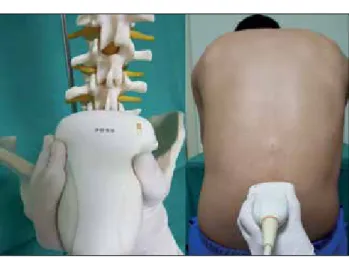
Grau et al. (16) emphasise that a paramedian sagittal oblique view seems to be the optimum window for an ultrasound image of the epidural space. The transducer is positioned lon-gitudinally 1-2 cm lateral to the spinous process, and the ul-trasound beam is directed towards the midline of the spine by tilting the transducer slightly medial (Figure 5). This position is also described as the parasagittal oblique view. The scanning could be performed either from the left or right. The exact intervertebral level can be determined using the paramedian sagittal approach by counting the lumbar vertebrae upwards, starting from the sacrum and moving the transducer cranially (5, 13). As also defined by Karmakar et al. (10), this approach is considered superior to the median transverse and median longitudinal (sagittal) axes for visualising the neuraxial anato-my because of the larger acoustic window (16, 17).
It is possible to identify the sacrum, laminae, ligamentum flavum, posterior dura, interlaminar space, intrathecal space, cauda equina and vertebral body/posterior longitudinal lig-ament/anterior dura complex using this approach. In this plane, the ligamentum flavum, epidural space and posterior dura mater is seen as a linear hyperechoic structure, which is called ‘the posterior complex’. The ’anterior complex’ is identified as linear hyperechoic structure, which consists of anterior dura mater, posterior longitudinal ligament and pos-terior surface of the vertebral body or the intervertebral disk. The intrathecal space (dural sac) appears as an anechoic space between the posterior and anterior complexes (Figure 6) (6, 8, 17).
Figure 1. Positioning of the ultrasound probe for the transverse median approach
Figure 3. Spinous process with underneath acoustic shadowing (transverse median approach)
Figure 2. Visualisation of the spinal structures in the transverse median approach
LF-D: ligamentum flavum-dura mater; VB-PLL: vertebral body-poste-rior longitudinal ligament; TP: transverse processes; AP: articular pro-cesses; DSac: dural sac; M: para-spinal muscles
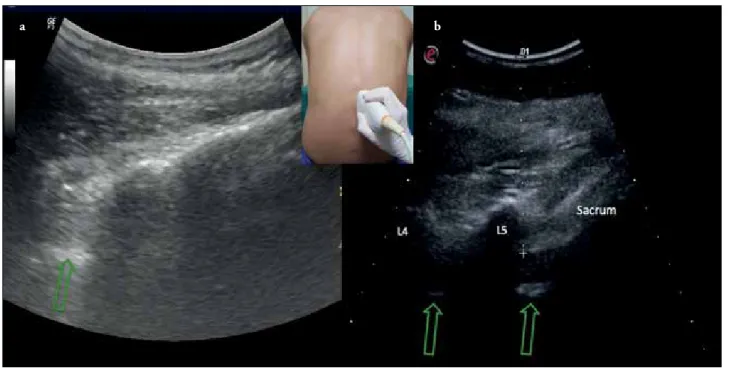
The sacrum is usually seen as a flat or wedge-shaped hy-perechoic structure (Figure 7a). At the cranial end of the sacrum, a gap between the lamina of L5 and the sacrum is identified.
When the transducer is moved cranially in the paramedian sagittal plane, a ‘saw-teeth’ pattern was observed (Figure 8a). The saw-teeth pattern indicates the laminae interrupted by intervertebral spaces. The lamina is seen as inclined white lines arranged one after another producing a pattern that im-itates the head and neck of a horse, which is determined as ‘horse head sign’ (3, 18).
The ligamentum flavum, dura, epidural space and anterior wall of spinal canal can be visualised through the ‘acoustic window’ between laminae, which represents the ultrasound reflections passing the interlaminar space (18). The ligamen-tum flavum is hyperechoic, but less than the lamina, and it often appears as a thick band across the adjacent lamina (10).
This visualisation could be achieved by tilting the transduc-er slightly medial to view the neuraxial structures (usually the ligamentum flavum-posterior dura complex) through the in-terlaminar space (acoustic window) (5). Each intervertebral space consists of two parallel hyperechoic bands. The outer (superficial) band represents the ligamentum flavum/posterior dura complex, and the inner (deeper) band represents posterior surface of the vertebral body/posterior longitudinal ligament/ anterior dura complex. The spinal canal (thecal sac) with the cerebrospinal fluid is seen as an anechoic space between these complexes. Commonly, the anterior dura cannot be differenti-ated from the posterior longitudinal ligament and the posterior surface of the vertebral body. In few cases, the anterior epidural space can be identified as a hypoechoic area between the
pos-Figure 6. Visualisation of anatomical structures using the para-median sagittal oblique approach. PC: posterior complex, con-sists of ligamentum flavum, epidural space and posterior dura mater. AC: anterior complex, consists of anterior dura mater, posterior longitudinal ligament, and posterior surface of the vertebral body or intervertebral disk
L: lamina; DSac: dural sac (intrathecal space) Figure 5. Positioning of the ultrasound probe for paramedian
sagittal oblique scanning
Figure 4. a, b. Ultrasonographic measurement of epidural space depth using transverse median (a) and paramedian sagittal (b) appro-aches with the help of built-in calliper
a b
terior longitudinal ligament and the anterior dura (10). The cauda equina sometimes may appear as multiple horizontal hyperechoic shadows within the spinal canal (10).
In some individuals, the posterior epidural space may be seen as a small gap (2-3 millimetres wide hypoechoic space) be-tween two small parallel lines that consists of a smaller equal (=) sign, which represents the ligamentum flavum, the
epi-dural space and the posterior dura mater (6) (Figure 8b). The articular processes appear as one continuous hyperechoic line with no gaps in between, and the transverse process appears as a crescent shaped hyperechoic reflection with its concavity facing anteriorly (18). The facet joints are seen as near-con-tinuous wavy white structures with humps (described as the ‘camel hump sign’) (6).
Figure 7. a, b. (a) Sacrum is usually seen as a hyperechoic flat or wedge-shaped structure (b) The exact vertebral interspace can be determined by counting vertebrae caudad-to-cephalad direction starting from the sacrum. The arrows are showing the gaps (acoustic windows) between vertebrae where anterior and posterior complex could be seen. The images are obtained using the paramedian sagittal oblique approach
a b
Figure 8. a, b. (a) Saw-teeth pattern, which indicates the laminae interrupted by intervertebral spaces (b) The epidural space between the hyperechoic dura mater and the ligamentum flavum produces a thin hyperechoic parallel line seen as an equal sign. The images are obtained using the paramedian sagittal oblique approach
a b
The paramedian sagittal approach could be used to mea-sure the distance between the skin and epidural space (Figure 4b).
3. Longitudinal median approach
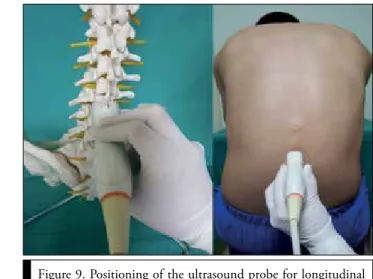
In this approach, the visibility of neuraxial structures is dif-ficult to achieve due to acoustic shadows of the spinous pro-cesses. This view has a limited current use compared to the transverse and paramedian sagittal approaches. The main objective of the longitudinal median approach is to visualise the spinous processes and to identify the vertebral levels (6). The transducer is placed on the midline along the spinous processes in the longitudinal direction (Figure 9). Under the skin, the tips of the spinous processes are seen as a series of hyperechoic lines with posterior acoustic shadowing (Figure 10). For identifying the vertebral level, identify the sacrum, which is seen as a hyperechoic wedge shape, and subsequently move the transducer cranially to locate the superior edge of
sacrum. Thereafter, continue counting the spinous processes and intervertebral spaces upwards to identify the exact inter-vertebral level (19).
Ultrasound-Guided Epidural Blocks
For epidural catheter placement, a preprocedural (pre-punc-ture) lumbar ultrasound scan can be implemented. A pre-puncture ultrasound scan was found beneficial for deter-mining the depth of the epidural space from the skin and for identifying the optimal needle insertion site. A good correla-tion between the skin to the epidural space depth and needle insertion depth was reported in many previous studies. (9, 17, 20). In pregnant women, US has been shown to decrease the number of needle insertion attempts and to improve anaes-thesia efficacy and patient satisfaction, even when difficulties are expected based on the presence of spinal deformities, obe-sity or prior difficult epidural needle placement (20-22). A realtime, ultrasound-guided, epidural needle insertion seems technically challenging, as the needle may impede optimal transducer positioning, and the operator must use one hand to hold the transducer. Of note, an ultrasound examination does not preclude the need for loss-of-resistance for epidural space localisation.
Ultrasound-Guided Spinal/Combined Spinal Epidural Blocks
An ultrasound-guided approach reduces the number of at-tempts to achieve a successful spinal block and reduces the procedure time, particularly in obese patients and those with technical difficulties. Ultrasound imaging facilitates spinal anaesthesia in adults with difficult surface anatomic landmarks and in an obese parturient (23, 24). The use of pre-puncture US before the insertion of epidural catheter in-creases the chance of a successful combined-spinal-epidural procedure on the first attempt and reduces the number of attempts during catheter insertion in obstetric patients (25). According to the study of Ansari et al. (26) in obstetric pa-tients with easily palpable spines, the use of ultrasound did not increase the success rate of spinal anaesthesia or reduce the procedure time or number of puncture attempts, when performed by anaesthetists experienced in both ultrasound and landmark techniques.
Lumbar Ultrasonography Interventions in Clinical Practice Pre-puncture lumbar US is commonly utilised for the landmarking of the epidural space by determining three components: determination of the exact intervertebral level for the desired puncture place, identification of the midline of the spine on the back of patients and estima-tion of the skin-to-epidural space depth for proper needle insertion.
1. Using lumbar ultrasonography to determine the exact intervertebral level
Selecting the appropriate intervertebral space is important to avoid spinal cord injuries. Incorrect determination of the puncture level is a known risk factor for medullary cone
Figure 9. Positioning of the ultrasound probe for longitudinal median scanning
Figure 10. Visualisation of the spinal structures in the longitu-dinal median approach. The tips of spinous processes are seen as a series of hyperechoic lines with posterior acoustic shadowing SP: spinous process; AS: acoustic shadow
injury in spinal blocks. The American Society of Regional Anaesthesia recommends that anaesthesiologists should be aware of the limitations of the physical exam to determine the neuraxial puncture level, especially in patients with difficult topographic anatomy including pregnant and obese patients (27). The intercristal line called the Tuffier’s line is conven-tionally used to identify the vertebral interspace used for spi-nal anaesthesia. Theoretically, the Tuffier’s line indicates the level of L4 or L4-L5 intervertebral space. Spinal anaesthesia
should be performed at or below the L3-L4 level to avoid the potential risk of spinal cord injury as the conus medullar-is reaches the upper part of the body of L2 vertebra in 48% of women. Moreover, the location of the conus medullaris may vary from the mid-third of T12 to the upper third of L3 vertebra (28, 29). The accuracy for identifying the lumbar intervertebral spaces by palpation among anaesthesiologists varies significantly. Mistaken identifications are usually in the cephalad direction, and the difference can be up to four inter-vertebral spaces (19, 30, 31).
The requested intervertebral level can be determined using the paramedian sagittal approach. An ultrasound accurately identifies a spinous process or interspace in 68-76% of cases; however, the accuracy could be possibly increased up to 90% with training (32, 33). Further research is necessary to assess whether the use of the lumbar ultrasound to determine the intervertebral level of the lumbar puncture in pregnant wom-en decreases the risk of medullary cone damage.
The intervertebral levels can be identified by counting the laminae to the cephalad direction starting from the sacrum using the paramedian sagittal oblique approach. This meth-od is more accurate than the landmark examination using the Tuffier’s line (8). First, the sacrum is visualised as a thin, wedge-shaped hyperechoic structure. There is a gap (acoustic window) between the sacrum and L5 vertebra where the
pos-terior and anpos-terior complexes could be seen (Figure 7a and 7b). The first lamina adjacent to the sacrum with an acoustic shadow underneath represents the L5 vertebra. The second lamina adjacent to L5 with its acoustic shadow represents the
L4 vertebra (Figure 7b). The probe should be slid to caudad direction to count the rest of the vertebrae at the upper level. 2. Lumbar ultrasonography for identifying the midline of the vertebral column
This modality is especially useful in obese patients with non-palpable spinous processes.
The US transducer is placed on the middle of the back in the transverse plane and moved left, right, caudal, cephalad and tilted slightly until the acoustic shadow of spinous processes is viewed. When the midline of the transducer and spinous processes intersects, the transducer is maintained still. Then, the midline of the vertebral column is marked as a longitu-dinal perpendicular line passing from the midpoint of the transducer. In addition, when replicated by the needle tra-jectory, the angle of the transducer (in the transverse plane
if a midline puncture is planned) may determine the needle insertion angle (5).
3. Using lumbar ultrasonography for determining the skin-to-epidural space depth
An excellent correlation between the US-estimated depth from the skin to the epidural space and the actual needle depth was found in previous studies (9, 14, 20). The epidural space depth could be determined by visualising the posteri-or dura/ligamentum flavum complex posteri-or the epidural space if seen between these. The length from the skin to the epidural space is measured by using the built-in calliper of the US de-vice. The transverse median approach is primarily suggested for measuring the skin to the epidural space distance, but the paramedian sagittal approach could be used as well (34). Of note, several studies have reported that the paramedian distance from the skin to the lamina and the posterior dura was significantly greater than that measured in the vertical axis (transverse approach). The paramedian sagittal oblique distance is recommended for predicting the needle insertion depth to the epidural space during a paramedian epidural ac-cess (18). Nevertheless, both distance measurements can be used alternatively for punctures in obese pregnant women (17). The main advantage of measuring the epidural space depth is the reduction of dural puncture rates by preventing the performer to exceed the depth measured using US be-fore the puncture (25). In obese women, slight differences are expected between the skin-to-epidural space distance (US depth) and the needle depth. In some cases, underestimation of the skin-to-epidural space depth by US is possible. This is attributed to increased soft tissue of obese pregnant and compression by the US probe (20). Also, the deviation of the needle trajectory from that of the US beam can result in differences between the estimated US and the actual needle depth (15, 17).
4. Real-time lumbar ultrasonography during the neuraxial procedure
In this technique, the loss of resistance could be either used or not. Single-or two-operator techniques can be im-plemented. In the single-operator technique, the operator performs both the US and needle insertion by one hand holding the US transducer and other hand holding the nee-dle-syringe assembly. A springloaded epidural syringe with automated loss-of-resistance (a syringe with an internal compression spring that applies constant pressure on the plunger) should be used in the single operator technique (10). In the two operator technique, one operator performs the US and the other operator performs the neuraxial proce-dure (35). Echogenic (ultrasound visible) Tuohy needles can be helpful for better visualisation of the needle. It is possi-ble to visualise the complete procedure of the needle place-ment through the paramedian acoustic window including the application of intrathecal drugs and flow of the local anaesthetic solution into the epidural space (10, 35). Since the epidural needle is inserted in the long axis (in-plane) of
263
the US transducer with its tip directed to the intervertebral space (in the plane of the US beam), it is possible to visu-alise the entire needle in real time while advancing (10). The real-time advancement of a spinal needle or a catheter placement at the tip of the Tuohy needle is relatively more difficult to view. The observed sonographic changes within the spinal canal at the level of needle insertion during the loss of resistance are the anterior displacement of the poste-rior dura and widening of the posteposte-rior epidural space and compression of the thecal sac (10, 35).
Discussion
Since its first reported use in 1980, accumulating evidence confirms that neuraxial a US can provide reliable information to facilitate neuraxial blockades among pregnant women. Advantages of lumbar ultrasonography
The information of the spinal anatomy acquired before neuraxial puncture enabled the anaesthetist to perform with more focus compared to the blind technique. In patients with difficult palpable surface landmarks (pregnant women and those with body mass index >35 kg m-2), US has been
shown to improve the success rates of spinal anaesthesia (24). A preprocedure scanning has shown to have an 85% positive predictive value for a successful dural puncture (36).
In three meta-analyses of randomised studies comparing ul-trasound-assisted to standard palpation techniques for lum-bar punctures, Shaikh et al. (3) found a reduced number of insertion attempts and a reduced risk of failed or traumatic procedures with ultrasound. Perlas et al. (9) concluded that ultrasound identifies lumbar intervertebral levels more accu-rately than landmark palpation, accuaccu-rately predicts the depth of the epidural or intrathecal space and decreases the risk of failure and the number of needle insertions in patients with normal or difficult anatomical landmarks. Regarding the obstetric population, Schnabel et al. (37) reported an im-provement in the efficacy and safety of neuraxial blocks in obstetrics with ultrasound guidance and concluded that an ultrasound may lower the rate of procedure-related adverse events. The pregnancy-induced softening of the tissues and ligaments may increase the false-positive rate when identify-ing the epidural space usidentify-ing the loss-of-resistance technique (20). This issue may be overcome by measuring the epidural space depth using US. A possible value of pre-puncture lum-bar US for improving the learning curves of obstetric epidur-al anaesthesia and success of residents was epidur-also demonstrated (38).
Vallejo et al. (15) reported that ultrasound scanning to con-firm the midline, determine the needle direction and measure the depth to the epidural space before epidural placement decreased the epidural analgesia failure rate and the number of epidural attempts among anaesthesia residents. In future, electromagnetic guidance for neuraxial needle placement by combining ultrasound and electromagnetic needle tracking
systems may be a promising method, which may facilitate tracking the tip of the needle (39-41).
Limitations of lumbar ultrasonography
Although the published data regarding ultrasound assis-tance in obstetric neuraxial blocks are promising, this tech-nique is far from being a standard of care (37). Because the baseline rate of failed procedures is low even when an ultrasound is not used, it is unclear whether ultrasound imaging should be used for all lumbar punctures and epi-dural catheterisations. The negative arguments for lumbar US include:
1. Additional time required for ultrasonography scanning, which can be time consuming in a busy clinic
The urgency to perform spinal anaesthesia in some patients indicates that clinically palpated landmarks are still used in the majority of cases. Obstetric anaesthesiologists have man-aged to perform neuraxial procedures with great success with-out using imaging techniques. The detection of the epidur-al space is primarily based on the sense of loss of resistance. There is a relatively difficult learning curve associated with lumbar ultrasound leading to long US times in the beginning (42). The impact of the ultrasound on the total required time is unknown, as it was not evaluated in previous studies. In our opinion, the additional time required for US before plac-ing an epidural catheter is not reasonably long. Accordplac-ing to many authors’ observations, it usually takes an average of 2-5 minutes for the ultrasound assessment of the spine (5, 21). Given the pre-puncture information, US leads to fewer insertion attempts for successful epidural placement, and it is likely to reduce the overall procedure time in patients with difficult anatomic landmarks.
2. Detailed training and proficiency are required
A previous study has suggested that the providers require a substantial amount of practice before they become compe-tent in lumbar US, particularly for determining the inter-space, optimal insertion point and the epidural space depth (43). Studies about the learning curve for ultrasound-guided obstetric epidural blocks are lacking. Even some anaesthesia providers do not believe in lumbar ultrasound guidance for epidural catheter insertion success during labour. It has been proposed that with increased clinical experience, many of these disadvantages can be overcome.
3. Technical difficulty in the visualisation of the spinal structures by ultrasound
The main disadvantage is related to the deep location of the structures of interest and requirement of the use of low-fre-quency transducers with limited image-resolution capabili-ties. Subcutaneous adipose tissues can hamper the US beam, which results in the difficulty of US of obese patients with less image quality (44). Another disadvantage is the difficulty of visualisation of the soft tissues that are shadowed by bony structures. The quality the ultrasonographic identification of the neuraxial structures is reduced during pregnancy. Among
264
pregnant women, the epidural space is located deeper under the skin. The soft tissue channel between the spinal processes is narrower by the end of pregnancy. The distance from the skin to the epidural space is longer and the epidural space is narrower as well (4). Elderly patients may have degenerative spinal diseases with narrowed interspinous and intervertebral spaces as a result of the ossification of the interspinous liga-ments and hypertrophy of the facet joints (44). The steady improvements in ultrasound technology and image quality can be expected to improve lumbar spine US in the near fu-ture, and it will be possible to increase the reliability of the method.
4. There is limited evidence on the role of lumbar ultrasonography in improving the success rate
Although majority of the studies confirm the efficacy of ultra-sound for neuraxial blocks, there are some studies reporting no improvement with the usage of a pre-puncture ultrasound for normal weight patients and has no effect on the rate of failed procedures (45, 46).
The use of a preprocedural spinal ultrasound by a cohort of anaesthesia trainees did not improve the ease of insertion of labour epidural catheters in patients with easily palpable lum-bar spines (47). These studies conclude that for experienced anaesthesiologists, it remains unclear whether a preprocedur-al ultrasound improves the epidurpreprocedur-al catheterisation technique in a parturient with palpable anatomical landmarks. It is difficult to conduct double-blind studies about ultrasound guidance for neuraxial blocks, which enhances the risk of bias (42).
Finally, the need for equipment, cost and possible require-ment of a second person trained about lumbar US may be other limitations of lumbar US for neuraxial blocks.
In conclusion, lumbar US for obstetric neuraxial blocks could be considered in two different modalities, one is pre-puncture US before neuraxial interventions and the other is real-time US during the neuraxial procedures. Pre-puncture lumbar US is used to estimate the skin-to-epidural space distance and for determining the correct needle insertion point. Pre-punc-ture lumbar US is relatively easier compared to the real-time technique. Real-time lumbar US may need sufficient experi-ence and professionalism in US for neuraxial blocks. Clini-cians have been hesitant in using this technique due to many reasons, and lumbar US for neuraxial blocks did not get into widespread use. Although there are studies that failed to show improvement in normal weight patients (45, 46), recent me-ta-analyses revealed that there is enough evidence to support lumbar US for neuraxial procedures in obstetric population (37). Especially in patients with anatomical difficulties, such as a previous lumbar operation or with nonpalpable anatom-ical landmarks, lumbar US is clearly beneficial. As for the ob-stetric population, anatomical difficulties are usually evident and lumbar US could be strongly recommended. We believe this technique will be more popular among anaesthetists in
the future by obtaining adequate education and training. Future advances in ultrasound technology will likely make lumbar US simpler, more accurate and widely applicable. With increased availability of this technology and increased awareness among the anaesthesia providers, obstetric lumbar US would become more feasible.
Peer-review: Externally peer-reviewed.
Author Contributions: Concept - T.Ş., O.B.; Design - T.Ş., O.B.; Supervision - T.Ş.; Resources - T.Ş., O.B.; Materials - T.Ş., O.B.; Data Collection and/or Processing - O.B.; Analysis and/or Interpre-tation - T.Ş.; Literature Search - O.B.; Writing Manuscript - T.Ş., O.B.; Critical Review - T.Ş.; Other - T.Ş., O.B.
Conflict of Interest: Authors have no conflicts of interest to declare.
Financial Disclosure: The authors declared that this study has re-ceived no financial support.
Hakem Değerlendirmesi: Dış bağımsız.
Yazar Katkıları: Fikir - T.Ş., O.B.; Tasarım - T.Ş., O.B.; Denetleme - T.Ş.; Kaynaklar - T.Ş., O.B.; Malzemeler - T.Ş., O.B.; Veri Toplan-ması ve/veya İşlemesi - O.B.; Analiz ve/veya Yorum - T.Ş.; Literatür Taraması - O.B.; Yazıyı Yazan - T.Ş., O.B.; Eleştirel İnceleme - T.Ş.; Diğer - T.Ş., O.B.
Çıkar Çatışması: Yazarlar çıkar çatışması bildirmemişlerdir.
Finansal Destek: Yazarlar bu çalışma için finansal destek al-madıklarını beyan etmişlerdir.
References
1. Cork RC, Kryc JJ, Vaughan RW. Ultrasonic localization of the lumbar epidural space. Anesthesiology 1980; 52: 513-6.
[CrossRef]
2. National Institute for Health and Clinical Excellence. Ultra-sound guided catheterisation of the epidural space: under-standing NICE guidance. January 2008. Available from: URL: http://www.nice.org.uk.
3. Shaikh F, Brzezinski J, Alexander S, Arzola C, Carvalho JC, Beyene J, et al. Ultrasound imaging for lumbar punctures and epidural catheterisations: systematic review and meta-analysis. BMJ 2013; 346: f1720. [CrossRef]
4. Grau T, Leipold RW, Horter J, Conradi R, Martin E, Motsch J. The lumbar epidural space in pregnancy: visualization by ul-trasonography. Br J Anaesth 2001; 86: 798-804. [CrossRef]
5. Balki M. Locating the epidural space in obstetric patients-ultra-sound a useful tool: continuing professional development. Can J Anaesth 2010; 57: 1111-26. [CrossRef]
6. Srinivasan KK, Lee PJ, Iohom G. Ultrasound for neuraxial blockade. Med Ultrason 2014; 16: 356-63.
7. Sahin Yildiz T, Balaban O, Sahin B, Solak M, Toker K. Ultra-sound guidance for subarachnoid block in parturient. Anestezi Dergisi 2011; 19: 224-7.
8. Chin KJ, Karmakar MK, Peng P. Ultrasonography of the adult thoracic and lumbar spine for central neuraxial blockade.
9. Perlas A, Chaparro LE, Chin KJ. Lumbar neuraxial ultrasound for spinal and epidural anesthesia: a systematic review and me-ta-analysis. Reg Anesth Pain Med 2016; 41: 251-60. [CrossRef]
10. Karmakar MK, Li X, Ho AM, Kwok WH, Chui PT. Real-time ultrasound-guided paramedian epidural access: evaluation of a novel in-plane technique. Br J Anaesth 2009; 10: 845-54.
[CrossRef]
11. Arzola C. Preprocedure Ultrasonography Before Initiating a Neuraxial Anesthetic Procedure. Anesth Analg 2017; 124: 712-3. [CrossRef]
12. Darrieutort-Laffite C, Hamel O, Glemarec J, Maugars Y, Le Goff B. Ultrasonography of the lumbar spine: sonoanatomy and practical applications. Joint Bone Spine 2014; 81: 130-6.
[CrossRef]
13. Carvalho JCA. Ultrasound-facilitated epidurals and spinals in obstetrics. Anesthesiology Clin 2008; 26: 145-58. [CrossRef]
14. Arzola C, Davies S, Rofaeel A, Carvalho JC. Ultrasound using the transverse approach to the lumbar spine provides reliable landmarks for labor epidurals. Anesth Analg 2007; 104: 1188-92. [CrossRef]
15. Vallejo MC, Phelps AL, Singh S, Orebaugh SL, Sah N. Ultra-sound decreases the failed labor epidural rate in resident train-ees. Int J Obstet Anesth 2010; 19: 373-8. [CrossRef]
16. Grau T, Leipold RW, Horter J, Conradi R, Martin EO, Motsch J. Paramedian access to the epidural space: the optimum win-dow for ultrasound imaging. J Clin Anesth 2001; 13: 213-7.
[CrossRef]
17. Sahota JS, Carvalho JC, Balki M, Fanning N, Arzola C. Ultra-sound estimates for midline epidural punctures in the obese par-turient: paramedian sagittal oblique is comparable to transverse median plane. Anesth Analg 2013; 116: 829-35. [CrossRef]
18. Karmakar MK, Li X, Kwok WH, Ho AM, Ngan Kee WD. Sonoanatomy relevant for ultrasound-guided central neuraxial blocks via the paramedian approach in the lumbar region. Br J Radiol 2012; 85: e262-9. [CrossRef]
19. Locks Gde F, Almeida MC, Pereira AA. Use of the ultrasound to determine the level of lumbar puncture in pregnant women. Rev Bras Anestesiol 2010; 60: 13-9. [CrossRef]
20. Balki M, Lee Y, Halpern S, Carvalho JC. Ultrasound imaging of the lumbar spine in the transverse plane: the correlation be-tween estimated and actual depth to the epidural space in obese parturients. Anesth Analg 2009; 108: 1876-81. [CrossRef]
21. Grau T, Leipold RW, Conradi R, Martin E. Ultrasound con-trol for presumed difficult epidural puncture. Acta Anaesthesiol Scand 2001; 45: 766-71. [CrossRef]
22. Grau T, Leipold RW, Conradi R, Martin E, Motsch J. Efficacy of ultrasound imaging in obstetric epidural anesthesia. J Clin Anesth 2002; 14: 169-75. [CrossRef]
23. Chin KJ, Perlas A, Chan V, Brown-Shreves D, Koshkin A, Vaishnav V. Ultrasound imaging facilitates spinal anesthesia in adults with difficult surface anatomic landmarks. Anesthesiolo-gy 2011; 115: 94-101. [CrossRef]
24. Sahin T, Balaban O, Sahin L, Solak M, Toker K. A random-ized controlled trial of preinsertion ultrasound guidance for spinal anaesthesia in pregnancy: outcomes among obese and lean parturients: ultrasound for spinal anesthesia in pregnancy. J Anesth 2014; 28: 413-9. [CrossRef]
25. Nassar M, Abdelazim IA. Pre-puncture ultrasound guided epi-dural insertion before vaginal delivery. J Clin Monit Comput 2015; 29: 573-7. [CrossRef]
26. Ansari T, Yousef A, El Gamassy A, Fayez M. Ultrasound-guided spinal anaesthesia in obstetrics: is there an advantage over the landmark technique in patients with easily palpable spines? Int J Obstet Anesth. 2014; 23: 213-6. [CrossRef]
27. Neal JM, Bernards CM, Hadzic A, Hebl JR, Hogan QH, Hor-locker TT, et al. ASRA Practice advisory on neurologic compli-cations in regional anesthesia and pain medicine. Reg Anesth Pain Med 2008; 33: 404-15. [CrossRef]
28. Soleiman J, Demaerel P, Rocher S, Maes F, Marchal G. Mag-netic resonance imaging study of the level of termination of the conus medullaris and the thecal sac: influence of age and gender. Spine 2005; 30: 1875-80. [CrossRef]
29. Srinivasan KK, Deighan M, Crowley L, McKeating K. Spinal anaesthesia for caesarean section: an ultrasound comparison of two different landmark techniques. Int J Obstet Anesth 2014; 23: 206-12. [CrossRef]
30. Schlotterbeck H, Schaeffer R, Dow WA, Touret Y, Bailey S, Di-emunsch P. Ultrasonographic control of the puncture level for lumbar neuraxial block in obstetric anaesthesia. Br J Anaesth 2008; 100: 230-4. [CrossRef]
31. Whitty R, Moore M, Macarthur A. Identification of the lum-bar interspinous spaces: palpation versus ultrasound. Anesth Analg 2008; 106: 538-40. [CrossRef]
32. Halpern SH, Banerjee A, Stocche R, Glanc P. The use of ultra-sound for lumbar spinous process identification: A pilot study. Can J Anaesth 2010; 57: 817-22. [CrossRef]
33. Watson MJ, Evans S, Thorp JM. Could ultrasonography be used by an anaesthetist to identify a specified lumbar interspace before spinal anaesthesia? Br J Anaesth 2003; 90: 509-11.
[CrossRef]
34. Furness G, Reilly MP, Kuchi S. An evaluation of ultrasound imaging for identification of lumbar intervertebral level. An-aesthesia 2002; 57: 277-80. [CrossRef]
35. Grau T, Leipold RW, Fatehi S, Martin E, Motsch J. Real-time ultrasonic observation of combined spinal-epidural anaesthe-sia. Eur J Anaesthesiol 2004; 21: 25-31. [CrossRef]
36. Chin KJ, Ramlogan R, Arzola C, Singh M, Chan V. The utility of ultrasound imaging in predicting ease of performance of spi-nal anesthesia in an orthopedic patient population. Reg Anesth Pain Med 2013; 38: 34-8. [CrossRef]
37. Schnabel A, Schuster F, Ermert T, Eberhart LH, Metterlein T, Kranke P. Ultrasound guidance for neuraxial analgesia and anesthesia in obstetrics: a quantitative systematic review. Ultra-schall Med 2012; 33: E132-7. [CrossRef]
38. Grau T, Bartusseck E, Conradi R, Martin E, Motsch J. Ultra-sound imaging improves learning curves in obstetric epidural anesthesia: a preliminary study. Can J Anaesth 2003; 50: 1047-50. [CrossRef]
39. Brinkmann S, Tang R, Sawka A, Vaghadia H. Single op-erator real-time ultrasound-guided spinal injection using SonixGPS: a case series. Can J Anaesth 2013; 60: 896-901.
[CrossRef ]
40. Niazi AU, Chin KJ, Jin R, Chan VW. Real-time ultrasound- guided spinal anesthesia using the SonixGPS ultrasound guid-ance system: a feasibility study. Acta Anaesthesiol Scand 2014; 58: 875-81. [CrossRef]
41. Wong SW, Niazi AU, Chin KJ, Chan VW. Real-time ultra-sound-guided spinal anesthesia using the SonixGPS(R) needle tracking system: a case report. Can J Anaesth 2013; 60: 50-3.
[CrossRef]
42. Gambling DR. Lumbar ultrasound: useful gadget or time-consum-ing gimmick? Int J Obstet Anesth 2011; 20: 318-20. [CrossRef]
43. Margarido CB, Arzola C, Balki M, Carvalho JC. Anesthesiol-ogists’ learning curves for ultrasound assessment of the lumbar spine. Can J Anaesth 2010; 57: 120-6. [CrossRef]
44. Darrieutort-Laffite C, Bart G, Planche L, Glemarec J, Maugars Y, Le Goff B. Usefulness of a pre-procedure ultrasound scan-ning of the lumbar spine before epidural injection in patients with a presumed difficult puncture: A randomized controlled trial. Joint Bone Spine 2015; 82: 356-61. [CrossRef]
45. Tawfik MM, Atallah MM, Elkharboutly WS, Allakkany NS, Abdelkhalek M. Does Pre-procedural Ultrasound Increase the
First-Pass Success Rate of Epidural Catheterization Before Ce-sarean Delivery? A Randomized Controlled Trial. Anesth Analg 2017; 124: 851-6. [CrossRef]
46. Balaban O, Sahin T, Sahin L, Solak M, Toker K. Does lumbar ultrasonography improve epidural catheterization for labor an-algesia? A randomized controlled study. Middle East J Anaes-thesiol In Press 2017.
47. Arzola C, Mikhael R, Margarido C, Carvalho JC. Spinal ul-trasound versus palpation for epidural catheter insertion in la-bour: A randomised controlled trial. Eur J Anaesthesiol 2015; 32: 499-505. [CrossRef]