• Sonuç bulunamadı

Effect of septoplasty on the mean platelet volume and inflammation markers

N/A
N/A
Protected

Academic year: 2021

Share "Effect of septoplasty on the mean platelet volume and inflammation markers"

Copied!
9
0
0

Yükleniyor.... (view fulltext now)

Tam metin

(1)

CLINICAL STUDY

EFFECT OF SEPTOPLASTY ON THE MEAN PLATELET VOLUME AND

INFLAMMATION MARKERS

Bülent ULUSOY

1

,

MD; Kazım BOZDEMİR

2

,

MD; Halil İbrahim MİŞE

3

,

MD; Ahmet

KUTLUHAN

2

,

MD; Mehmet Hakan KORKMAZ

2

,

MD

1Selçuk Üniversitesi Tıp Fakültesi KBB Anabilim Dalı, KBB, KonyA, Turkey2Yıldırım Beyazıt Üniversitesi Tıp Fakültesi KBB

Anabilim Dalı, KBB, Ankara, Turkey 3Kaçkar Devlet Hastanesi, KBB, Rize, Turkey

SUMMARY

Objective: Nasal septal deviation is an obstructive pathology and can be related to inflammation. This study aimed to investigate the relationship between nasal septum deviation leading to obvious nasal obstruction and certain variables including mean platelet volume (MPV), platelet distribution width (PDW), neutrophil/lymphocyte ratio (NLR), and platelet/lymphocyte ratio (PLR). The study will also examine the effect of this operation on this relationship.

Materials and Methods: This is a prospective study. The study sample included 29 patient group participants that had septoplasty indications, and 31 control group participants that were healthy volunteers. Control and patient groups were compared with regard to MPV, PDW, NLR, PLR, platelet count and white blood cell (WBC) counts. The significance level was accepted as 0.05 in the relevant statistical analyses.

Findings: The preoperative MPV and PDW levels were found to be higher in the patient group than the control group. The difference between them was statistically significant (p < 0.05). In the patient group, the researchers observed a decrease of 0.72 units in the MPV value and 0.91 units in the PDW value, and an increase of 6.72 units in the platelet count at the end of the month following the operation compared with the relevant values in the preoperative period (p < 0.05).

Result: The MPV and PDW values were high in the patients with nasal septum deviation requiring surgery. Moreover, these high MPV and PDW values were found to normalize after the operation.

Keywords: Inflammation, mean platelet volume, nasal airway obstruction, nasal surgical procedures, platelet activation

SEPTOPLASTİNİN MEAN PLATELET VOLUME VE İNFLAMASYON MARKERLARI ÜZERİNE ETKİSİ ÖZET

Amaç: Nazal septum deviasyonu, obstruktif bir patoloji olmakla birlikte inflamasyon ile de ilişkili olabilen bir durumdur. Bu çalışmamızda bariz burun tıkanıklığına yol açan septum nazi deviasyonu olan hastalarda mean platelet volüm (MPV), platelet distrubution width (PDW), nötrofil lenfosit oranı (NLR) ve platelet lenfosit oranı (PLR) ile septum deviasyonu arasındaki ilişkiyi ve ameliyattan sonra bunun ne şekilde etkilendiğini araştırmayı amaçladık.

Gereç Yöntem:< Bu prospektif çalışmada, septoplasti endikasyonu konulan 29 birey hasta grubu olarak ve 31 sağlıklı gönüllü birey ise kontrol grubu olarak çalışmaya dahil edildi. Kontrol ve hasta grubları MPV, PDW, NLR, PLR, Platelet sayısı ve White blood cell (WBC) değerleri açısından karşılaştırıldı. İstatistiksel değerlendirmelerde anlamlılık sınırı 0.05 olarak kabul edildi.

Bulgular:< Septum deviasyonu olan hastaların ameliyat öncesi ile kontrol grubu karşılaştırıldığında MPV ve PDW düzeylerinin hasta grubunda istatistiksel olarak anlamlı oranda yüksek olduğu görüldü (p<0.05). Hasta grubunda ameliyat öncesi ve sonrası değerler karşılaştırıldığında ise operasyon öncesine göre operasyondan 1 ay sonra Platelet sayısında 6,72 birimlik artış tespit edilirken MPV değerinde 0,47 birimlik, PDW değerinde ise 0,91 birimlik bir azalma olduğu görüldü (p<0.05).

Sonuç: Ameliyat gerektiren nazal septum deviasyonu olan hastalarda MPV ve PDW değerlerinin yüksek olduğu görüldü. Buna ek olarak MPV ve PDW değerlerindeki bu yüksekliğin ameliyattan sonra düzeldiği tespit edildi.

Anahtar Sözcükler: Burun tıkanıklığı, İnflamasyon, mean platelet volüm, platelet aktivasyonu, nazal cerrahi prosedürler

INTRODUCTION

Nasal septum deviation is one of the most common causes of nasal obstruction. This operation optimizes the smoothness of the nasal septum for gas exchange by ensuring the formation of laminar airflow and facilitating the heating, humidification, and cleaning of the inhaled air 1.

Corresponding Author: Bülent Ulusoy MD Selçuk Üniversitesi Tıp Fakültesi KBB Anabilim Dalı, KBB, Konya, Turkey, E-mail: ulusoybulent@hotmail.com

Received: 30 December 2017, revised for: 27 May 2018, accepted for publication: 27 May 2018

Nasal septum deviation causing nasal obstruction may lead to chronic hypoxia and a number of cardiovascular complications including alveolar hypoventilation, cor pulmonale, and pulmonary arterial hypertension 2-4. The relationship

between hypoxia and platelet activation has been reported in many studies 5-8. In addition, MPV has

been proved to increase in hypoxic conditions 5-8.

MPV is a marker that is easily revealed by a routine complete blood count. Mean platelet volume indicates the size of platelets. An increase in MPV value suggests that larger platelets were released

(2)

from bone marrow into bloodstream. Platelets with larger volume have been reported to be more active metabolically and enzymatically, and more inclined to aggregate than the small ones 7-10.

This study aimed to examine the patients with nasal septum deviations that require obvious nasal obstruction with a consideration of neutrophil/lymphocyte ratio (NLR) and platelet/lymphocyte ratio (PLR) values 11,12 which have been published in the recent literature as new inflammatory markers, and the mean platelet volume (MPV) and platelet distribution width (PDW) values10,13,14 which have been reported to indicate a tendency for thrombosis 10

MATERIAL and METHODS

This is a prospective study. The study was conducted with 60 individuals: 29 individuals had septoplasty indications and were included in the patient group, and 31 individuals were healthy volunteers and included in the control group. Nasal septal deviation was diagnosed by anterior rhinoscopy and nasal endoscopy. Their physicians had requested preoperative paranasal sinus computed tomography for the patients. All patients underwent septoplasty surgery under general anesthesia. CBC test was performed twice for the patient group and only once for the control group before and 1 month after the operation. The patient and control groups were compared regarding NLR, PLR, MPV and PDW values.

The researchers excluded from the study the patients with chronic inflammatory disease, acute infection, acute or chronic renal failure, chronic obstructive pulmonary disease, coronary artery disease, collagen tissue disease, allergic rhinitis, liver disease, and also additional diseases (such as concha hypertrophy, chronic sinusitis, and adenoid hypertrophy) that could require obstruction in the upper respiratory tracts.

For the patient group, the absolute neutrophil count was divided by the absolute lymphocyte count to obtain NLR, while the absolute platelet count was divided by the absolute lymphocyte count to obtain PLR in complete blood examination. The relevant MPV and PDW values were also determined from the complete blood examination. NLR, PLR, MPV, and PDW values of the control group were calculated in the same way. The analysis was made using an automated blood cell counter (Sysmex XE-2100, Kobe, Japan).

The present study was carried out with the permission (No. 03.2015/77) Ankara Yıldırım

Beyazıt University’s Clinical Research Ethics Committee.

The study data were statistically analyzed using the SPSS for Windows 11.5 package program (SPSS, IL, USA). Variables in both control and patient groups were found to be normally distributed using the Shapiro–Wilks normality test. The study compared the quantitative variables of the control and patient groups using independent samples t test. The chi-square test was used for comparison by gender. Also, the preoperative values of the patient group were compared with the relevant values using paired samples t test at the end of the month following the operation. The significance level was accepted as 0.05.

RESULTS

The mean age of the participants in the patient group was 34.78 ± 11.02 years, and it was 32.03 ± 8.54 years for the control group participants. There was no significant difference between the groups by age (p > 0.05). Furthermore, 65.5% of the cases were male and 34.5% were female in the patient group, whereas 67.7% of the cases were male and 32.3% were female in the control group. No significant differences were found between the groups by gender (p > 0.05) (Table 1).

The MPV value was 10.34 ± 0.68 fl in the control group, whereas the corresponding preoperative and postoperative values in the patient group were 10.93 ± 0.87 fl and 10.46 ± 0.83 fl, respectively. The MPV value increased significantly in the patients with septum deviations and led to nasal obstruction (p = 0.005), which was similar to the value in the control group in the postoperative period (p > 0.05) (Table 2). Moreover, the MPV value in the patient group decreased significantly after the operation compared to its preoperative level (p = 0.001) (Table 3)(Figure 1).

The PDW value was measured as 11.97 ± 1.21 fl in the control group, whereas the preoperative and postoperative values in the patient group were 13.23 ± 1.51 fl and 12.32 ± 2.45 fl, respectively. The PDW value increased significantly in the patient group (p = 0.001), and it was close to the value in the control group in the postoperative period (p > 0.05) (Table 2). Moreover, the decrease in the PDW value of the patient group after the operation was statistically significant compared with its preoperative level (p = 0.023) (Table 3) (Figure 2).

No statistically significant difference was found between the groups in NLR (p > 0.05) (Table 2). Similarly, no difference was found between

(3)

The WBC count was 7.35 ± 1.11 (× 109 / l) in the control group. In the patient group, the values were 7.09 ± 1.92 (× 109/l) in the preoperative period and 6.79 ± 1.46 (× 109/l) in the postoperative period. There was no significant difference between control group’s preoperative and postoperative results (p>0.05) (Table 2). Similarly, there was no significant difference between the preoperative and postoperative results of the patient group (p> 0.05) (Table 3).

preoperative and postoperative NLR values of the groups (p > 0.05) (Table 3).

Moreover, while the PLR values were similar in both the patient and control groups, the increase in the postoperative period PLR value was found to be statistically significant in the present study (p < 0.05) (Tables 2 and 3).

The platelet count was 252.19 ± 51.22 (× 109/l) in the control group. In the patient group, 239.55 ± 43.98 (× 109/l) in the preoperative period and 246.28 ± 41.64 (× 109/l) in the postoperative period were detected. There was no significant difference between control group’s preoperative and postoperative results in platelet count (p> 0.05) (Table 2). However, the increase in postoperative platelet count was found statistically significant in the patient group (p <0.05) (Table 3).

Table 1: Comparison of the control and patient groups by gender and age

Control

(n = 31)

NSD (n = 29)

p

Gender

Male

Female

21 (67.7%)

10 (32.3%)

19 (65.5%)

10 (34.5%)

0.855

Age (mean ± SD)

32.03 ± 8.54

34.78±11.02

0.359

(4)

Table 2: Comparison of the results of control and patient groups

NSD operation

Control vs

before

Control vs

after

Control

Before

After first

month

Mean ± SD

Mean ± SD

Mean ± SD

p value*

p value*

PLR

101.96 ±

16.02

116.02 ±

45.13

125.51 ± 45.16

0.122

0.012

NLR

1.68 ± 0.42

1.92 ± 0.79

2.00 ± 0.81

0.167

0.073

MPV(fl)

10.34 ± 0.68

10.93 ± 0.87

10.46 ± 0.83

0.005

0.542

Platelet

count

252.19 ±

51.22

239.55 ±

43.98

246.28 ± 41.64

0.311

0.627

PDW(fl) 11.97 ± 1.21

13.23 ± 1.51

12.32 ± 2.45

0.001

0.482

WBC

7.35 ± 1.11

7.09 ± 1.92

6.79 ± 1.46

0.517

0.094

*t test for independent samples.

NSD: Nasal Septal Deviation, SD: Standard deviation, PLR: platelet-to-lymphocyte ratio, NLR: neutrophil-to-lymphocyte ratio, MPV: mean platelet volume, PDW: platelet distribution width, WBC: white blood cell

(5)

Table 3: Comparison of the preoperative and postoperative results of the patient group.

NSD operation

Difference

(before and after the

first month)

Before

After the first

month

Mean ± SD

Mean ± SD

Mean ± SD

p value**

PLR

116.02 ±

45.13

125.51 ± 45.16

-9.49 ± 23.98

0.042

NLR

1.92 ± 0.79

2,00 ± 0,81

-0.08 ± 0.49

0.407

MPV(fl) 10.93 ± 0.87

10.46 ± 0.83

0.47 ± 0.56

0.001

Platelet

count

239.55 ±

43.98

246.28 ± 41.64

–6.72 ± 13.93

0.015

PDW

(fl)

13.23 ± 1.51

12.32 ± 2.45

0.91 ± 2.05

0.023

WBC

7.09 ± 1.92

6.79 ± 1.46

0.31 ± 1.10

0.146

** Paired-sample t test.

NSD: Nasal Septal Deviation, SD: Standard deviation, PLR: platelet-to-lymphocyte ratio, NLR: neutrophil-to-lymphocyte ratio, MPV: mean platelet volume, PDW: platelet distribution width, WBC: white blood cell

(6)

Figure 2: MPV value in preoperative and postoperative patients.

(7)

Figure 4: PDW value preoperative and postoperative patients.

DISCUSSION

The most important finding of the present study was that the MPV and PDW values increased in the patients with nasal septum deviations leading to nasal obstruction while there was no increase in the control group. These values decreased to the levels close to those of the control group after the operation. The MPV and PDW values are the indicators of platelet activation. PDW is used to assess variation in the size of blood platelets in circulation, while MPV is used to indicate the size of platelets 10,13,14. The increase in these parameters

suggests that larger platelets are released from the bone marrow into circulation. It has been reported that large blood platelets are metabolically and enzymatically more active and tend to aggregate more compared with the smaller ones 10,14,15. The use

of MPV and PDW markers together has been reported to be more valuable in assessing coagulation tendency 14,15. Microvascular endothelial function and fibrinolysis deteriorated whereas hematocrit, blood viscosity, platelet activity, and blood coagulation increased in hypokalemic diseases 16,17. Nasal septal deviations have been reported as one of the most common causes of nasal obstruction, which can lead to many cardiovascular complications and chronic hypoxia 1-4,18. Fidan et al. pointed out that the mean

pulmonary arterial pressure increased in the patients with obvious septum deviations, and that the relevant pressure dropped after septoplasty 2. Similarly,

Ozkececi et al. reported that septum deviation increased mean pulmonary artery pressure and

caused an adverse effect on right ventricular function. They also reported a decrease in pulmonary artery pressure and a recovery tendency in right ventricular function during the evaluation made three months after septoplasty 3. Another study demonstrated that

septoplasty in the patients with hypertension, particularly those younger than 35 years, had a positive effect on treatment 4. Derin et al. stated that nasal septum deviation might be related to cardiac

arrhythmia 18. The increase in the MPV and PDW

values in the patients with obvious septum deviation may lead to hypercoagulopathy. It has been suggested that this may be related to possible cardiovascular pathologies in the patients with septum deviation 5-8. Sagit et al. reported that the

MPV value increased in the patients with obvious septum deviation and that this situation improved after the operation 7. Sahin et al. separated the patients with septum deviation into two groups according to the Epworth sleepiness scale (ESS), and stated that the increase in the MPV and PDW values was significant only in the patient group since they had an ESS value of 10 or higher 8. Ulu et al reported that the MPV value increased in the patients with septum deviation and that this increase was related to the severity of obstruction 6. Unlu et al. conducted a

retrospective study, and grouped the patients with septum deviation according to the duration of nasal obstruction. They reported that the MPV and PDW values increased, whereas contralateral platelet count decreased, with an increase in the duration of nasal obstruction. Therefore, they asserted that the risk of cardiopulmonary complications in these patients

(8)

might increase in direct proportion with the duration of nasal obstruction, although it is unclear when they would develop 5.

Increased NLR and PLR values have been reported to be an indication of systemic inflammation

11,12. Seo et al. have showed that the NLR and PLR

values increased in patients with sudden hearing loss, suggesting that this is probably the result of systemic stress activating inflammation 11. Although no change was observed in terms of the NLR value, an increase in the PLR value was detected after the operation. This may be an effect of the postoperative stress according to the hypothesis of Seo et al. However, lack of increase in the NLR value, which is also an indication of systemic inflammation, weakens this hypothesis. This hypothesis might have been more valid if there was a significant increase in both values.

The present study also found that the MPV and PDW values increased in the patients requiring septoplasty, and that these values decreased to the level of the control group within the month following the operation. These findings are consistent with the relevant literature. An analysis of the comparative studies in the literature that were conducted in preoperative and postoperative revealed that postoperative measurements were made 3 months or 6 months after the operation 7,8. In these studies, the

increase in MPV and PDW values in the patient group was corrected in the postoperative period 7,8.

This study found that this correction, which was detected in patients who underwent septoplasty, occurred earlier (postoperative 1st month). This finding is not consistent with the relevant literature.

CONCLUSION

The MPV and PDW values were high in the patients with nasal septum deviation requiring surgery. Moreover, these high MPV and PDW values were corrected down after the operation. Although this situation may be an indicator of a prothrombotic state as mentioned in the literature, there is a need for further studies confirming this hypothesis by the examinations that are routinely made to reveal the thrombotic tendency.

Acknowledgments

We would like to thank Associate Professor Doctor Serdal Kenan Köse for the statistical analysis of the study.

Ethical approval

The present study was carried out with the permission no. 03.2015/77 of the Clinical Research Ethics Committee of Ankara Yıldırım Beyazıt

University. Informed consent was obtained from all individual participants included in the study.

Conflicts of Interest and Source of Funding

There is no conflict of interest between the authors. No financing support was received from any place related to this work.

REFERENCES

1. Aziz T, Biron VL, Ansari K, Flores-Mir C. Measurement tools for the diagnosis of nasal septal deviation: a systematic review. J Otolaryngol Head Neck Surg 2014; 43:11.

2. Fidan V, and Aksakal E. Impact of septoplasty on pulmonary artery pressure in patients with markedly deviated septum. J Craniofac Surg 2011; 22:1591-1593.

3. Ozkececi G, Akci O, Bucak A, Ulu S, Yalim Z, Aycicek A, Onrat E, Avsar A. The effect of septoplasty on pulmonary artery pressure and right ventricular function in nasal septum deviation. Eur Arch Otorhinolaryngol 2016; 273:3747-3752. 4. Smitha SG, Jagannath B, and Mathew AS. Impact of Septal

Correction on the Blood Pressure of Hypertensive Patients with Deviated Nasal Septum. Indian J Otolaryngol Head Neck Surg 2016; 68:46-51.

5. Unlu I, Kesici GG, Onec B,Yaman H, Guclu E. The effect of duration of nasal obstruction on mean platelet volume in patients with marked nasal septal deviation. Eur Arch Otorhinolaryngol 2016; 273:401-405.

6. Ulu S, Ulu MS, Bucak A, Kahveci OK, Yucedag F, Aycicek A. Evaluating the relationship between nasal obstruction and mean platelet volume by using acoustic rhinometry in patients with septum deviation. Rhinology 2013; 51:249-252. 7. Sagit M, Korkmaz F, Kavugudurmaz M, Somdas MA.

Impact of septoplasty on mean platelet volume levels in patients with marked nasal septal deviation. J Craniofac Surg 2012; 23:974-976.

8. Sahin MS and Kizilirmak D. Changes at Mean Platelet Volume and Platelet Distribution Width Levels After Septoplasty and Its Correlation With Epworth Sleepness Scale. J Craniofac Surg 2017; 28:71-73.

9. Ulusoy B, Bozdemir K, Akyol M, Mişe HI, Kutluhan A, Korkmaz MH. Investigation of neutrophil-to-lymphocyte ratio, platelet-to-lymphocyte ratio and mean platelet volume in patients with tinnitus. J Laryngol Otol 2018;132:129-132. 10. Martin JF, Trowbridge EA, Salmon G, Plumb J. The

biological significance of platelet volume: its relationship to bleeding time, platelet thromboxane B2 production and megakaryocyte nuclear DNA concentration. Thromb Res 1983; 32:443-460.

11. Seo YJ, Jeong JH, Choi JY, Moon IS. Neutrophil-to-lymphocyte ratio and platelet-to-Neutrophil-to-lymphocyte ratio: novel markers for diagnosis and prognosis in patients with idiopathic sudden sensorineural hearing loss. Dis Markers 2014; 2014:702807.

12. Ulu S, Ulu MS, Bucak A, Ahsen A, Yucedag F, Aycicek A. Neutrophil-to-lymphocyte ratio as a new, quick, and reliable indicator for predicting diagnosis and prognosis of idiopathic sudden sensorineural hearing loss. Otol Neurotol 2013; 34:1400-1404.

(9)

13. Chen Y, Xiao Y, Lin Z, Xiao X, He C, Bihl JC, Zhao B, Ma X, Chen Y. The Role of Circulating Platelets Microparticles and Platelet Parameters in Acute Ischemic Stroke Patients. J Stroke Cerebrovasc Dis 2015; 24:2313-2320.

14. Vagdatli E, Gounari E, Lazaridou E, Katsibourlia E, Tsikopoulou F, Labrianou I. Platelet distribution width: a simple, practical and specific marker of activation of coagulation. Hippokratia 2010; 14:28-32.

15. Sevuk U, Bahadir MV, Altindag R, Baysal E, Yaylak B, Ay N, Ayaz F, Demirtas E. Value of serial platelet indices measurements for the prediction of pulmonary embolism in patients with deep venous thrombosis. Ther Clin Risk Manag 2015; 11:1243-1249.

16. Liak C and Fitzpatrick M. Coagulability in obstructive sleep apnea. Can Respir J 2011; 18:338-348.

17. Buchner NJ, Quack I, Woznowski M, Stahle C, Wenzei U, Rump LC. Microvascular endothelial dysfunction in obstructive sleep apnea is caused by oxidative stress and improved by continuous positive airway pressure therapy. Respiration 2011; 82:409-417.

18. Derin S, Akin F, Sahan M, Altun I, Sahin C, Elicora SS. Impact of markedly nasal septal deviation on 24-hour rhythm Holter findings. Kulak Burun Bogaz Ihtis Derg 2015; 25:284-288.

Şekil

Table 1: Comparison of the control and patient groups by gender and age
Table 2: Comparison of the results of control and patient groups  NSD operation  Control vs  before  Control vs after   Control  Before  After first  month  Mean ± SD  Mean ± SD  Mean ± SD
Table 3: Comparison of the preoperative and postoperative results of the patient group
Figure 2: MPV value in preoperative and postoperative patients.
+2

Referanslar

Benzer Belgeler

GCS: Glasgow Coma Score; HR: Heart Rate; RR: Re- spiratory Rate; WBC: White blood cell count; CRP: C-reactive protein; PLR: Platelet-lymphocyte ratio; LMR: Lymphocyte-monocyte

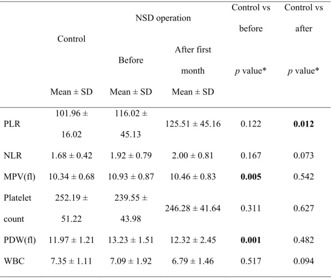
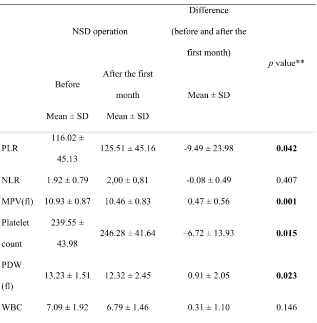
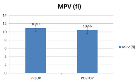
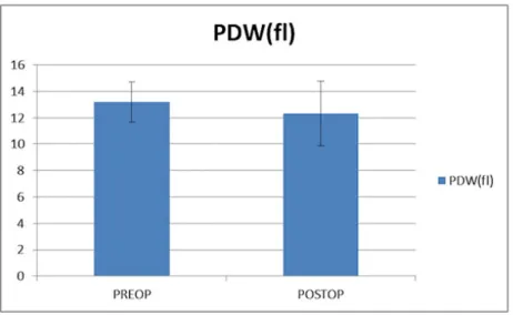
When the pre-operative and post-operative values of the patients in the study group were compared, a statistically significant difference was found between the two groups in terms

[6] The neutro- phil-to-lymphocyte ratio (NLR), calculated by dividing the number of neutrophils in a sample of peripheral blood by the lymphocyte count, is now used as a simple

The aim of this study was to evaluate the changes in hematolo- gic inflammatory markers such as neutrophil /lymphocyte ratio (NLR), platelet/ lymphocyte ratio (PLR), red

Accordingly, when patients with cellulitis were divided into two groups as ≥65 years and &lt;65 years, a statistically sig- nificant difference was noted among the WBC, NLR, and

Elevated C-re- active protein levels and increased cardiovascular risk in patients with obstructive sleep apnea syndrome. Szkandera J, Pichler M, Gerger A, et al (2013b)

Comparison of Liver Histopathology with Non-invasive Inflammation Markers as Neutrophil- lymphocyte Ratio, Platelet-lymphocyte Ratio and Mean Platelet Volume in Chronic Hepatitis

The aim of the present study was to show the NLR and MPV values in patients who have COM with or without ossicular/bony destruction or cholesteatoma, to compare the results