enign lung tumors are rare tumors; consist of less than 1% of body tu-mors. Among these, hamartomas are the most common and found in the range of 0.02% to 0.3% incidence.1Hamartoma may locate into
pulmonary parenchyma; these hamartomas are generally asymptomatic. Some hamartomas may be located in the trachea or bronchi, and present with result of tracheal or bronchial obstruction as cough, expectoration, and dyspnea.2Endobronchial hamartomas (EH) often grow and obstruct the
bronchial lumen substantially without symptoms such as dyspnea, and wheezing. Without an apparent radiologic sign, the symptoms secondary to bronchial obstruction may lead to misdiagnosis of asthma.1Because of
the benign nature of these tumors, bronchoscopic removal is generally re-commended, but in patients with irreversible lung destruction surgical tre-atment should be performed.3We herein report a case of a bulky EH placed
in the left main bronchus, diagnosed by bronchoscopy and extracted by en-dobronchial treatment via rigid bronchoscope.
CASE REPORT
A 52-year-old female patient with a 5-year history of asthma was admitted to the hospital complaining of chest pain and progressive shortness of bre-ath. The patient was a nonsmoker and had no other significant medical
his-Endobronchial Management of
an Endobronchial Hamartoma
AABBSS TTRRAACCTT Endobronchial hamartoma (EH) is a rare benign tumor of the bronchial tree. This tumor can obstruct the bronchus or cause bleeding. The treatment method is chosen according to location and extent of the tumor. Endobronchial hamartoma can be resected via bronchoscopic ap-proach; surgical therapy, is alternative only for cases where the tumor cannot be resected com-pletely through endoscopy, or if irreversible pulmonary changes secondary to obstruction of the bronchus occurred. We herein describe a 52-year-old female presented with progressive dyspnea and treated with diagnosis of asthma. In this patient, EH which is located in the left main lobe bronchus was found in radiologic assement. Bronchoscopy revealed a broad-based mass obstruct-ing the bronchus. The tumor was resected after coagulation of the tumor with argon plasma coag-ulation, by the rigid bronchoscope. Pathologic diagnosis was reported as EH. This report highlights the success of rigid bronchoscopic approach in the management of EH.
KKeeyywwoorrddss:: Endobronchial hamartoma; endobronchial management; argon plasma coagulation Ekrem Cengiz SEYHANa,
Mehmet Zeki GÜNLÜOĞLUb,
Mustafa DÜGERc,
Mehmet Akif ÖZGÜLa,
Elif ÇALIŞd,
İrem ÖZÖVERd aClinic of Chest Diseases, Yedikule Chest Diseases and Thoracic Surgery Training and
Research Hospital, Departments of bThoracic Surgery, cChest Diseases, dPathology,
İstanbul Medipol University Faculty of Medicine, İstanbul, TURKEY Re ce i ved: 23 Apr 2019 Ac cep ted: 08 Jul 2019 Available online: 09 Jul 2019 Cor res pon den ce:
Ekrem Cengiz SEYHAN Yedikule Chest Diseases and Thoracic Surgery Training and Research Hospital, Clinic of Chest Diseases, İstanbul, TURKEY [email protected]
Cop yright © 2019 by Tür ki ye Kli nik le ri
DOI: 10.5336/caserep.2019-66572
FIGURE 1: Thorax computed tomography scan showing a, b) the presence of a solid endobronchial lesion subtotally occluding the main left bronchus, c, d) to-tally opened left main bronchus after endobronchial treatment.
tory. Physical examination revealed decreased bre-ath sounds in the lower part of the left hemitho-rax, and no pathological finding in other systems. Chest x-ray, biochemical blood analysis and com-plete blood counts were normal.
A computed tomography (CT) of the thorax scan revealed the presence of a solid endobronchial lesion subtotally occluding the left main bronchus (Figure 1 a, b). The patient was performed bronc-hoscopy that confirmed the presence of a broad-based tumor arising from anterolateral wall of the left main bronchus covered with smooth mucosa, mobile during breathing and near totally occluding the bronchial lumen. The procedure was termina-ted by planning rigid bronchoscopy under general anesthesia to obtain large samples under safe con-ditions or with the hope of completely removing the tumor. Rigid bronchoscopy showed near com-plete obstruction of the proximal of the left main bronchus caused by a broad-based tumor (Figure 2 a, b). Using a biopsy forceps and argon plasma
coa-gulation (APC), the lesion was totally removed with no residual tissue (Figure 2 c). After removel of the tumor, cryotherapy was applied to the bronchial wall under the the tumor. Lumen of the bronchus was seen fully opened (Figure 2 d). Hi-stopathological evaluation revealed mature carti-lage and fat tissue in the deeper layer of the tumor, and the tumor was diagnosed as EH (Figure 3 a, b). Computerized tomography of the thorax taken im-mediately after the endobronchial therapy showed that the left lobe bronchus was entirely patent ( Fi-gure 1 c, d).
DISCUSSION
Hamartomas are rarely at endobronchial location.2
Endobronchial hamartoma is accounts for 1.4-19.5% of hamartomas.2Symptoms vary according
to the tumor location. The most of parenchymal hamartoma patients are asymptomatic. Unlike, most patients with EH had respiratory symptoms secondary to bronchial obstruction such as cough,
dyspnea, and obstructive pneumonia symptoms; fever and purulent sputum.3Patients with EH are
often misdiagnosed as asthma due to bronchial obstruction symptoms and signs, and these patients can remain undiagnosed for months or years as in our patient.1-4
Conventional radiography often shows chan-ges related to bronchial obstructions, such as ate-lectasis, pneumonia, and bronchiectasis in EH patients. However, chest X-ray of most of the pati-ents shows no pathological changes due to partial obstruction of the bronchus as in our case. Usual CT appearance of pulmonary hamartomas (PH) is a soft tissue including fat alternating with foci of cal-cification. Endobronchial hamartoma typically in-cludes more fat tissue than parenchymal ones.5On
CT scan, the EH appears as an endobronchial mass with or without signs of obstructive pneumonia or atelectasia.6At careful revision of the pertinent
li-terature, specific radiological features of EH were seen not to be reported; generally, they appear as ovoid low-density endobronchial lesions, with a smooth edge, the lesion includes focal fat, or fat al-ternating with calcification.7In our case, CT image
FIGURE 2: Rigid bronchoscopic view; a) the mass occluding left main lobe bronchus, b) the tumor covered with smooth mucosa, c) mass removal by using argon plasma coagulation, d) view of bronchial lumen after the removal of the mass.
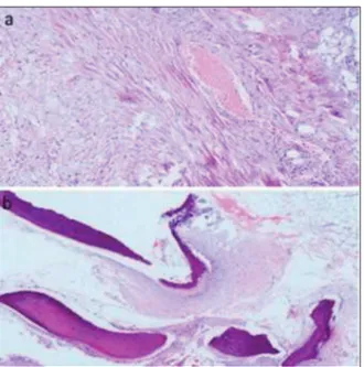
FIGURE 3: Histological examination shows endobronchial hamartoma: a) fib-roblast cell mesenchymal proliferation and vascular structure (hematoxylin and eosin, x100). b) mature adipose tissue, cartilage and occasionally la-mellar bone formation (hematoxylin and eosin, x20).
of the tumor was a low attenuation endobronchial mass in the left main bronchus.
Early endoscopic examination is important for detection of EHs before the lung distal to the obs-truction is irreversibly destroyed. EH at bronchos-copic view appears as a polypoid or pedunculated, well-circumscribed neoplasm, with a smooth sur-face, no signs of submucosal infiltration are seen.8
The diagnosis may be suspected by endoscopic ap-pearance, but EH cannot be reliably distinguished from the other benign bronchial tumors or typical carcinoid tumors, even with small biopsies. Consi-dering these difficulties, we did not prefer to re-ceive a small biopsy via flexible bronchoscope and the patients underwent rigid bronchoscophic pro-cedure.
Pulmonary hamartoma is composed of bronc-hial primitive mesenchymal tissue; this tissue can turn into various mesenchymal components.4So,
on histopathology, PH has various components like fat, cartilage, fibrous tissue, and rarely bone mar-row. Endobronchial hamartomas usually have more fat content than pulmonary intraparenchy-mal hamartomas.1For differential diagnosis from
the other benign endobronchial tumors and from carcinoid tumor, biopsy is generally required. In our case, we did not obtain a biopsy but final hi-stopathological evaluation showed mature cartilage and also adipose tissue in the deeper layer of the tumor, and diagnosis of EH was confirmed.
Endobronchial hamartoma can be succesfully removed both endoscopically and surgically. Cur-rently, bronchoscopic removal has been recom-mended for both diagnosis and also curative treatment, in the first line.8Bronchoscopic
resec-tion techniques such as Nd-YAG or snare laser, electrocautery, cryotherapy, and APC can be used. Surgical resection is preserved for the cases which
an EH cannot be resected through bronchoscopy, or cases needed lung resection secondary to irre-versible pulmonary parenchymal destruction from long-standing airway obstruction.3 In our case,
there were no irreversible pulmonary parenchymal changes and the tumor was accessible via bronc-hoscope, so we preferred to resect the the tumor using endobronchial treatment modalities such as APC and cryotherapy.
In conclusion, atypical bronchial asthma symptoms may be related to endobronchial tumors like an EH. According to our experience, bronchos-copic resection of an EH with cryoablation and APC is a safe and effective option for selected patients.
I
Innffoorrmmeedd CCoonnsseenntt
Written informed consent was obtained from the patient who par-ticipated in this case report.
S
Soouurrccee ooff FFiinnaannccee
During this study, no financial or spiritual support was received neither from any pharmaceutical company that has a direct connection with the research subject, nor from a company that provides or produces medical instruments and materials which may negatively affect the evaluation process of this study.
C
Coonnfflliicctt ooff IInntteerreesstt
No conflicts of interest between the authors and / or family members of the scientific and medical committee members or members of the potential conflicts of interest, counseling, ex-pertise, working conditions, share holding and similar situati-ons in any firm.
A
Auutthhoorrsshhiipp CCoonnttrriibbuuttiioonnss
I
Iddeeaa//CCoonncceepptt:: Ekrem Cengiz Seyhan; DDeessiiggnn:: Mustafa Düger;CCoonnttrrooll//SSuuppeerrvviissiioonn:: Mehmet Zeki Günlüoğlu; DDaattaa C
Coolllleeccttiioonn aanndd//oorr PPrroocceessssiinngg:: Mehmet Akif Özgül; AAnnaallyyssiiss a
anndd//oorr IInntteerrpprreettaattiioonn:: Ekrem Cengiz Seyhan; LLiitteerraattuurree RRee--v
viieeww:: Elif Çalış; WWrriittiinngg tthhee AArrttiiccllee:: Ekrem Cengiz Seyhan; C
Crriittiiccaall RReevviieeww:: İrem Özöver; RReeffeerreenncceess aanndd FFuunnddiinnggss:: Mus-tafa Düger; MMaatteerriiaallss:: Mehmet Akif Özgül.
1. Cosío BG, Villena V, Echave-Sustaeta J, de Miguel E, Alfaro J, Hernandez L, et al. Endo-bronchial hamartoma. Chest. 2002;122(1): 202-5. [Crossref] [PubMed]
2. Gjevre JA, Myers JL, Prakash UB. Pulmonary hamartomas. Mayo Clin Proc. 1996;71(1):14-20. [Crossref] [PubMed]
3. Kaya S, Karalezli A, Balkan E, Cakiroğlu E, Hasanoğlu HC. Endobronchial hamartoma re-moved by flexible fiberoptic bronchoscopy via electrocautery. Tuberk Toraks. 2006;54(3): 273-6. [PubMed]
4. Bernabéu Mora R, García Martín E, Polo Gar-cía LA, Méndez Martínez P, Abellán Martínez MC, Sánchez Gascón F. [Endobronchial chondroid hamartoma presenting as asthmatic attack]. An Med Interna. 2003;20(5):271-2.
[Crossref] [PubMed]
5. Ahn JM, Im JG, Seo JW, Han HS, Yoon HK, Kim WS, et al. Endobronchial hamartoma: CT findings in three patients. AJR Am J Roentgenol. 1994;163(1):49-50. [Crossref] [PubMed]
6. Karabulut N, Bir F, Yuncu G, Kiter G. Endo-bronchial lipomatous hamartoma: an unusual
cause of bronchial obstruction (2007: 7b). Eur Radiol. 2007;17(10):2687-90. [Crossref] [PubMed]
7. Suut S, Al-Ani Z, Allen C, Rajiah P, Durr-E-Sabih, Al-Harbi A, et al. Pictorial essay of ra-diological features of benign intrathoracic masses. Ann Thorac Med. 2015;10(4):231-42.
[PubMed]
8. Stey CA, Vogt P, Russi EW. Endobronchial lipomatous hamartoma: a rare cause of bronchial occlusion. Chest. 1998;113(1):254-5. [Crossref] [PubMed]