Can Tissue Factor, A Multifactorial Molecule
of the Hemostasis, be used As A Biomarker
for Thrombosis, Inflammation and Cancer?
Acta Pharm. Sci. Vol 55 No: 3. 2017DOI: 10.23893/1307-2080.APS.05521
*Corresponding author: Nesrin Emekli E-mail address: [email protected]
Nesrin Emekli
1*1Division of Biochemistry Faculty of Medicine, Medipol University, Istanbul, Turkey
INTRODUCTION
Hemostasis, which allows blood to circulate without clotting, allows bleeding to
stop through a rapid response to tissue damage. A clot is formed through
reac-tions involving blood vessels, thrombocytes, coagulation and fibrinolytic system,
which then bleeding stops and dissolves the fibrin clot.
1-5The work of hemostasis in a vascular damage is summarized as follows: 1-The artery
in the region is vasoconstricted, the purpose is to slow the blood flow through the
re-ABSTRACT
Tissue factor (TF) is a transmembrane protein found in many tissues and is active in various biological reactions. It is a member of the cytokine receptor superfam-ily and is referred to as CD 142 because of this feature. TF is also known as Factor III in the coagulation system and binds FVII/VIIa. The TF and FVIIa complex has both procoagulant and signaling activities. It functions in many biological pro-cesses, including hemostasis, thrombosis, inflammation and cancer. TF is essential for haemostasis but increased TF expression within atherosclerotic plaques and TF positive microparticles were detected in thrombotic conditions. TF increases inflammation by enhancing intravascular fibrin deposition and activates protease-activated receptors (PARs). TF and FVIIa complex also contribute to tumor growth by activating PAR2. Recent retrospective studies have shown that TF positive mi-croparticles increase in the plasma of cancer patients. Therefore TF may be sug-gested to be used as a biomarker. However further studies are required to reveal the availability of TF as a biomarker to identify cancer and risk of thromosis and inflammation.
gion, 2-Circulating thrombocytes recognize endothelial damage, apply adhesion,
secretion and aggregation, therefore rapidly forming a weak clot, 3-The
coagula-tion proteins in the circulacoagula-tion recognize that they’ve encountered an environment
different than the endothelium, and intact clots form in the interrupted region,
4-The fibrinolytic system helps repair the injured area, allowing the fibrin to
break down and the blood flow in the area to return to normal. In this system,
clotting is simultaneously created and prevented. The blood running in a
ves-sel does not coagulate but immediately it coagulates when encounters a foreign
surface such as tissue factor and collagen. Endothelial cells play a major role in
stopping a bleeding and preventing thrombus formation.
6-11The mechanism of partial thromboplastin time (PTT) and prothrombin time
(PT) tests used in the follow-up of diagnosis and treatment of bleeding and
coagulation-related diseases is clotting when blood encounters non endothelial
surface.
12This mechanism is important for arterial thrombus formation. In
sum-mary, it is not wrong to say that the formation of blood clots in endothelial
dys-function is not well managed.
The hemostatic system parameters continue to be at the center of scientific
re-search with unknown aspects, while providing opportunities to facilitate
diag-nosis and treatment on bleeding and thrombosis events. As in the past, the
in-cidence of thrombosis in arterial and venous circulation is currently very high.
Atherosclerotic cardiovascular and cerebrovascular diseases are still major
caus-es of morbidity and mortality worldwide.
13-17Tissue factor, which is present as Factor III in the clotting pathway and also
known as “CD 142” and “thromboplastin”, is a transmembrane protein which
enables thrombin formation and is multifunctional. Parallel to today’s
technol-ogy, there are many studies aiming to better know the structure of tissue factor,
while reporting that this protein is not only effective in the clotting system, but
also in inflammation and cancer formation.
18-33The purpose of this article is to draw attention to how some phases of hemostasis
developed historically and encourage young researchers to shine a light on the
unknown aspects of this subject.
Historical development of and current knowledge on hemostasis
The hypothesis that the German physician Rudolph Virchow, who lived between
1820 and 1902, which corresponds to the thrombosis aspect of present-day
he-mostasis;
1-Intravenous wall change (thrombosis due to atherosclerotic change,
inflam-matory change)
2-Change of blood components (hypercoagulability, thrombocyte activation,
an-ticoagulant insufficiency)
3- It is in the form of blood flow alteration (Deep vein thrombosis and
pulmo-nary embolism)
Numerous additions have been made to this hypothesis through scientific
stud-ies in parallel with the development of technology, but Wirschoff’s thrombus
formation hypothesis still remains valid (13,18-13).
Thrombocytes in Hemostasis
1842 is considered as the year of birth for thrombocytes. Because in those years,
four different researchers, unaware of each other, reported that a different
par-ticle was circulating differently from erythrocytes and leukocytes. In 1846,
Zim-merman noticed that these particles formed aggregates. The following rapid
developments in these studies have led to a better understanding of the role of
thrombocytes in hemostasis. In 1956, Ulutin and Karaca first announced to the
world the the mechanisms of secretion of thrombocytes, and that disorders in
this mechanism would cause a disease
1-2,34-42Coagulation Proteins and Tissue Factor (CD 142)
The foundations of the coagulation mechanism were laid in 1834. In those years,
brain tissue suspensions were found to be immediately lethal when
adminis-tered intravenously to animals. This finding led to the understanding that tissue
extracts formed clots in blood. Tissue factor had a place in Schmidt’s works in
1892 and in Morawitz’s work in 1904 and tissue factor, prothrombin and
fibrino-gen were all portrayed in the simple mechanism of the coagulation system. After
the year 1900, the studies focused on the tissue factor of clotting mechanism for
a long time. Works to purify tissue factor started in 1912 with Howell, and in
1944 with Chargaff et al. Studies on this purification of tissue factor continued
until the beginning of 1980s. Later studies were rather on the genetics of tissue
factor. At the end of the 1980s the tissue factor gene was isolated and cloned.
From the 1990s onwards, tissue factor was considered to be a real initiator in
the coagulation system. Among 13 clotting proteins, only the tissue factor is an
integral membrane protein. Clotting factors exhibit structural homology, while
only the tissue factor exhibits homology with type 2 cytokine receptors. For this
reason, tissue factor was also named as “CD 142”.
18-33, 43Tissue factor, a transmembrane protein, is present in various proportions in all
tissues, mostly in the brain, lung, and uterus. The tissue factor contains protein,
phospholipid and carbohydrate in varying proportions according to the tissue
from which it is obtained. The carbohydrate portion of the tissue factor is added
by posttranslational modification. Tissue factor is a cofactor for Factor FVII in
cell membranes. It is comprised of 263 amino acids in total. 219 of these amino
acids are located in the extracellular region, 23 amino acids in transmembranal
regions and 21 amino acids are in intracellular regions. The extracellular region
of the tissue factor contains the binding site for Factor VII.
18-33In 1947 Owren reported that Factor V was necessary for the formation of the clot
and this invention was followed by other coagulation proteins. Concerning the
conversion of prothrombin to thrombin, Prof.Dr. Walter H. Seegers’ intensive
studies and contributions from other researchers led to significant advances in
clotting mechanisms between 1905-1950.
The identification and
mechanism-de-scription of the 13 clotting proteins present in the present coagulation pathway,
expressed as “clotting factors”, were carried out by Davie and Ratnoff in 1964
(36).The current flow chart of the intractable coagulation system has emerged
from the examination of patients with bleeding findings with clotting defects.
13, 43-46The Prothrombin time test, administered by Quick in 1935, remains among
to-day’s gold standards in the control of the clotting system
4In our faculty, Medical
and Dental Practice students in the 1st grade isolete tissue factor from the bovine
lung and use it in the prothrombin time test. The students presented this study
in İstanbul Medipol University “Student Scintific Days” in 2015 and received a
runner-up prize
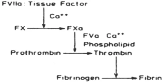
47Figure 1 shows the prothrombin time test’s flow diagram.
Figure 1.Biochemical reactions take place in the prothrombin time test (Ref.12)
Tissue Factor and Thrombosis
Adverse changes leading to thrombus formation in the blood stream begin with
hypercoagulation and endothelial activation. Wirschoff explains the reasons for
the thrombus that still remain valid today with the possibilities of the 1800’s.
One of the most important aspects of the present invention is the
demonstra-tion of the presence of microparticles bearing a tissue factor. Dimensions of
mi-croparticules carrying TF vary between 30-1000nm. Microparticles are released
from activated leukocytes and thrombocytes. Hatemi
11reported that in diabetic
and especially type 2 diabetic patients, thrombophilia is observed and that these
patients have increased coagulation factors, especially fibrinogen, and also
in-creased thrombocyte adhesion and aggregation, while fibrinolytic activities were
observed to be in a decrease. Today, contrary to previous knowledge, the
pres-ence of biocompatible tissue-factor-bearing microparticles has been
demon-strated and it is predicted that the tissue factor may be a biomarker in this area
since the importance of arterial thrombus formation is emphasized.
5-17,26,27,31,48-54Is Tissue Factor a Proinflammatory Agent?
Endothelial cells are a common point between coagulation and inflammation.
Inflammatory diseases are known to increase tendency to thrombus. In 1936,
life-threatening thromboembolic events were seen in ulcerative colitis cases,
which were thought to be associated with increased tissue factor. Fibrin
depos-its are seen in synoviums of patients with rheumatoid arthritis. The
extracel-lular portion of the tissue factor obtained with the recombinant technique was
injected into the articulations of healthy mice and 80% of the mice developed
arthritis. Studies on which factor to remove for the purpose of preventing the
relation between inflammation and coagulation have not yet reached a point of
clarity; this subject is still yet to be resolved.
32, 33, 55-61Tissue Factor and Cancer
Patients with cancer are more likely to have thrombosis. The relationship
be-tween thrombosis and cancer first began with the observations of Armand
Trousseau in 1865. Various studies have shown that tissue factor expression
increases in cancerous cells. In physiological conditions, the amount of micro
particles bearing tissue factor is low. These particles are seen to increase in
cas-es of cancer. TF plays an important role in development of physiological and
pathological angiogenesis. Since TF-deficient transgenic mice had deteriorating
vascular integrity, embryogenic death occurred in a short time and abnormal
development of embryonic development was observed. Similar histopathologic
results were seen in VEGF-deficient embryos. Here it is understood that TF and
VEGF act similarly. Proangiogenic and antiangiogenic factors are essential for
vessel growth and development. TF shows its effect on tumor growth,
metasta-sis and angiogenemetasta-sis independently of the clotting system but dependently on
clotting. TF indirectly increases angiogenesis and tumor growth in
coagulation-independent mechanisms by forming fibrin directly or via thrombin. In the
coagulation-dependent case, it is known that increasing TF expression leads to
increased fibrin formation.
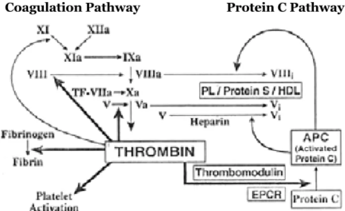
28-30, 62-68Control in Hemostatic System
Hemostatic system is internally controlled. Minimal thrombin coagulation,
which forms out of very small amounts of tissue factor, activates the system
by activating proteins and thrombocytes in the coagulation cascade. These
ef-fects of thrombin on procoagulants are positive feedback reactions in the
sys-tem. Thrombin, on the other hand, converts the protein C to the active protein
C (formerly autoprotrombin II-A), which is an anticoagulant, with the effect of
the protein C receptor (EPCR) and thrombomodulin found in the endothelium.
Figure 2 shows positive and negative feedback reactions of the clotting system.
That is, the clot formed with minimal tissue factor is neutralized with minimal
tissue factor.
Protein C complex, which exhibits anticoagulant activity by inhibiting strong
cofactors such as Factor VIII and Factor V in physiological conditions, has not
yet been successfully used in therapeutic applications such as heparin. However,
both protein C pathway and tissue factor inhibitor pathway (TFPI) are vital in
hemostatic control. This anticoagulant, first identified as autoprotrombin II-A
by Seegers and colleagues because of its relevance to prothrombin, has then
been referred to as “protein C”. Emekli and Ulutin reported that protein C
inhib-ited fibrin production in disseminated intravascular coagulation induced rabbits
and the animal model used in this study was the first in the literature.
69-84Coagulation Pathway Protein C Pathway
Figure 2: Effect of thrombin on anticoagulant system driven by procoagulant and protein C in the clotting mechanism (Ref.84)
Heparin is a heterogeneous polysaccharide with glucose derivatives and
sul-phates in its structure. It has been researched since 1800s and is still used as an
anticoagulant in treatments.
85-861953 and maintained it throughout the years, demanded this Journal be
pub-lished by İstanbul Medipol University from this year onwards.
87-90In conclusion, vascular, thrombocyte, coagulation and fibrinolytic systems of
he-mostasis are a complex reaction sequence containing negative-positive feedback
reactions, multienzymes systems, humoral and cellular procoagulant and
anti-coagulants. Tissue factor (CD 142), which is included in this system and has
ver-satile functions, is a current topic in today’s studies in that it can be a biomarker
for arterial-venous thrombus, inflammation and cancer.
REFERENCES
1. Ulutin ON, Şeştakof D. Coagulability of the blood in ischaemic heart disease. Lancet. 1958,
(Feb), 324-28.
2. Ulutin ON. The Plateletes: Fundamentals and Clinical Application Kağıt ve Basım İşleri AŞ. İstanbul, 1974.
3. Emekli N. Biochemical Aspects of Haemostasis. In: Basic and Applied Biochemistry. p.341-417, Marmara Üniversitesi Yayınları, Teknik Eğitim Fakültesi Matbaası, İstanbul, 1994. 4. Demir M.Geleneksel pıhtılaşma testleri ve klinik uygulamalardaki yeri, In: Klinik Biyokimya, Editörler: N.Emekli&T.Yiğitbaşı, Akademi Basın Yayın-Yayımcı: Nobel Tıp Kitapevleri Tic.Ltd. Şti. İstanbul, 2015; pp 323-331.
5. Ferhanoğlu B. Hemostaz mekanizması. In: Kanamaya ve Tromboza Eğilim. Sürekli Tıp
Eğitimi Etkinlikleri. Deomed Medikal Yayıncılık, İstanbul, 2003; pp 9-16.
6. ten Cate H, Hemker HC. Thrombin Generation and Atherothrombosis: What Does the Evi-dence Indicate?. Journal of the American Heart Association.5(8). e003553, 2016.
7. Tatsumi K, Mackman N. Tissue factor and atherothrombosis. Journal of Atherosclerosis and
Thrombosis. 2015, 22(6). 543-549.
8. Vural Ö. Damar endotelinin antienflamatuar ve antitrombojenik özellikleri 4.Ulusal Trom-boz, Hemostaz ve Anjioloji Kongresi Kitabı, May Matbaacılık Sanayi A.Ş. İstanbul, 2003; pp 71-75.
9. Arıkan E, Güldiken S.Endotel disfonksiyonu ve PPAR’lar ile ilişkisi 6. Ulusal Tromboz, He-mostaz ve Anjioloji Kongresi Kitabı, Cem Ofset Matbaacılık Sanayi A.Ş. İstanbul, 2006; pp 163-170.
10. Diamant M, Nieuwland R, Pablo RF, Sturk A, Smit JW, Radder JK. Elevated numbers of tissue-factor exposing microparticles correlate with components of the metabolic syndrome in uncomplicated type 2 diabetes mellitus. Circulation. 2002, 106(19) 2442-2447.
11. Hatemi H. Diyabet ve Hemostaz. 6. Ulusal Tromboz, Hemostaz ve Anjioloji Kongresi Kitabı, Cem Ofset Matbaacılık Sanayi A.Ş. İstanbul, 2006; pp 223-229.
12. Emekli N.Basic and Applied Biochemistry, Nobel Tıp Kitabevleri A.Ş.İstanbul, 2004; pp 384-385.
13. Emekli N. Hemostatik sistemin dünü bugünü. İçinde: Temel ve Uygulamalı Biyokimya. Cem Ofset Matbaacılık San A.Ş. İstanbul, 2004; pp 445-458.
factor activity in patients with acute unprovoked deep vein thrombosis and during the course of one year. Thrombosis Research. 2014, 134(5). 1093-1096.
15. Iacoviello L, Di Castelnuovo A, De Curtis A, Agnoli C, Frasca G, Mattiello A,Tumino R. Circu-lating Tissue Factor Levels and Risk of Stroke Findings From the EPICOR Study. Stroke. 2015,
46(6), 1501-1507.
16. van Es N, Bleker S, Sturk A, Nieuwland R. Clinical Significance of Tissue Factor–Exposing Microparticles in Arterial and Venous Thrombosis. In Seminars in Thrombosis and Hemosta-sis. Thieme Medical Publishers. 41(07), 718-727, 2015.
17. Camera M, Toschi V, Brambilla M, Lettino M, Rossetti L, Canzano P, Tremoli E. The role of tissue factor in atherothrombosis and coronary artery disease: insights into platelet tissue factor. Seminars in Thrombosis and Hemostasis. Vol. 41. No. 07. Thieme Medical Publishers, 2015.
18. Yarat A.Trombopllastik aktivite ve önemi. 4. Ulusal Tromboz, Hemostaz ve Anjioloji Kon-gresi Kitabı, May Matbaacılık Sanayi A.Ş. İstanbul, 2003; pp 97-105.
19. Cohen S, Chargaff E. Studies on the chemistry of blood coagulation IX. The thromboplastic protein from lungs. Journal of Biological Chemistry. 1940, 136(1). 243-256.
20. Cohen S, Chargaff E. Studies on the chemistry of blood coagulation XIII. The phosphatide constituents of the thromboplastic protein from lungs. Journal of Biological Chemistry. 1941,
139(2). 741-752.
21. Chargaff E, Bendich A, Cohen SS. The thromboplastic protein: structure, properties, disin-tegration. Journal of Biological Chemistry. 1944, 156(1), 161-178.
22. E, Bendich A, Cohen SS. The thromboplastic protein: structure, properties, disintegration.
Journal of Biological Chemistry. 1944, 156(1), 161-178.
23. Ulutin ON, Mammen EF, Seegers WH. Purified platelet cofactor I and purified autopro-thrombin II in thromboplastin generation test. Thrombos Diathes Haemorrh. 1962, 5, 456-462. 24. Mammen EF. Physiology and Biochemistry of Blood Coagulation. in: Thrombosis and Bleed-ing Disorders Theory and Methods. Eds. Bang NU,Beller FK,Deutsch E, Mammen EF.Academic Press, Newyork-London, 1971; pp 24-26.
25. Antoniak S, Tatsumi K, Hisada Y, Milner JJ, Neidich SD, Shaver CM, Mackman N.Tissue factor deficiency increases alveolar hemorrhage and death in influenza A virus-infected mice.
Journal of Thrombosis and Haemostasis. 14: 1238–1248, 2016.
26. Brambilla M, Facchinetti L, Canzano P, Rossetti L, Ferri N, Balduini A, Toschi V. Human megakaryocytes confer tissue factor to a subset of shed platelets to stimulate thrombin genera-tion. Thrombosis and haemostasis. 2015, 114(3), 579-592.
27. Arderiu G, Peña E, Badimon L. Angiogenic microvascular endothelial cells release micropar-ticles rich in tissue factor that promotes postischemic collateral vessel formation.
Arteriosclero-sis, thromboArteriosclero-sis, and vascular biology. 35(2). 348-357, 2015 Publishers. 41.07. 708-717, 2015.
28. Yarat A. Doku Faktörü ve Kanser 5. Ulusal Tromboz, Hemostaz ve Anjioloji Kongresi Kitabı, Tasarım Baskı: İkite Reklam Hizmetleri İstanbul, 2004; pp 197-203.
29. Emekli N. Kanserde hemostatik değişimler 6. Ulusal Tromboz, Hemostaz ve Anjioloji Kon-gresi Kitabı, Cem Ofset Matbaacılık Sanayi A.Ş. İstanbul, 2006; pp 25-36.
Throm-bosis and Hemostasis. Thieme Medical Publishers. 41.07, 747-755, 2015.
31. D’Andrea D, Ravera M, Golino P, Rosica A, De Felice M, Ragni M, Gargiulo A. Induction of tissue factor in the arterial wall during recurrent thrombus formation. Arteriosclerosis,
Throm-bosis and Vascular Biology. 2003, 23(9), 1684-1689.
32. Tatsumi K, Mackman N. Tissue factor and atherothrombosis. Journal of atherosclerosis and Witkowski M, Landmesser U, Rauch U. Tissue factor as a link between inflammation and coagulation. Trends in cardiovascular medicine. 2016, 26(4), 297-303.
33. Rondina MT, Tatsumi K, Bastarache JA, Mackman N. Microvesicle Tissue Factor Activity and Interleukin-8 Levels are Associated with Mortality in Patients with Influenza A/H1N1 In-fection. Critical care medicine. 44.7, 2016.
34. Ulutin ON, Karaca M. A study on the pathogenesis of thrombopathia using the platelet os-motic resistance test. Brit J Haemat. 1959, 5, 302-306.
35. Ulutin ON, Becit N, Ulutin ŞB. A study on the procoagulant activity of platelets. Med Bull
İstanbul Univ. 1969, 2, 83-88.
36. Batırel S, Yarat A, Emekli N. Effects of short term streptozotocin-induced diabetes and vitamin C on platelet Non-enzymatic glycation. Pathophysiol. Haemost. Thromb. 2010,
37(2-4): 72-76.
37. Aktulga AZ, Ulutin ON. Platelet phospholipids in atherosclerotics and in normals before and after release. Eds. Neri Serneri GG, CRM Prentice. In: Haemostasis and Thrombosis. Academic Press, London, 1979.
38. Gall A, Kaliman J, Sinzinger H. Platelet sensitivity to prostaglandins (PGE1,D2, I2) in patients with coronary heart disease at young and older age. In: Proccedings of the 1981 International İstanbul Symposium on Haematology. Sermet Matbaası, İstanbul, 1982; pp 63-70.
39. Çizmeci G, Aytiş Ş and Ulutin ON. Transport of arachidonic acid across platelet membrane and PG metabolism in atherosclerosis. In: Proccedings of the 1981 International İstanbul Sym-posium on Haematology. Sermet Matbaası, İstanbul, 1982; pp 55-62.
40. Yardımcı TU. Alterations observed in the platelet glucose permease system fo atheroscle-rotic subjects and the effect of release inducers on theis system. In: Proccedings of the 1981 In-ternational İstanbul Symposium on Haematology. Sermet Matbaası, İstanbul, 1982; pp 37-48. 41. Emekli N and Ulutin ON. The effect of cyclooxygenase inhibitors on the glycoprotein syn-thesis. In: Proccedings of the 1981 International İstanbul Symposium on Haematology. Sermet Matbaası, İstanbul, 1982; pp 185-191.
42. Emekli N.Galactose transport and glycoprotein changes in the platelets of atherosclerot-ics. In: Proccedings of the 1981 International İstanbul Symposium on Haematology. Sermet Matbaası, İstanbul, 1982; pp 80-89.
43. Drake TA, Ruf W, Morrissey JH, Edgington TS. Functional tissue factor is entirely cell sur-face expressed on lipopolysaccharide-stimulated human blood monocytes and a constitutively tissue factor-producing neoplastic cell line. The Journal of Cell Biology. 1989, 109(1), 389-395. 44. Tenekeciğil A. Koyun ve kuzu akciğerinden izole edilen doku faktörü aktivitesinde lipitlerin etkisi. Yüksek Lisans tezi, Danışman: Prof. Dr. Nesrin Emekli İstanbul Medipol Üniversitesi Sağlık Bilimleri Enstitüsü, 2016.
45. Davie EW and Ratnoff. Waterfall sequence for intrinsic blood clotting. Science. 1964, 145, 1310-1312.
46. Cimmino G, Ciccarelli G, Golino P. Role of tissue factor in the coagulation network. In Semi-nars in Thrombosis and Hemostasis. Thieme Medical. 2014, 99-105.
47. Aydemir AB, Zengin OM, Eroğlu M, Özcan C, Tenekecigil A. Kuzu akciğerinden elde edilen doku faktörünün protrombin zamanı testinde kullanılması. İstanbul Medipol Üniversitesi Tıp Fakültesi, Bilim Şenliği, 2015.
48. Kılıç B, Batirel S, Akdeste Z, Emekli N. Deneysel Hiperlipidemi ve Apo-B Konjugatı Uygulamasının Kan Doku Faktörü Seviyesi ve Karaciğer Doku Faktörü Aktivitesi Üzerine Et-kileri. MÜSBED. 2012, 2(2):64-71.
49. Emekli Alturfan E, Basar I, Malali E, Elemek E, Oktay S, Ayan F, Emekli N, Noyan U: Plasma Tissue Factor Levels and Salivary Tissue Factor Activities of Periodontitis Patients with and without Cardiovascular Disease. Pathophysiol. Haemost Thromb. 2010, 37(2-4), 77-81. 50. Emekli Alturfan E, Kasikci E, Yarat A: Tissue factor activities of streptozotocin induced dia-betic rat tissues and the effect of peanut consumption. Diabetes Metab Res Rev. Nov. 2007,
23(8), 653-658.
51. Emekli Alturfan E, Basar I, Alturfan AA, Ayan F, Koldas L, Balci H, Emekli N. The relation between plasma tissue factor and oxidized LDL levels in acute coronary syndromes.
Patho-physiol. Haemost. Thromb. 2007, 36(6), 290-297.
52. Emekli Alturfan E, Kasikci E, Yarat A: Peanuts improve blood glutathione, HDL-cholesterol level and change tissue factor activity in rats fed a high-cholesterol diet. Eur J Nutr. 2007,
46(8), 476-482.
53. Reichman-Warmusz E, Reichman-Warmusz E, Domal-Kwiatkowska D, Matysiak N, Kurek J, Spinczyk D, Dudek D, Wojnicz R. Tissue factor is unregulated in microvascular endothelial cells of patients with heart failure. Journal of Clinical Pathology. 2016, 69(3), 221-225. 54. Mathieu E, van Dreden P, Aulagnier J, Grusse M, Dreyfus JF, Francois D, Vasse M. De-creased levels of procoagulant phospholipids in bleeding patients treated by vitamin K antago-nists. Thrombosis Research. 2016, 137, 36-40.
55. Witkowski M, Landmesser U, Rauch U. Tissue factor as a link between inflammation and coagulation. Trends in cardiovascular medicine. 2016, 26(4), 297-303.
56. Bakarewa MI, Morrissey JH, Tarkowski A. Tissue factor as aproinflammatory agent.
Arthri-tis es. 2002, 4, 190-195.
57. Alturfan A. A, Eralp L, Emekli N: Investigation of inflammatory and hemostatic parameters in female patients undergoing total knee arthroplasty surgery. Inflammation. 2008, 31(6):414-421.
58. Emekli N. Koagulasyon ve inflamasyon 5. Ulusal Tromboz, Hemostaz ve Anjioloji Kongresi Kitabı, Tasarım Baskı: İkite Reklam Hizmetleri İstanbul, 2004; pp 69-77.
59. Libby P, Simon DI. Inflammation and thrombosis. Circulation. 2001, 103, 1718-1720. 60. Xue M, Sun Z, Shao M, Yin J, Deng Z, Zhang J, Han Y. Diagnostic and prognostic utility of tissue factor for severe sepsis and sepsis-induced acute lung injury. Journal of Translational
Medicine. 2015, 13(1), 1.
61. Xue M, Sun Z, Shao M, Yin J, Deng Z, Zhang J, Han Y. Diagnostic and prognostic utility of tissue factor for severe sepsis and sepsis-induced acute lung injury. Journal of Translational
62. Berkarda B. Introduction to cancer and blood coagulation. In: Proceedings of the Interna-tional İstanbul Symposium on Hematology. Sermet Matbaası İstanbul 1982; pp 210-214. 63. Woei-A-Jin FJSH, Tesselaar MET, Rodriguez PG, Romijn FPHTM, Bertina RM, Osanto S. Tissue factor-bearing microparticles and CA19.9: two players in pancreatic cancer-associated thrombosis & quest. British Journal of Cancer, 2016.
64. Nomura S, Niki M, Nisizawa T, Tamaki T, Shimizu M. Microparticles as Biomarkers of Blood Coagulation in Cancer. Biomarkers in cancer. 2015, 7, 51.
65. Falanga A, Schieppati F, Russo D. Cancer tissue procoagulant mechanisms and the hyper-coagulable state of patients with cancer. In Seminars in Thrombosis and Hemostasis. Thieme Medical Publishers. 41.07.756-764, 2015.
66. Motoori M, Yano M, Tomita Y, Takahashi H, Tanaka K, Sugimura K, Goto K. Tissue factor predicts response to chemotherapy in esophageal cancer. Journal of Surgical Research. 2014,
191(1), 99-105.
67. Arderiu G, Peria E, Badimon L. Angiogenic microvascular endothelial cells release micro-particles rich in tissue factor that promotes post ischemic collateral vessel formation. Arterio-sclerosis, Thrombosis and Vascular Biology. 2015 35(2), 348-357.
68. Unruh D, Sagin F, Adam M, Van Dreden P, Woodhams BJ, Hart K, Bogdanov VY. Levels of alternatively spliced tissue factor in the plasma of patients with pancreatic cancer may help predict aggressive tumor phenotype. Annals of Surgical Oncology. 2015, 22(3), 1206-1211. 69. Tunalı T, Yarat A, Bulut M, Emekli N. 6,7-Dihydroxy-3-phenylcoumarin inhibits thrombo-plastin induced disseminated intravascular coagulation. Br J Haematol. 2004, 126(2): 226-230.
70. Esmon CT. Protein C: Biochemistry, physiology and clinical implications. Blood. 1983, 62, 171-155-1162.
71. Stenflo J, Fernlund P.Amino acid sequence of the heavy chain of bovine protein C. J Biol
Chem. l982, 257, 121-128.
72. Comp PC. Hereditary disorders predisposing to thrombosis. In: Progress in Hemostasis and Thrombosis, volume 8. Ed.Coller BS. Grune & Stratton Inc, Harcourt Brace Jovanovich Pub-lishers, New York, 1986; pp 71-99.
73. Emekli NB, Ulutin ON. The protective effect of autoprothrombin II-A anticoagulant on ex-perimental DIC formed animals. Haematologia. 1980, 65, 544-551.
74. Emekli NB, Ulutin ON. Some properties of autoprothrombin II-A anticoagulant, Recent Progress in Blood Coagulation and Thrombosis Research. Biblith Haem. 44, 15-20,1978. 75. Emekli NB, Ulutin ON. Sığır plazmasından protrombin ve inhibitörün elde edilmesi.
Cerrahpaşa Tıp Fakültesi Dergisi. 1978, 19, 235-248.
76. Emekli NB, Ulutin ON. Otoprotrombin II- A antikoagulantın yaygın damar içi pıhtılaşması meydana getirilen deney hayvanlarına etkisi. Cerrahpaşa Tıp Fakültesi Dergisi. 1978, 19, 249-259.
77. Seegers WH, Novoa RL, Henry HI: Relationship of “new” vitakin K dependent protein C and “old” Autoprothrombin II-A. Thrombosis Research. 1976, 8, 543-552.
78. Emekli N, Ulutin ON. International Reg. Of animal models of thrombus and hemorrhagic disease. National Academy Press USA. pp 41, 1981.
79. Ulutin ON, Emekli NB. Prothrombin C and autoprothrombin II-A anticoagulant. New İstanbul Contr Clin Sci. 1982, 13, 171-185.
80. Rao LVM, Esmon CT, Pendurthi UR. Endothelial cell protein C receptor: a multiliganded and multifunctional receptor. Blood. 2014, 124(10), 1553-1562.
81. Akar N. Endotelyal protein C reseptörü. 6. Ulusal Tromboz, Hemostaz ve Anjioloji Kongresi Kitabı, Cem Ofset Matbaacılık Sanayi A.Ş. İstanbul, 2006; pp 91-93.
82. Demir M.Antikoagulan tedavinin dünü bugünü ve yarını 5. Ulusal Tromboz, Hemostaz ve Anjioloji Kongresi Kitabı, Tasarım Baskı: İkite Reklam Hizmetleri, İstanbul, 2004; pp 247-258. 83. Özbay G.Akut miyokard infarktüsünde trombolitik tedavi. 4. Ulusal Tromboz, Hemostaz ve Anjioloji Kongresi Kitabı, May Matbaacılık Sanayi A.Ş. İstanbul, 2003; pp 255-261.
84. Griffin JH. Control of coagulation reactions, In: Williams Hematology 6thEdition, Mc Graw-Hill Medical Publication Division, New York, 2001; pp 1435-1449.
85. Haliloğlu E, Usta S, Özkan M, Sayıl Ö. Akut derin ven trombozunun uzun süreli tedavisinde düşük molekül ağırlıklı heparin ile oral antikoagulanların karşılaştırılması. Turkish J Thoracic
and Cardiovascular Surgery. 2011, 19(4), 551-558.
86. Badak Mİ, Kurtoğlu T, Özkısacık EA, Boğa M, Gürcün U, Sirek N, Köseoğlu K, Dişcigil B. Derin ven trombozunda standart heparin tedavisi sonuçlarımız. Adnan Menderes Üniversitesi
Tıp Fakültesi Dergisi. 2005, 6(2), 19-22.
87. Demirayak Ş. Aims and scope of Acta Pharmaceutica Sciencia. Acta Pharm. Sci. 2016,
54(1), 5.
88. Özsoy Y. Biography of former editor Prof. Dr. Kasım Cemal Güven. Acta Pharm. Sci. 2016,
54(1), 6.
89. Güven KC, Özsoy Y, Ulutin ŞB, Ulutin ON. Anticoagulant and antithrombin and fibrinolytic activities of heparin fractions. Acta Pharmaceutica Turcica. 1990, 32, 107-111.
90. Güven KC, Toulemonde F, Koç H. Molecular weight and glycosamine glycane content of raparin, a heparinoid obtained from Rapana venosa. Acta Pharmaceutica Turcica 2003, 45, 77-80.