Original Article
Cryptogenic and Secondary Organizing Pneumonia:
Clinical Presentation, Radiological and Laboratory
Findings, Treatment, and Prognosis in 56 Cases
INTRODUCTION
Organizing pneumonia (OP) is defined histopathologically by intra-alveolar buds of granulation tissue, consisting of in-termixed myofibroblasts and connective tissue. This condition can be cryptogenic OP (COP) or secondary OP (SOP) to other known causes [1].
The bronchiolitis obliterans with OP (BOOP) terminology was abandoned because the main event is OP, and bronchiolitis oblit-erans is only a minor finding [1]. The presenting symptoms, radiographic findings, and laboratory data are usually non-specific [2]. SOP has a characteristic pathological pattern, but it is associated with known diseases or situations. Some of these entities include connective tissue diseases, infections, malignancies, drugs, radiation, transplantation, and aspiration. COP is diagnosed in the appropriate clinical, radiographic, and pathological setting after excluding situations associated with SOP [3].
The aim of the present study was to examine the etiological factors; clinical, laboratory, and radiological features; treat-ment response; and prognosis in patients with COP and SOP.
MATERIALS AND METHODS
The medical records of the patients from 2010 to 2016 were retrospectively reviewed. Demographic characteristics, radiological examinations, laboratory data, pulmonary function tests (PFTs), and follow-up data were collected retrospec-tively through the hospital information management system.
The diagnosis of OP was based on the following criteria:
1. Abnormal chest radiograph and/or thorax high-resolution computed tomography (HRCT) ranging from multiple acinar/ nodular shadows,
Ayşe Baha1 , Fatma Yıldırım2 , Nurdan Köktürk3 , Züleyha Galata3 , Nalan Akyürek4 , Nilgün Yılmaz Demirci3 , Haluk Türktaş3
1Department of Pulmonary Medicine, Ufuk University School of Medicine, Ankara, Turkey
2Clinic of Pulmonary Disease and Intensive Care Unit, Dışkapı Yıldırım Beyazıt Training and Research Hospital, Ankara, Turkey 3Department of Pulmonary Medicine, Gazi University School of Medicine, Ankara, Turkey
4Department of Pathology, Gazi University School of Medicine, Ankara, Turkey
Address for Correspondence: Ayşe Baha, Department of Pulmonary Medicine, Ufuk University School of Medicine, Ankara, Turkey
E-mail: [email protected]
©Copyright 2018 by Turkish Thoracic Society - Available online at www.turkthoracj.org
201
Cite this article as: Baha A, Yıldırım F, Köktürk N, Galata Z, et al. Cryptogenic and Secondary Organizing Pneumonia: Clinical Presentation,
Radiological and Laboratory Findings, Treatment, and Prognosis in 56 Cases. Turk Thorac J 2018; 19(4): 201-8.
OBJECTIVES: Organizing pneumonia is an important disease that is associated with non-specific clinical findings and radiographic
appearance. Our aim was to examine the clinical and radiological features, laboratory findings, diagnostic approach, and response to therapy in subjects with cryptogenic (COP) and secondary organizing pneumonia (SOP).
MATERIALS AND METHODS: Patients’ medical records were retrospectively reviewed between 2010 and 2016 in our hospital. We
analyzed the symptoms, radiological features, pulmonary function tests, laboratory data, bronchoalveolar lavage findings, treatment, and prognosis.
RESULTS: Thirty-seven patients were diagnosed with COP and 19 patients with SOP. The most common causes of SOP were determined
as rheumatologic diseases. The most common symptoms were cough (71.4%) and dyspnea (66.1%). Bilateral symmetrical consolidations were the most prominent radiological appearance in both COP and SOP. The general radiographic findings were not different in COP and SOP. However, pulmonary lesions were located rather in the central (p=0.023) and middle (p=0.001) zones in patients with SOP. Corticosteroid (CS) therapy was administered to 34 (60.7%) patients. Two patients showed deterioration despite CS therapy.
CONCLUSION: The clinical and radiographic findings, treatment response, prognosis were similar in patients with COP and SOP. KEYWORDS: Cryptogenic organizing pneumonia, secondary organizing pneumonia, clinical radiological laboratory features, prognosis
Abstract
2. Histopathologically, the presence of intraluminal fibrotic buds within the alveoli and alveolar ducts with or without bronchiolar involvement and infiltration of chronic inflam-matory cells in the alveolar septa with preservation of the alveolar structure,
3. Negative microbiological analysis on bronchoalveolar la-vage (BAL) fluid, and
4. A well-documented improvement that was either sponta-neous or after exclusive corticosteroid (CS) treatment. Multidisciplinary approach was used in the diagnosis, treat-ment, and follow-up of the patients. Patients who were not diagnosed histopathologically were diagnosed according to clinical and radiological features. The diagnosis of OP was supported by the response to CS treatment in these patients. No pathogen was detected in the BAL examination of pa-tients.
PFTs (SensorMedics Vmax Series 20C Respiratory Analyzer; SensorMedics Corp., Yorba Linda, CA, USA) were performed according to the American Thoracic Society guidelines. Arte-rial blood gases were measured at rest (Radiometer ABL 735 blood gas analyzer; Radiometer, Copenhagen, Denmark). Fiberoptic bronchoscopy (FOB) (Olympus EVIS LUCERA CV-260; Olympus, Tokyo, Japan) was performed to obtain BAL and transbronchial biopsy (TBB). BAL was applied accord-ing to the guidelines. HRCT was used to detect the most af-fected area. The right middle lobe or lingula was used in the presence of diffuse involvement. At least three aliquots of 40 mL sterile saline at room temperature were instilled through FOB and gently retrieved by mechanical suction. Only the second aliquot was used for BAL analysis [4]. The following technique was used for biopsy. With the bronchoscope in the appropriate segmental bronchus, the forceps, with a biopsy
cup of 2×4 mm, is passed into the bronchus and advanced until resistance is met. It is withdrawn at 2 cm, opened, and again advanced until resistance is met. The patient breathes out, and the forceps is closed and withdrawn with the bi-opsy specimen.After fixation in formol saline, the tissue is prepared by the Millipore filter technique and processed by conventional methods [5].
Patients were evaluated at 1, 3, and 6 months and 1 year of diagnosis and examined in four categories according to their follow-up status: stable, remission, progression, and exitus. Stable patient was defined as a patient whose symptoms, func-tional status, and radiological findings remain unchanged. Remission was defined as a patient whose symptoms, func-tional status, and radiological findings remain recovered. Progression was defined as a patient whose symptoms, func-tional status, and radiological findings remain worsened. Approval of the ethics committee was not obtained because the study was designed retrospectively. The authors declare that there is no conflict of interests regarding the publication of this paper.
Statistical Analysis
The SPSS (Statistical Packege for Social Sciences) version 21.0 (IBM Corp.; Armonk, NY, USA) software was used for statis-tical analysis. Continuous data for normal distribution are expressed as mean±standard deviation (SD). A p<0.05 was considered as significant. In descriptive statistics, frequency and percentage were used for discrete data, and mean±SD were used for continuous variables. The normality test was performed by the Kolmogorov-Smirnov and Shapiro-Wilk methods. The Mann-Whitney U test and t-test were used to compare the differences between the groups.
RESULTS
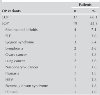
Table 1 shows the clinical types of OP and the associated diseases of SOP. The most common causes of SOP were de-termined as rheumatologic diseases and malignant diseases, respectively.
Table 2 shows the demographic features and symptoms. The most common symptoms were cough (71.4%), dyspnea (66.1%), and malaise (64.3%). Cough was usually non-pro-ductive. Dyspnea lasted from 10 days to 2 years. Both COP and SOP did not differ with regard to demographic findings and symptoms.
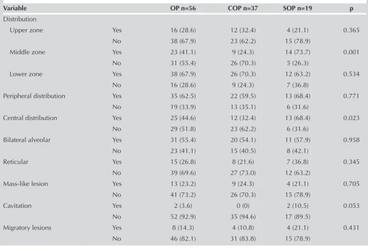
X-ray findings included consolidation in 29 (51.8%) patients that was bilateral in 68.0% and unilateral in 32.0% of the pa-tients. Migratory alveolar infiltrates were observed in 8 (14.3%) patients. A diffuse reticulonodular pattern was present in 15 (26.8%) patients and mass-like lesions in 13 (23.2%) patients. There was no difference between the groups in consolida-tion, migratory infiltraconsolida-tion, reticulonodular pattern, and mass-like lesion. The SOP group had more middle zone infiltration (p=0.025) and central localization (p=0.049) in chest X-ray. On HRCT scan, a reverse halo sign was not detected in pa-tients. None had honeycomb changes. The distribution of the infiltrates was more frequent in the lower and peripheral
202
Table 1. Clinical variants of OP in the study population (n=56) Patients OP variants n % COP 37 66.1 SOP 19 33.9 Rheumatoid arthritis 4 7.1 SLE 1 3.6 Sjogren syndrome 3 5.4 Lymphoma 2 3.6 Ovary cancer 1 1.8 Lung cancer 2 3.6 Nasopharynx cancer 1 1.8 Psoriasis 1 1.8 HBV 1 1.8 Stevens-Johnson syndrome 1 1.8 POEMS 1 1.8
OP: organizing pneumonia; COP: cryptogenic organizing pneumonia; SOP: secondary organizing pneumonia; SLE: systemic lupus erythematosus; HBV: hepatitis B virus; POEMS: polyneuropathy, organomegaly, endocrinopathy, M protein, and skin changes
zones. Middle zone involvement (p=0.001) and central local-ization (p=0.023) in the SOP group were significantly higher than those in the COP group (Table 3).
Erythrocyte sedimentation rate (ESR) was >20 mm/h in 38 (67.8%) patients. The mean C-reactive protein (CRP) level in
the total patient population was 34.5±32.6 mg/L. There was no difference between the groups in CRP levels (p=0.868). The leukocyte count was >10,000/mm3 in 18 (32.1%)
tients. A slight eosinophilia was observed in 18 (32.1%) pa-tients. There was no difference between the groups with re-gard to laboratory parameters (Table 4).
203
Table 2. Clinical characteristics of patients with COP and SOPVariable OP n=56 COP n=37 SOP n=19 p
Age, mean±SD, years 57.09±12.68 57.38±12.04 56.53±14.15 0.789 Age, range, years 28-83 35-83 28-75 -Male 29 (51.8%) 20 (54.1%) 12 (63.2%) 0.267 Female 27 (48.2%) 17 (45.9%) 7 (36.8%) Smoking Smokers 33 (58.9%) 22 (59.5%) 11 (57.9%) 0.775 Non-smokers 21 (37.5%) 13 (35.1%) 8 (42.1%) Missing data 2 (3.6%) 2 (5.4%) 0 (0%)
Antibiotic use Yes 31 (55.4%) 20 (54.1%) 11 (57.9%) 0.948 Before diagnosis No 22 (44.6%) 14 (37.8%) 8 (42.1%) Cough Yes 40 (71.4%) 25 (67.6%) 15 (78.9%) 0.747 No 14 (25.0%) 10 (27%) 4 (21.1%) Sputum Yes 20 (35.7%) 12 (32.4%) 8 (42.1%) 0.769 No 34 (60.7%) 23 (62.2%) 11 (57.9%) Hemoptysis Yes 4 (7.1%) 3 (8.1%) 1 (5.3%) 0.657 No 47 (83.9%) 20 (81.1%) 17 (89.5%) Dyspnea Yes 37 (66.1%) 23 (62.2%) 14 (73.7%) 0.760 No 17 (30.4%) 12 (32.4%) 5 (26.3%) Fever Yes 24 (42.9%) 16 (43.2%) 8 (42.1%) 0.775 No 28 (50.0%) 17 (45.9%) 11 (57.9%)
Chest pain Yes 7 (12.5%) 5 (13.5%) 2 (10.5%) 1.000 No 46 (82.1%) 30 (81.1%) 16 (84.2%)
Loss of appetite Yes 25 (44.6%) 16 (43.2%) 9 (47.4%) 0.939 No 27 (48.2%) 17 (45.9%) 10 (52.6%)
Malaise Yes 36 (64.3%) 24 (64.9%) 12 (63.2%) 0.541 No 16 (28.6%) 9 (24.3%) 7 (36.8%)
Weight loss Yes 14 (25.0%) 8 (21.6%) 6 (31.6%) 0.515 No 39 (69.6%) 27 (73.0%) 12 (63.2%) Crackles Yes 31 (55.4%) 20 (54.1%) 11 (57.9%) 0.869 No 24 (42.9%) 16 (43.2%) 8 (42.1%) Wheezing Yes 2 (3.6%) 2 (5.4%) 0 (0%) 0.539 No 53 (94.6%) 34 (91.9%) 19 (100%) Clubbing Yes 1 (1.8%) 0 (0%) 1 (5.3%) 0.333 No 53 (94.6%) 37 (100%) 17 (89.5%) Cyanosis Yes 5 (8.9%) 2 (5.4%) 3 (15.8%) 0.327 No 50 (89.3%) 34 (91.9%) 16 (84.2%)
Bronchial breath sounds Yes 2 (3.6%) 2 (5.4%) 0 (0%) 0.539 No 53 (94.6%) 34 (91.9%) 19 (100%)
NPEF Yes 23 (41.1%) 15 (40.5%) 8 (42.1%) 0.975 No 32 (57.1%) 21 (56.8%) 11 (57.9%)
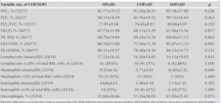
PFT was available for 49 patients, and diffusion capacity for carbon monoxide (DLCO) test was available for 43 pa-tients (Table 5). Eleven (22.4%) papa-tients had pure-restric-tive defect, and 7 (14.3%) patients had obstrucpure-restric-tive defects. DLCO was reduced (<60%) in 15 (34.9%) of 43 patients. Normal PFT was determined in 30 (61.2%) of 49 patients (Table 5).
BAL analysis was completed in 38 (67.8%) patients. Neutro-philia >5% in BAL fluid was observed in 20 (52.6%) patients. The BAL neutrophil count was higher in the SOP group than in the COP group (16.85±5.35% vs 5.71±5.01) (p=0.044). Lymphocytes were 28.6% of the total cells. A lymphocytosis of >25% was identified in 12 (31.6%) patients. There was no difference between the groups in terms of spirometric
mea-204
Table 3. CT scan and HRCT findings in 56 patients with COP and SOP
Variable OP n=56 COP n=37 SOP n=19 p
Distribution
Upper zone Yes 16 (28.6) 12 (32.4) 4 (21.1) 0.365 No 38 (67.9) 23 (62.2) 15 (78.9)
Middle zone Yes 23 (41.1) 9 (24.3) 14 (73.7) 0.001 No 31 (55.4) 26 (70.3) 5 (26.3)
Lower zone Yes 38 (67.9) 26 (70.3) 12 (63.2) 0.534 No 16 (28.6) 9 (24.3) 7 (36.8)
Peripheral distribution Yes 35 (62.5) 22 (59.5) 13 (68.4) 0.771 No 19 (33.9) 13 (35.1) 6 (31.6)
Central distribution Yes 25 (44.6) 12 (32.4) 13 (68.4) 0.023 No 29 (51.8) 23 (62.2) 6 (31.6)
Bilateral alveolar Yes 31 (55.4) 20 (54.1) 11 (57.9) 0.958 No 23 (41.1) 15 (40.5) 8 (42.1)
Reticular Yes 15 (26.8) 8 (21.6) 7 (36.8) 0.345 No 39 (69.6) 27 (73.0) 12 (63.2)
Mass-like lesion Yes 13 (23.2) 9 (24.3) 4 (21.1) 0.705 No 41 (73.2) 26 (70.3) 15 (78.9)
Cavitation Yes 2 (3.6) 0 (0) 2 (10.5) 0.053 No 52 (92.9) 35 (94.6) 17 (89.5)
Migratory lesions Yes 8 (14.3) 4 (10.8) 4 (21.1) 0.431 No 46 (82.1) 31 (83.8) 15 (78.9)
Table 4. Laboratory data in patients with COP and SOP
Variable (no. of COP/SOP) OP±SD COP±SD SOP±SD p
WBC, 103/mL (26/14) 7.960±2.631 6.959±4.449 6.959±4.449 0.207 Hb, g/dL (26/14) 12.8±1.86 12.61±1.91 13.16±1.76 0.309 Plt, 103/mL (26/14) 270.0±1.48 295.45±1.39 223.00±1.57 0.089 ESR, mm/h (26/14) 48.97±37.71 50.56±40.05 45.95±33.65 0.666 CRP, mg/dL (26/14) 34.57±24.56 35.32±23.24 33.20±28.40 0.868 Eosinophilia, % (26/14) 2.8±1.4 3.38±2.48 1.75±1.70 0.178 ANA 3/56 0/37 3/19 -Anti-dsDNA 2/39 0/37 2/19 -RA 2/39 0/37 2/19 -ANCA 1/39 0/37 1/19 -Anti-Ro 3/39 0/37 3/19 -Anti-La 2/39 0/37 2/19
-Viral hepatitis markers 3/40 1/37 2/19
-SD: standard deviation; WBC: white blood cell; Hb: hemoglobin; Plt: platelet; ESR: erythrocyte sedimentation rate; CRP: C-reactive protein; ANA: antinuclear antibody; RA: rheumatoid arthritis; ANCA: antineutrophil cytoplasmic antibody
surement and eosinophil and lymphocyte counts. Table 5 shows the other BAL findings.
Thirteen (23.2%) patients were diagnosed with OP (COP: 8 (14.3%) patients and SOP: 5 (8.9%) patients) clinically and radiologically, after exclusion of all other possible etiologies. Clinical and radiological improvement in patients using cor-ticosteroid (CS) treatment with OP pre-diagnosis supported the diagnosis of OP. Twenty-four (42.9%) patients were di-agnosed with OP (COP: 17 (30.4%) patients and SOP: 7 (12.5%) patients) using TBB. Seventeen (30.4%) patients were diagnosed with OP (COP: 10 patients and SOP: 7 patients) using video-assisted thoracoscopy (VATS). Two (3.6%) pa-tients were diagnosed with COP using CT-guided percutane-ous transthoracic needle biopsy (PTNB).
Oral CS was administered to 37.5% (n=21) of the patients with COP and 23.2% (n=13) of the patients with SOP. Eight (14.3%) patients were not treated because of the lack of spe-cific symptoms or functional and physical limitations. In ad-dition, 6 (10.7%) patients (4 (7.1%) with COP and 2 (3.6%) with SOP) underwent surgery for removal of a solitary pul-monary nodule due to suspected carcinoma. Treatment of patients with COP and SOP was similar.
In-hospital mortality and a 1-year mortality in patients with OP were 2.5% and 0%, respectively. One patient (who had SOP-Hodgkin lymphoma) who used CS treatment had a rap-idly progressive respiratory failure requiring mechanical ven-tilation; the patient died.
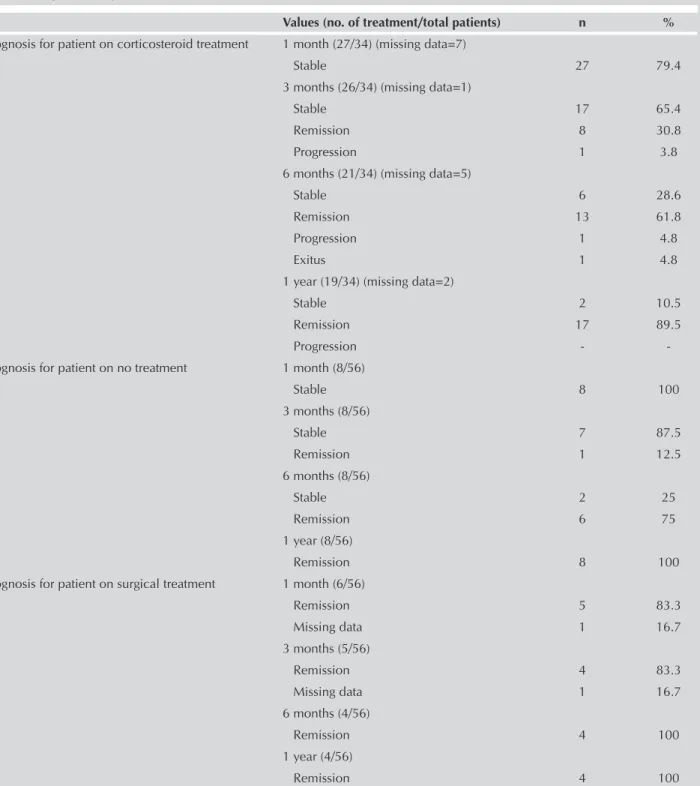
Overall, 34 patients underwent CS therapy. Table 6 shows the information about the prognosis of the disease at 1, 3, and 6 months and 1 year of follow-up of the patients. Eight patients were followed up without any treatment, and 5 patients were treated surgically. Table 6 shows the features of these patients. The response to CS treatment was not different between those with lymphocyte dominance and those with neutrophil
dominance (p=0.6). However, the response to CS treatment was different between those with ground glass opacity and those without ground glass opacity patterns in high resolution computed tomography (HRCT) (p=0.002). In the ground glass opacity group, 9 of the patients with CS therapy at 3 months were in remission, and 12 at 6 months were in remission. However, none of the patients without ground glass opacity in HRCT were in remission at 3 months of CS treatment, and 3 were in remission at 6 months.
DISCUSSION
The classification of OP is very important because the treat-ment and follow-up of patients with SOP include not only the treatment of OP but also the management of underlying diseases. The most common causes of SOP include drugs, infections, rheumatologic diseases, malignancies, and their treatments [6]. In our study, 37 patients were COP, and 19 were SOP. Rheumatologic diseases and malignancies were the most common causes of SOP.
OP is most common in the 5-6 decades of life [7,8]. In our study, patients were between 28 and 83 (mean age: 57.09±12.68) years. Studies that examined the distribution of patients with COP and SOP according to gender have shown no significant difference between the two groups [6,8]. Our results were similar to previous studies.
The association of smoking with OP has been controver-sial [9]. In our study, 58.9% of all patients had a smoking background, and there was no statistically significant dif-ference between COP and SOP (p=0.775). These results were similar to studies by Sveinsson [6] and Drakopanag-iotakis [2]. In the study by Lazor et al. [9], 71% of the patients were non-smokers, and most of the patients who were non-smokers were women. Researchers thought that smoking could be protective against COP development in
women [9].
205
Table 5. PFTs and BAL findings in patients with COP and SOP
Variable (no. of COP/SOP) OP±SD COP±SD SOP±SD p
FEV1, % (32/17) 82.77±19.32 81.50±20.21 85.18±17.88 0.526 FVC, % (32/17) 86.11±18.59 83.76±19.55 90.12±16.63 0.263 FEV1/FVC, % (32/17) 77.81±9.06 76.65±8.97 80.06±9.07 0.220 DLCO, % (26/17) 67.73±13.98 68.11±13.29 67.06±15.56 0.817 DL ADJ, % (26/17) 68.79±14.84 69.24±13.76 68.00±17.13 0.802 DLCO/VA, % (26/17) 80.18±13.60 77.44±13.34 85.07±13.12 0.093 DL/ADJ/VA, % (26/17) 80.31±14.57 78.28±14.36 84.23±14.73 0.232 Lymphocytes (mean±SD) (24/14) 17.52±16.42 16.58±14.85 19.15±19.03 0.643 Lymphocytes (>20% of total BAL cells, n) (24/14) 16 (28.6%) 10 (41.67%) 6 (42.86%) 1.000 Neutrophils (mean±SD) (25/14) 9.52±6.16 5.71±5.01 16.85±5.35 0.044 Neutrophils (>5% of total BAL cells) (25/14) 19 (33.91%) 12 (50%) 7 (50%) 1.000 Eosinophils (mean±SD) (25/14) 4.68±4.63 5.48±4.18 3.15±2.35 0.305 Eosinophils (>2% of total BAL cells) (25/14) 14 (25%) 10 (41.67%) 4 (28.57%) 1.000 Macrophages, % (25/14) 51.68±26.66 57.24±26.05 41.00±25.41 0.075
OP is characterized by non-specific symptoms, such as flu-like illness [10-13]. Most of our patients had flu-like symp-toms. Non-specific symptoms of malaise, cough, fever, and dyspnea occurred in more than two-thirds of the patients [9,14]. Hemoptysis was previously described to be uncom-mon in many studies. The hemoptysis rate in our patients was 7.1%. Our results were similar to previous studies [8,15,16]. Hemoptysis could occur as a result of underlying diseases, such as malignancies but not OP.
The most common radiological findings in patients with OP are consolidation and ground glass opacities, and these are usually bilateral-peripheral [1]. Our findings were consistent
with the literature; however, centrally located lesions were more frequent in SOP than in COP (p=0.023). A previous study showed a predominance of lesions in the lower lung areas in 55% of the patients [1]. Another study showed a predominance of lesions in the middle zone in 91.7% of the patients [7]. We detected involvement in the lower lobes in the general patient population, but the most common middle lobe was affected in SOP (p=0.001). Only 14.3% of the cases had migratory infiltrates. This number is significantly lower than previous reports [13,17]. Although a solitary opacity is an uncommon presentation in OP that is known as focal OP, and 10%-15% of the patients are focal OP [15]. We found 23.4% of mass-like lesions.
206
Table 6. Prognosis for patients
Values (no. of treatment/total patients) n %
Prognosis for patient on corticosteroid treatment 1 month (27/34) (missing data=7)
Stable 27 79.4
3 months (26/34) (missing data=1)
Stable 17 65.4
Remission 8 30.8
Progression 1 3.8 6 months (21/34) (missing data=5)
Stable 6 28.6
Remission 13 61.8 Progression 1 4.8
Exitus 1 4.8
1 year (19/34) (missing data=2)
Stable 2 10.5
Remission 17 89.5
Progression -
-Prognosis for patient on no treatment 1 month (8/56)
Stable 8 100 3 months (8/56) Stable 7 87.5 Remission 1 12.5 6 months (8/56) Stable 2 25 Remission 6 75 1 year (8/56) Remission 8 100
Prognosis for patient on surgical treatment 1 month (6/56)
Remission 5 83.3 Missing data 1 16.7 3 months (5/56) Remission 4 83.3 Missing data 1 16.7 6 months (4/56) Remission 4 100 1 year (4/56) Remission 4 100
An elevated ESR was common in patients. This was similar to previous studies [1,14,18,19]. The majority of cases have been reported to have elevated CRP [20]. The cause of el-evated acute phase reactants (APRs; such as CRP and ESR) in patients with OP is not well known. Elevated APRs have been defined in several studies. However, no explanation was given as reason [6,20]. APRs are synthesized from liver cells during inflammation, most often with the effect of cytokines (especially interleukin 6). They are elevated in acute infec-tions and autoimmune, rheumatologic, and granulomatous diseases and are used in the course of active disease [21,22]. Previous studies have shown increased inflammatory cyto-kines in OP [23,24]. APRs may be elevated by the increase of cytokine in OP. In addition, most of the known causes of SOP are associated with acute inflammation. It is thought that elevated APRs may develop secondary to these diseases. OP normally presents a restrictive pattern on PFT [25]. How-ever, our results and the study by Kavakli et al. [26] differed from previous studies. In the study by Kavakli et al. [26], nor-mal PFT was detected in 58% of the patients, and we ob-served normal PFT in 33 (58.9%) patients. Nine (16.1%) of our cases had isolated restrictive defects. The mechanism of the restrictive pattern in OP is not well established yet. BAL examination usually shows expansion of all cell lines [27]. In the study by Drakopanagiotakis et al. [2], BAL was performed in 32 of 61 patients. In 43% of the 32 patients, BAL lymphocyte was found >20%. However, in the present study, there were 21 patients in the SOP group, and only 5 had BAL lymphocyte. Of these 5 patients, 4 (80%) had a BAL lympho-cyte level >20%. Possibly, the rates could have changed if there were more patients whose BAL lymphocyte levels were examined in the SOP group. In our study, a significant part of patients with SOP had BAL neutrophilia, and this was statisti-cally significant (p=0.044). These findings were inconsistent with previous studies [1,6]. Costabel et al. [27] analyzed BAL findings in 10 patients with BOOP syndrome. All 10 of them had lymphocytosis >20%, 8 (80%) exhibited neutrophilia (>5%), and 5 (50%) exhibited eosinophilia (>5%). There have been almost no explanation about the cause of the cel-lular distribution in previous studies.
Open lung biopsy, VATS, and CT-guided PTNB are preferred in the diagnosis of OP, whereas TBB often fails to obtain a large and adequate piece of lung tissue [28]. However, by the proper clinical and radiographic findings, TBB and BAL may be diagnosed with OP [1,27]. Moreover, OP can be di-agnosed by clinical findings and compatible imaging (espe-cially in patients who are too frail) [1]. Cazzato et al. [29] investigated the clinical and radiological features at onset, outcome, and diagnostic approach in subjects with OP. They found that although clinical and radiological findings usu-ally suggest the diagnosis, a definitive confirmation requires TBB and BAL. In our study, 13 (23%) patients were diagnosed with OP by clinical and radiological findings. Of these pa-tients, 10 were >65 years old, and the patients did not ac-cept TBB or VATS. TBB has been used for diagnosis in many studies [1,14,29,30]. According to Cazzato et al. [29], from a diagnostic perspective, TBB (together with BAL) should be the first diagnostic step. They diagnosed 74% of the patients
with TBB and determined that although the sensitivity of BAL was found to be lower than that of TBB, the combination of the two procedures improved the diagnostic yield (sensitivity 86%) [29]. In our study, approximately 43% of the patients had pathological diagnosis by TBB.
VATS allow biopsy of the lung in well conditions of security. Currently, VATS is a safe procedure that may be used in many pa-tients [30]. Seventeen (30%) papa-tients were diagnosed by VATS. Two patients were diagnosed by CT-guided PTNB. These patients had peripheral located consolidation in thorax CT. PTNB is a rare diagnostic method in the literature [26]. There are no sufficient studies available to make recommen-dation CS, and length of treatment is not known [6]. In ac-cordance with previous studies, we applied CS treatment to most of our patients (34 of 56 patients) [1,7]. Of the patients who were followed up for 1 year, 89% were fully recovered with CS therapy. Only one patient relapsed, and one patient died. All patients with focal lesions underwent surgery for both diagnosis and treatment, and there was no relapse in any patient. Relapses were frequently reported in the litera-ture, but in our patients, it was negligible. However, approxi-mately half of our patients were lost during follow-up. The response to CS treatment has not been evaluated in sub-groups, such as neutrophil predominance group and lym-phocyte predominance group, in HRCT pattern in previous studies [6,7]. These subgroups were evaluated in our study. We found that in patients with ground glass opacity in HRCT, the response to CS treatment is better. Ground glass opacities also respond better to CS treatment in other interstitial lung diseases. However, we do not know how to respond to CS treatment in nodular OP because these patients are usually patients with surgical resection with malignancy pre-diagno-sis and did not use CS therapy.
In conclusion, the clinical and radiological findings in patients with both COP and SOP are similar. The most common com-plaints were cough and dyspnea. The most common radio-logical appearance was peripheral consolidation area. Lesions tended to predilect the central part of the middle zone in SOP. COP and SOP have similar treatment response and prognosis.
Ethics Committee Approval: Authors declared that the research was
conducted according to the principles of the World Medical Associa-tion DeclaraAssocia-tion of Helsinki “Ethical Principles for Medical Research Involving Human Subjects” (amended in October 2013).
Informed Consent: N/A
Peer-review: Externally peer-reviewed.
Author contributions: Concept - A.B., N.K.; Design - A.B., N.K., N.D.;
Supervision - H.T., N.A., N.K.; Resource - N.A., Z.G., N.D.; Materials - A.B., Z.G., N.A., N.D.; Data Collection and/or Processing - A.B., Z.G., N.A., N.D.; Analysis and/or Interpretation - A.B., N.K., H.T.; Literature Search -A.B.,N.K., Z.G.; Writing - A.B., N.K., N.D.; Critical Reviews - N.K., H.T.
Conflict of Interest: The authors have no conflicts of interest to
Financial Disclosure: The authors declared that this study has
re-ceived no financial support.
REFERENCES
1. Cordier JF. Cryptogenic organising Pneumonia. Eur Respir J 2006;28:422-46. [CrossRef]
2. Drakopanagiotakis F, Paschalaki K, Abu-Hijleh M, et al. Crypto-genic and secondary organizing pneumonia: clinical presenta-tion, radiographic findings, treatment response, and prognosis. Chest 201;139:893-900.
3. Drakopanagiotakis F, Polychronopoulos V, Judson MA. Organiz-ing pneumonia. Am J Med Sci 2008;335:34-9. [CrossRef]
4. Costabel U, Guzman J. Bronchoalveolar lavage in interstitial lung disease. Curr Opin Pulm Med 2001;7:255-61. [CrossRef]
5. Grech P, Gray PB, Lambourne CA, et al. Aspiration needle bi-opsy for solitary, peripheral lung lesions: a review of 50 exami-nations. Br J Dis Chest 1978;72:235-41. [CrossRef]
6. Sveinsson OA, Isaksson HJ, Sigvaldason A, et al. Clinical fea-tures in secondary and cryptogenic organising pneumonia. Int J Tuberc Lung Dis 2007;11:689-94.
7. Faria IM, Zanetti G, Barret MM, et al. Organizing pneumonia: chest HRCT findings. J Bras Pneumol 2015;41:231-7. [CrossRef]
8. Basarakodu KR, Aronow WS, Nair CK, et al. Differences in treat-ment and in outcomes between idiopathic and secondary forms of organizing pneumonia. Am J Ther 2007;14:422-6. [CrossRef]
9. Lazor R, Vandevenne A, Pelletier A, et al. Cryptogenic orga-nizing pneumonia. Characteristics of relapses in a series of 48 patients. The Groupe d’Etudes et de Recherche sur les Maladles “Orphelines” Pulmonaires (GERM”O”P). Am J Respir Crit Care Med 2000;162:571-7. [CrossRef]
10. Epler GR. Bronchiolitis obliterans organizing pneumonia. Se-min Respir Infect 1995;10:65-77.
11. Colby TV. Pathologic aspects of bronchiolitis obliterans organis-ing pneumonia. Chest 1992;102:38-43. [CrossRef]
12. Dogan D, Ocal N, Yucel O, et al. Cryptogenic organizing pneu-monia diagnosed with transbronchial parenchymal biopsy: a case report with accompanying histopathological images. J Tur-gut Ozal Med Cent 2016;23:81-3. [CrossRef]
13. Oymak FS, Demirbas HM, Mavili E, et al. Bronchiolitis obliter-ans organizing pneumonia. Clinical and roentgenological fea-tures in 26 cases. Respiration 2005;72:254-62. [CrossRef]
14. Chang J, Han J, Kim DW, et al. Bronchiolitis obliterans organiz-ing pneumonia: clinicopathologic review of a series of 45 Ko-rean patients including rapidly progressive form. J KoKo-rean Med Sci 2002;17:179-86. [CrossRef]
15. Lohr RH, Boland BJ, Douglas WW, et al. Organizing pneumo-nia. Features and prognosis of cryptogenic, secondary, and focal variants. Arch Intern Med 1997;157:1323-9. [CrossRef]
16. Henriet AC, Diot E, Marchand-Adam S, et al. Organising pneumonia can be the inaugural manifestation in connective tissue diseases, including Sjogren’s syndrome. Eur Respir Rev 2010;19:161-3. [CrossRef]
17. Vasu TS, Cavallazzi R, Hirani A, et al. Clinical and radiologic distinctions between secondary bronchiolitis obliterans orga-nizing pneumonia and cryptogenic orgaorga-nizing pneumonia. Respir Care 2009;54:1028-32.
18. Nizami IY, Kissner DG, Visscher DW, et al. Idiopathic bronchi-olitis obliterans with organizing pneumonia. An acute and life-threatening syndrome. Chest 1995;108:271-7. [CrossRef]
19. Yamamoto M, Ina Y, Kitaichi M, et al. Clinical features of BOOP in Japan. Chest 1992;102:21-5. [CrossRef]
20. Yoshinouchi T, Ohtsuki Y, Kubo K, et al. Clinical and patho-logical studies of organizing pneumonia. Nihon Kyobu Shikkan gakkai Zasshi 1993;31;951-8.
21. Hardison CS. The sedimentation rate. Erythrocyte sedimentation rate; blood sedimentation rate. JAMA 1968;204:257. [CrossRef]
22. Hutchinson WL, Koenig W, Fröhlich M, et al. Immunoradiomet-ric assay of circulating C-reactive protein: Age-related values in the adult general population. Clin Chem 2000;46:934-8. 23. Radzikowska E, Roży A, Jaguś P, et al. Cryptogenic Organizing
Pneumonia: IL-1β, IL-6, IL-8, and TGF- β1 Serum Concentra-tions and Response to Clarithromycin Treatment. Adv Exp Med Biol 2016;911:77-85. [CrossRef]
24. Radzikowska E, Roży A, Jagus P, et al. Clarithromycin De-creases IL-6 Concentration in Serum and BAL Fluid in Patients with Cryptogenic Organizing Pneumonia. Adv Clin Exp Med 2016;25:871-8. [CrossRef]
25. Silva CI, Marchiori E, Souza Júnior AS, et al. Comissão de Ima-gem da Sociedade Brasileira de Pneumologiae Tisiologia. Illus-trated Brazilian consensus of terms and fundamental patterns in chest CT scans. J Bras Pneumol 2010;36:99-123. [CrossRef]
26. Kavakli K, Ocal N, Dogan D, et al. Is there any superior-ity among diagnostic methods in organizing pneumonia in terms of clinical features of the patients? Ther Clin Risk Manag 2016;12:1473-9. [CrossRef]
27. Costabel U, Teschler H, Guzman J. Bronchiolitis obliterans organizing pneumonia (BOOP): the cytological and immu-nocytological profile of bronchoalveolar lavage. Eur Respir J. 1992;5:791-7.
28. Schlesinger C, Koss MN. The organizing pneumonias: an update and review. Curr Opin Pulm Med 2005;11:422-30. [CrossRef]
29. Cazzato S, Zompatori M, Baruzzi G, et al. Bronchiolitis obliter-ans-organizing pneumonia: an Italian experience. Respir Med 2000;94:702-8. [CrossRef]
30. Cordier JF. Cryptogenic organising pneumonia. Clin Chest Med 2004;25: 727-38. [CrossRef]