ABSTRACT
Introduction: There is limited knowledge about the use of bioresorbable scaffolds (BRSs) in long segment coronary artery lesions. We aimed to evaluate the clinical outcomes of BRS-BRS and drug eluting stents (DESs)-BRS overlapping applications.
Patients and Methods: Cross-sectional, single-center study between 2013 and 2016 enrolled 97 patients and 100 lesions scheduled for BRS placement in long segment lesions (> 28 mm). BRS-BRS overlap was performed in 30 patients and 30 lesions, DES-BRS overlap was performed in 67 patients and 70 lesions. Acute procedural success and major adverse cardiac events (MACE) (death, stent thrombosis, and target lesion reintervention) were assessed.
Results: Acute procedural success was 97.1% in the overall group. MACE was observed in 6 patients (6.2%) in the entire group, 4 (5.9%) in the DES-BRS group, and 2 (6.6%) in the BRS-BRS group.
Conclusion: BRS use might be a safe and effective option for the treatment of long segment lesions. Both BRS-BRS overlap and BRS-DES overlap may be performed with short overlap segment.
Key Words: Bioresorbable scaffolds; overlapping; hybrid strategy
Uzun Segment Koroner Lezyonlarda Biyoeriyebilen Stentlerin Güvenlik ve Etkinlik Sonlanımları
ÖZET
Giriş: Uzun segment lezyonlarda biyoeriyebilen stentlerin (BRS) kullanımı ile ilgili sınırlı bilgimiz oldu-ğundan hem BRS-BRS hem de İlaç salınımlı metal stent (DES)-BRS overlap uygulanan hastaların klinik sonuçlarını değerlendirmeyi planladık.
Hastalar ve Yöntem: Tek merkezli, kesitsel planlanan; Ocak 2013-Haziran 2016 tarihleri arasında uzun segment lezyonlara (> 28 mm) BRS yerleştirilmesi planlanan 97 hasta ve 100 lezyon çalışmaya alındı. Otuz hasta ve 30 lezyonda BRS-BRS overlap uygulanırken , 67 hasta 70 lezyona DES-BRS overlap uygulandı. Akut işlem başarısı ve MACE (ölüm, stent trombozu, hedef damara yeniden girişim) değerlendirildi. Bulgular: Akut işlem başarısı %97.1 idi. Tüm hasta grubunda toplam MACE 6 (%6.2) hastada gelişirken, DES-BRS grubunda 4 (%5.9), BRS-BRS grubunda 2 (%6.6) hastada MACE gerçekleşti.
Sonuç: Uzun segment lezyonlarda BRS kullanımı seçenek olarak değerlendirilebilinir. BRS veya BRS-DES overlap yapacak şekilde yerleştiriken kısa segment overlap olacak şekilde gerçekleştirilmelidir. Anahtar Kelimeler: Biyoeriyebilen stentler; overlap; hibrit tekniği
Hacı Murat Güneş, Tayyar Gökdeniz, İbrahim Oğuz Karaca, Gültekin Günhan Demir, Filiz Kızılırmak, Ekrem Güler, Gamze Babür Güler, Mehmet Onur Omaygenç, Umeyir Savur, Ersin İbişoğlu, Bilal Boztosun
University of İstanbul Medipol, Faculty of Medicine, Department of Cardiology, İstanbul, Turkey
Safety and Efficacy Outcomes of Bioresorbable
Scaffolds in Long Segment Coronary Lesions
Hacı Murat Güneş
E-mail: [email protected] Submitted: 10.09.2017
Accepted: 04.11.2017
© Copyright 2018 by Koşuyolu Heart Journal. Available on-line at
www.kosuyoluheartjournal.com
Correspondence
INTRODUCTION
Bioresorbable coronary stents have been developed to overcome biocompatibility problems associated with drug-eluting stents through the advantage of gradual degradation (1). Capability of treating coronary lesions without permanent scaffolds and restoration of reactive vasomotion offer no constriction on any probable future surgical revascularization(2). Despite initial high expectations for bioresorbable scaffolds (BRSs), reports have shown improved efficacy and safety outcomes limited to uncomplicated, short, and stable lesions(3).
In current daily interventional practice, a second stent is used in almost 10% of percutaneous coronary interventions (PCI) due to inadequate coverage of lesion or edge dissections(4). Studies related with drug-eluting stents (DESs) demonstrated that increased amount of drug released
at the site of overlap versus segments without overlapping sites and strut thickness stimulated increased neutrophil and eosinophil release and fibrin deposition, resulting in enhanced inflammation, impaired healing, increased late lumen loss, and stent restenosis rates when compared with single stent use (4,5). Nevertheless, studies with second-generation DESs showed that overlapping technique can safely be used in contrast with the results of first-generation DESs(6).
Clinical studies using ABSORB, the first commercially available BRS, excluded lesions requiring overlapping; however, the UNDERDOGS trial demonstrated that overlapping of BRSs is as safe as second-generation DESs(7,8).
Here we aimed to evaluate clinical outcomes of hybrid het-erogeneous DES-BRS and BRS-BRS overlapping applications in long segment coronary lesions.
PATIENTS and METHODS
An approval of the İstanbul Medipol University Ethics Committee was obtained as ID 10840098-604.01.01.E.3982.
Study Population
In this single center study with cross-sectional design, we enrolled 100 lesions in 97 patients who underwent PCI for long-segment lesions (> 28 mm) that could not be covered with single BRS and therefore overlapping with BRS-BRS or BRS-DES was performed in our clinic between 2013 and 2016. Patients were treated with everolimus-eluting BRS device (Absorb BVS; Abbott Vascular, SantaClara, CA, USA) or novolimus-eluting BRS device (DESolve, Elixir Medical Corporation) and everolimus-eluting DES (XIENCE PRO, Abbott Vascular, Santa Clara, CA, USA). Baseline clinical characteristics, angiographic variables, and procedural characteristics were defined. Informed consent was obtained from all patients before the procedures.
Both patients with stable coronary artery disease and acute coronary syndrome were recruited. Native coronary artery lesions with stenosis > 50% and length > 28 mm and reference vessel diameter (RVD) ≥ 2.5 mm were included. Major exclusion criteria were left ventricular ejection fraction less than 35%, lesions located in the left main coronary artery, arterial or saphenous vein graft lesions, acute ST-elevation myocardial infarction, and lesions extending to coronary ostium.
After the index procedure, the follow-up data were obtained from outpatient clinic visits or emergency department admissions and monitoring of patients was also done by regular telephone calls. When patients decided to undergo coronary angiography or PCI during follow-up visit or emergency department admission, details about control angiography and PCI were recorded.
Procedures and Medications
Procedures were performed in accordance with current PCI standards. All patients received upstream 300 mg acetylsalicylic
acid (ASA) plus a loading dose of 300 to 600 mg clopidogrel or 180 mg ticagrelor or 60 mg prasugrel during the procedure. Unfractionated heparin (100 U/kg) was used for anticoagulation in all patients during interventions; additional bolus might be required to achieve an active clotting time of 250 s. None of the patients received glycoprotein IIb/IIIa inhibitors.
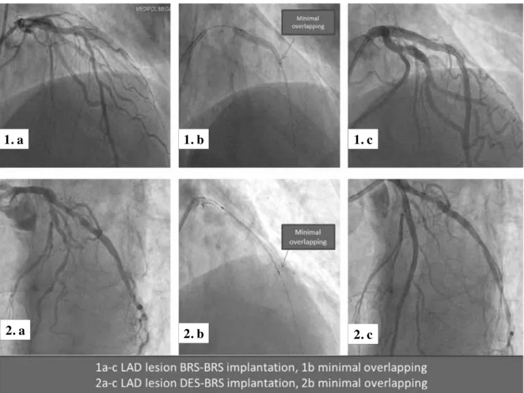
Predilation was performed before all interventions, scaffold implantation was achieved by gradually increased (1 atm increase per 5 s) pressure, which did not exceed the rated burst pressure. In the first place, BRS was implanted at the distal site followed with overlapping proximal BRS or DES in all patients. Stent overlap was defined as angiographically 1 mm overlapping of 2 stents (Figure 1)(4). Postdilation that would not exceed the BRS diameter of 0.5 mm was performed in all patients to stent overlapping region.
Angiographic Parameters and Quantitative Coronary Angiographic Analysis
Bending more than 45 degrees proximal to the lesion was defined as tortuosity. Single bend between 45 and 90 degrees proximal to the lesion was defined as mild tortuosity, while 3 or more bends between 45 and 90 degrees or one or more bends over 90 degrees were defined as severe tortuosity. Bendings out of these criterias (mild and severe tortuosity) were defined as moderate tortuosity(8).
Calcification was defined as overt radiopacity of the vessel wall across the lesion site. It was classified as moderate (radiopacity noted only during the cardiac cycle before contrast injection) and severe (radiopacity noted across both sides of the vessel wall before contrast injection and independently from cardiac motion)(9).
Quantitative coronary angiography (QCA) was evaluated at 1 angiographic core laboratory with use of CAAS 5.9 (Pie Medical Imaging) in the hospital’s angiographic analysis center. Baseline and postprocedural minimal lumen diameter (MLD), mean lumen diameter (MnLD), RVD, residual diameter stenosis (%DS), lesion length and acute gain were measured. Angiographic measurements were made by contrast-filled standard calibration. These images were analyzed from same angiographic projection to minimize foreshortening.
Study Endpoints and Definitions
Acute procedural success was defined as final angiographic residual stenosis of < 30% with thrombolysis in myocardial infarction flow grade 3. categorization of complications was made. Instent restenosis was defined as reduction in the percent DS > 50% within the stented segment.
Primary endpoints of this study were major adverse cardiac events (MACE) including cardiac death, myocardial infarction (MI), target vessel revascularization (TVR), target lesion revascularization (TLR), and stent thrombosis.
MLD was defined as the minimum lumen diameter of lesion area, and MnLD was defined as the mean lumen diameter
of lesion area. TLR was defined as repeat PCI or surgical revascularization within the index procedure stent or 5 mm edge, meanwhile TVR was defined as any revascularization procedure occurring within the treated vessel outside the margins of the stent or 5 mm.
MI was defined as elevation of cardiac troponin values (> 5 x 99th percentile) with symptoms of ischemia or new ECG changes in the periprocedural period or elevation > 99th percentile in the postprocedural period or new echocardiographic changes suggestive of ischemia.
Minimum follow-up duration was 6 months, although patients with 2-year follow-up were also present. Last follow-up visit was performed in January 2017.
Statistical Analysis
SPSS 23.0 statistical software (SPSS Inc., Chicago, IL, USA) was used for performing statistical analysis. Continuous variables were expressed as mean ± standard deviation or median and interquartile range as appropriate. Categorical variables were
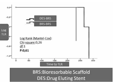
expressed as percentages. The Kolmogorov-Smirnov test was used to test normality of distribution of continuous variables. Group means for continuous variables were compared with Student t-test, the Mann-Whitney U test, ANOVA, or Kruskal-Wallis test, as appropriate. Categorical variables were compared with the use of chi-square test. Descriptive statistics for MACE was presented as percentage and minimum, maximum, and median follow-up time of patients. Kaplan-Meier curves were generated to analyze the impact of BRS-BRS versus DES-BRS overlapping on the endpoint of TLR during the follow-up. The results were expressed as Log rank and p values and demonstrated with a graphic. A p value of ≤ 0.05 was considered statistically significant.
RESULTS
In the first place, 100 patients with a total of 103 lesions to be treated with overlapping were planned for enrollment; however, 3 patients and 3 lesions could not be recruited due to lack of acute procedural success (97.1%); thus, 97 patients with 100
1. a
1. b
1. c
2. a
2. b
2. c
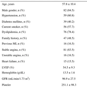
lesions were enrolled (Figure 2). Inability to advance BRSs to the lesion area due to severe calcification and tortuosity was the common characteristic in 3 patients with acute procedural failure; the scaffold stripped off during advancement in one of them, stent could not be crossed to the lesion area in another, whereas residual stenosis > 30% was present despite advancement to the lesion area in the last patient. Mean age of the patients were 57.8 ± 10.4 years, and 82 of them (84.5%) were men. When risk factors of the patients were evaluated, 59 (60.8%) had hypertension, 39 (40.2%) had diabetes mellitus, 56 (57.7%) were active smokers, and 76 (78.4%) had dyslipidemia. Sixteen (16.5%) patients underwent PCI for unstable angina pectoris, while mean left ventricular ejection fraction (LVEF) and glomerular filtration rate (GFR) of the patients were 54.5 ± 9.3 ml/min and 96.9 ± 27.5 ml/min, respectively (Table 1).
There were no significant differences in demographic characteristics between patients in the DES-BRS Hybrid overlapping group and BRS-BRS overlapping group (Table 2). Femoral access was used in 81 (83.5%) patients, and new P2Y12 inhibitors were preferred in combination with ASA for dual antiplatelet therapy in 45 (46.4%) patients. Thirty (44.8%) patients in the DES-BRS group and 15 (50%) patients in the BRS-BRS group received new P2Y12 inhibitors (p= 0.398). Of the scaffolds, DESolve BRS was preferred in 36 (51.4%) and ABSORB-BRS was preferred in 34 (48.6%) DES-BRS group lesions, whereas DESolve BRS and ABSORB BRS were the preferred scaffolds in 32 (53.3%) and 28 (46.7%) lesions in the BRS-BRS group, respectively.
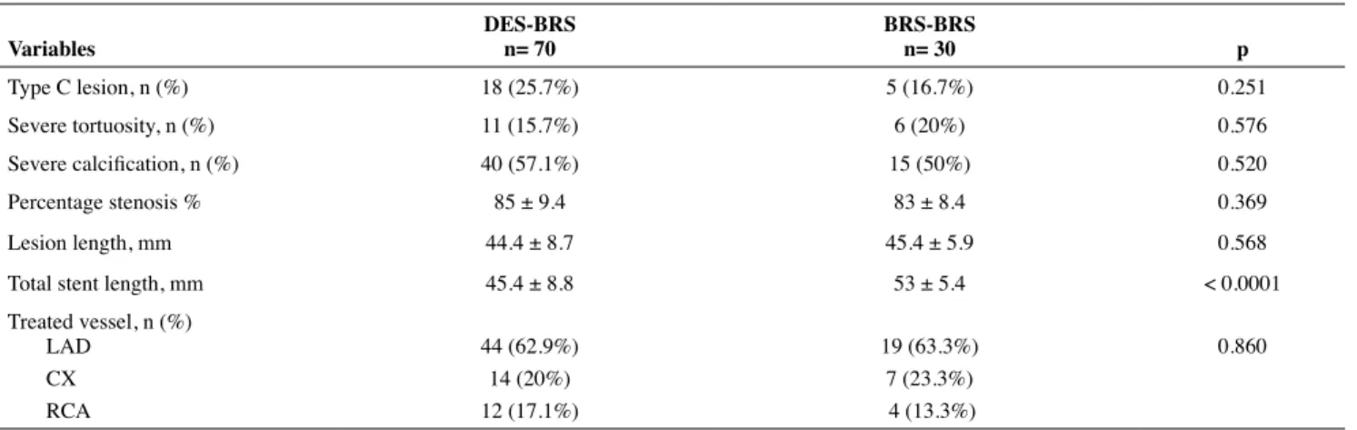
Baseline angiographic characteristics such as lesion type, severe calcification, severe tortuosity, treated vessel, and lesion length were similar between the two groups; however, implanted
stent length was significantly different between the DES-BRS group (45 mm ± 9 mm) and the BRS-BRS group (53 mm ± 5 mm) (p< 0.0001) (Table 3). BRS was implanted distally in both groups and the length of BRS was not different between the DES-BRS (27 mm ± 3 mm) and BRS-BRS (27 mm ± 2 mm) groups (p= 0.141). Furthermore, diameters of distal BRSs were not different between the DES-BRS (3 ± 0.4) and BRS-BRS (2.9 ± 0.4) groups (p= 0.440).
Predilation and postdilation were applied in all patients and the two groups were similar in terms of PTCA balloon size (Table 4). As shown in Table 4, parameters measured by QCA method were not different between the two groups. When stents used for proximal overlapping, BRS or DES, were compared, diameters were not different (DES: 3 ± 0.4; BRS: 2.9 ± 0.5; p= 0.268), whereas BRSs were longer than DESs (BRS: 25.8 ± 41 mm; DES: 18.9 ± 7.8 mm; p< 0.0001) (Table 4).
Procedural complications occurred in 2 (2.1%) patients. Coronary rupture after postdilation at high pressure due to inadequate expansion of the calcified site of overlap developed in one patient that was controlled with extended balloon inflation, whereas other patient experienced a cerebrovascular event during postprocedural follow-up period in hospital which recovered without any sequela.
During the in-hospital stay period, none of the patients had death, reintervention, or adverse cardiac events. The mean duration of follow-up of the patients was 492 days
Table 1. Baseline clinical characteristics of the patients
Age, years 57.8 ± 10.4 Male gender, n (%) 82 (84.5) Hypertension, n (%) 59 (60.8) Diabetes mellitus, n (%) 39 (40.2) Current smoker, n (%) 56 (57.7) Dyslipidemia, n (%) 76 (78.4) Family history, n (%) 47 (48.5) Previous MI, n (%) 16 (16.5) Stable angina, n (%) 81 (83.5) Unstable angina, n (%) 16 (16.5) Heart failure, n (%) 15 (15.5) LVEF (%) 54.5 ± 9.3 Hemoglobin (g/dL) 13.5 ± 1.6 GFR (mL/min/1.73 m2) 96.9 ± 27.5 Platelet 251.1 ± 98.3
LVEF: Left ventricular ejection fraction, GFR: Glomerular filtration rate. MI: Mycardial infarction.
Table 2. Clinical differences between BRS-BRS and DES-BRS hybrid groups Variables DES-BRS n= 67 BRS-BRSn= 30 p Age 57.7 ± 10.7 58 ± 9.8 0.870 Male gender, n (%) 56 (83.6%) 26 (86.7%) 0.772 Hypertension, n (%) 39 (58.2%) 20 (66.7%) 0.503 Diabetes mellitus, n (%) 25 (37.3%) 14 (46.7%) 0.502 Dyslipidemia, n (%) 49 (73.1%) 27 (90%) 0.069 Current smoker, n (%) 38 (56.7%) 18 (60%) 0.826 Family history, n (%) 33 (49.3%) 14 (46.7%) 0.830 Previous MI, n (%) 10 (14.9%) 6 (20%) 0.562 Unstable angina, n (%) 11 (16.64%) 5 (16.7%) 0.999 Heart failure, n (%) 7 (10.4%) 8 (26.7%) 0.066 LVEF 55.7 (47.6-59.3) 54.9 (47.9-58.12) 0.828 Hemoglobin 12.8 (12.1-13.4) 12.9 (12.1-13.7) 0.744 Platelet 244 (175-299) 255 (166-291) 0.382 GFR 95.2 (77.4-112.8) 93.8 (75.3-122.1) 0.644
BRS: Bioresorbable scaffold, DES: Drug eluting stent, LVEF: Left ventricular ejection fraction, GFR: Glomerular filtration rate.
Table 3. Angiographic characteristics of BRS-BRS and DES-BRS lesions
Variables DES-BRS n= 70 BRS-BRSn= 30 p Type C lesion, n (%) 18 (25.7%) 5 (16.7%) 0.251 Severe tortuosity, n (%) 11 (15.7%) 6 (20%) 0.576 Severe calcification, n (%) 40 (57.1%) 15 (50%) 0.520 Percentage stenosis % 85 ± 9.4 83 ± 8.4 0.369 Lesion length, mm 44.4 ± 8.7 45.4 ± 5.9 0.568
Total stent length, mm 45.4 ± 8.8 53 ± 5.4 < 0.0001
Treated vessel, n (%) LAD CX RCA 44 (62.9%) 14 (20%) 12 (17.1%) 19 (63.3%) 7 (23.3%) 4 (13.3%) 0.860
BRS: Bioresorbable scaffold; DES: Drug eluting stent.
(minimum, 212 days; maximum, 755 days). One patient in the DES-BRS group experienced subacute stent thrombosis 15 days after implantation that ended-up with failed attempt for revascularization and the patient was discharged with medical therapy. Three patients in the DES-BRS group and 2 in the BRS-BRS group were revascularized for TLR. As shown with the Kaplan-Meier curves in Figure 3, no statistical significance was observed between two groups in terms of TLR during the follow-up. In the whole study population, MACE were reported in 6 (6.2%) patients, of whom 4 (5.9%) were in the DES-BRS group and 2 (6.6%) were in the BRS-BRS group (Figure 2).
DISCUSSION
We report procedural success and clinical outcomes in patients with long segment coronary lesions that required more than one stent and treated with distal BRS (everolimus-eluting ABSORB or novolimus-(everolimus-eluting DESOLVE scaffolds) and proximal BRS or DES (everolimus-eluting XIENCE) for overlapping. Our findings can be shortly described as follows:
1. When procedural success and clinical outcomes are considered, BRS-BRS or DES-BRS hybrid overlapping technique is effective and safe.
Figure 3. Kaplan-Meier curve to determine TLR between the BRS-BRS and the DES-BRS overlapping groups.
Table 4. Procedural and QCA related features in DES-BRS and BRS-BRS groups and differences between proximal DES and BRS characteristics
Variables DES-BRS n= 70 BRS-BRS n= 30 p
Predilatation, PTCA, mm 2.80 ± 0.3 2.78 ± 0.3 0.787
Postdilatation, PTCA, mm 3.13 ± 0.5 3.15 ± 0.4 0.851
Pre min diameter, mm 0.91 ± 0.5 0.88 ± 0.4 0.745
Pre mean diameter, mm 1.76 ± 0.6 1.73 ± 0.6 0.858
Final min diameter, mm 2.56 ± 0.4 2.53 ± 0.4 0.712
Final mean diameter, mm 2.84 ± 0.4 2.83 ± 0.4 0.904
Reference diameter, mm 3.07 ± 0.4 3.11 ± 0.4 0.647 %DS 17.8 (12.3-24.1) 18.0 (12.5-23.8) 0
.
195 Proximal DES n= 70 Proximal BRS n= 30 Stent diameter, mm 3 ± 0.4 2.9 ± 0.5 0.268 Stent length, mm 18.9 ± 7.8 25.8 ± 4.1 < 0.0001QCA: Quantitative calculation angiography, BRS: Bioresorbable scaffold, DES: Drug eluting stent, PTCA: Percutaneous transluminal coronary angioplasty, DS: Diameter stenosis (residual stenosis), Pre min: Minimum basal diameter of native vessel at lesion point.
2. Some of the BRSs used for overlapping were DESolve; hence, the efficacy and safety of DESolve in this lesion group have been evaluated for the first time.
3. Although clinical outcomes for DES-BRS and BRS-BRS were similar, shorter total stent length, lower total strut thickness at the site of overlapping, and easier reimbursement by insurance companies in the DES-BRS group make hybrid technique a more favorable option for long lesions when BRSs are preferred.
Advantages associated with BRS including late lumen enlargement, freedom from permanent cage, cyclical strain, restoration of vasomotion and thus having no restriction on any future percutaneous or surgical revascularization has made BRS an attractive option in recent years(2). Previous reports largely suggested BRS use in simple lesions; however, recent studies have confirmed the safety and efficacy of BRS in chronic total occlusions, bifurcation lesions, and acute coronary syndromes(3,10-12).
Approximately 10% of PCIs (in particular, complex lesions) may require overlapping technique due to lesion length or edge dissections(4). Edge-to-edge implantation of 2 stents without an overlapping technique is associated with an increased risk of stent thrombosis and restenosis due to gap possibility; nevertheless, increased thrombogenicity and thrombus formation, as well as delayed reendothelialization, were associated with overlapping as well(13,14). Rikthegar et al. assessed hemodynamics of coronary arteries with stent overlap and demonstrated that overlap geometry could lead to adverse clinical outcomes through unfavorable flow condition(15).
In their study about overlapping everolimus-eluting BRS in a porcine coronary artery model, Farooq et al. demonstrated increased neointimal hyperplasia and delayed stent coverage because of thicker strut configuration, which might serve a key role for scaffold restenosis and thrombosis(16). In addition, with the hypothesis that minimalization of overlap area could reduce the risk of adverse clinical outcomes, Farooq et al. reported increased risk of geographical miss due to both poor visibility of scaffold edge markers and difficulty of positioning scaffold edges in the same plane in their in-vitro phantom study(17). In our study, use of minimal overlapping technique might have limited disadvantages associated with BRS use such as thicker struts by avoiding both long overlapping segment and gap formation (Figure 1).
In the first study comparing BRS-BRS overlapping and DES-DES overlapping in long-segment lesions, Bigaclia et al. detected lower rates of acute/subacute stent thrombosis and increased periprocedural myocardial injury in the BRS group and similar 1-year device-oriented endpoint in the DES group (7). Besides, Robert et al. demonstrated that DES-ABSORB BRS hybrid overlapping can be safely used in patients with long segment lesions with high procedural success and low MACE and complication rates(18). We investigated BRS-BRS and DES-BRS hybrid overlapping together in this study, which provided similar results with previous studies, thus suggesting that both techniques can safely be used for overlapping.
One of the reasons underlying our preference for DES-BRS overlapping was unavailability of DES-BRS size longer than 28 mm. Implantation of BRS at the distal site allows proximal implantation of DESs, which have longer size options (38 mm, 48 mm) in diffuse long coronary lesions (> 55 mm), which saves the patient from more BRS use and overlapping segments. Kuan Leong Yew suggested implantation of DES at the first place and advancement of BVS through DES and keeping BVS over the overlap site; otherwise, when BRS is implanted at the under site of overlapping, degradation of scaffold could cause disruption and malapposition of DES struts(19). When eventual degradation of scaffolds is taken into account, it might seem reasonable to place DES under the overlap site; however, both bulky, thick
structure and low delivery profile of BRS may reduce procedural success rate. Moreover, we detected high procedural success and acceptable adverse clinical event rates when we placed distal BRS first and second BRS for overlapping.
In a recent study, Serruys et al. demonstrated that ABSORB BRS did not show superior vasomotor reactivity or non-inferior late luminal loss and detected greater MACE rates in the ABSORB group(20). This condition indicates the need for development of new-generation resorbable scaffolds. Ideal BRS should have thinner struts, adequate radial force, and capability of degradation in short period. Similarly, Kitabata et al. reported significantly improved outcomes of stent overlap with second-generation DESs (everolimus-eluting stent) that have thinner struts in comparison with first-generation stents and suggested that the use of DES is effective and safe for overlapping stents(6). Performance of BRS implantation procedures by a single, senior operator with extensive experience with BRS may have been associated with improved outcomes in our study. Appropriate predilation and postdilation in all patients may be another factor that contributed to procedural success.
A meta-analysis by Polimeni et al. detected increased stent thrombosis associated with BRS in the first 30 days and 2-year outcomes (very late stent thrombosis) and concluded that follow-up duration longer than 2 years would provide more accurate information about the safety of BRS(21). Taking this recommendation into account, limitation of our follow-up at 2 years (despite favorable outcomes) shows that further studies with larger sample size and longer follow-up periods are required for guidance of BRS overlapping in daily clinical practice.
The incidence of MACE in our study was comparatively low and stent thrombosis was not observed. This instance was explained by utilization of novel potent P2Y12 inhibitors in the majority of the patients and complying strictly with BRS deployment during the procedures, which were performed by the same experienced operator.
CONCLUSION
Patients with long segment coronary lesions might be treated with ABSORB or DESolve scaffolds and DESs by performing BRS-BRS or DES-BRS hybrid overlap with good safety and efficacy in short/mid-term outcomes. Development of new BRSs with characteristics including thinner struts, adequate radial force, faster degradation, improved delivery profile, and more visible edges under fluoroscopy should help better clinical outcomes in long segment lesions.
Study Limitations
Our study had a cross-sectional design. Angiographic procedural success was assessed by QCA, which is a practical method providing substantial information about lesion characteristics; however, use of techniques such as IVUS or OCT
might have provided more accurate data. Similarly, assessment of overlapping site by means of OCT or IVUS could have better elucidated the presence of non-endothelialized areas under DES after degradation of BRS. Another limitation of our study was lack of routine control angiography. We could therefore have missed non-clinical scaffold restenosis because we only evaluated clinical outcomes in short/mid-term follow-up. Small sample size and lack of follow-up for longer periods are other limitations of the present study.
CONFLICT of INTEREST
The authors reported no conflict of interest related to this article.
AUTHORSHIP CONTRIBUTIONS
Concept/Design: HG Analysis/Interpretation: GG Data Acquisition: HG, Eİ Writting: HG, TG Critical Revision: TG, BB Final Approval: All of authors
REFERENCES
1. Ormiston JA, Serruys PWS. Bioabsorbable coronary stents. Circ Cardio-vasc Interv 2009;2:255-60.
2. Serruys PW, Garcia-Garcia HM, Onuma Y. From metallic cages to transi-ent bioresorbable caffolds: change in paradigm of coronary revasculariza-tion in the upcoming decade? Eur Heart J 2012;33:16-25.
3. Onuma Y, Dudek D, Thuesen L, Webster M, Nieman K, Garcia-Garcia HM, et al. Five-year clinical and functional multislice computed tomog-raphy angiographic results after coronary implantation of the fully resor-bable polymeric everolimus-eluting scaffold in patients with de novo coro-nary artery disease: the ABSORB cohort A trial. JACC Cardiovasc Interv 2013;6:999-1009.
4. Räber L, Jüni P, Löffel L, Wandel S, Cook S, Wenaweser P, et al. Impact of stent overlap on angiographic and long-term clinical outcome in pa-tients undergoing drug-eluting stent implantation. J Am Coll Cardiol 2010;55:1178-88.
5. Finn AV, Kolodgie FD, Harnek J, Guerrero LJ, Acampado E, Tefera K, et al. Differential response of delayed healing and persistent inflammation at sites of overlapping sirolimus- or paclitaxel-eluting stents. Circulation 2005;112:270-8.
6. Kitabata H, Loh JP, Pendyala LK, Badr S, Dvir D, Barbash IM, et al. Sa-fety and efficacy outcomes of overlapping second-generation everolimus-eluting stents versus first-generation drug-everolimus-eluting stents. Am J Cardiol 2013;112:1093-8.
7. Biscaglia S, Ugo F, Ielasi A, Secco GG, Durante A, D’Ascenzo F, et al. Bioresorbable scaffold vs. second generation drug eluting stent in long co-ronary lesions requiring overlap: a propensity-matched comparison (the UNDERDOGS study). Int J Cardiol 2016;208:40-5.
8. Ho HH, Jafary FH, Loh KK, Tan JK, Ooi YW, Ong PJ. Deliverability of in-tegrity coronary stents in severely tortuous coronary arteries: a preliminary experience. J Invasive Cardiol 2012;24:650-4.
9. Mintz GS, Popma JJ, Pichard AD, Kent KM, Satler LF, Chuang YC, et al. Patterns of intravascular ultrasound and coronary angiography in 1155 lesions. Circulation 1995;91:1959-65.
10. Wiebe J, Liebetrau C, Dörr O, Most A, Weipert K, Rixe J, et al. Feasibi-lity of everolimus- eluting bioresorbable vasculars caffolds in patients with chronic total occlusion. Int J Cardiol 2015;179:90-4.
11. Džavík V, Colombo A. The absorb bioresorbable vascular scaffold in co-ronary bifurcations: insights from bench testing. JACC Cardiovasc Interv 2014;7:81-8.
12. Gori T, Schulz E, Hink U, Wenzel P, Post F, Jabs A, et al. Early outcome after implantation of absorb bioresorbable drug-eluting scaffolds in pati-ents with acute coronary syndromes. Euro Intervention 2014;9:1036-41. 13. Kolandaivelu K, Swaminathan R, Gibson WJ, Kolachalama VB,
Nguyen-Ehrenreich KL, Giddings VL, et al. Stent thrombogenicity early in high-risk interventional settings is driven by stent design and deployment and protected by polymer-drug coatings. Circulation 2011;123:1400-9. 14. Murasato Y, Hikichi Y, Nakamura S, Kajiya F, Iwasaki K, Kinoshita Y, et
al. Recent perspective on coronary bifurcation intervention: statement of the “Bifurcation Club in KOKURA”. J Interv Cardiol 2010;23:295-304. 15. Rikhtegar F, Wyss C, Stok KS, Poulikakos D, Müller R, Kurtcuoglu V, et
al. Hemodynamics incoronary arteries with overlapping stents. Journal of Biomechanics 2014;47:505-11.
16. Farooq V, Serruys PW, Heo JHW, et al. Intracoronary optical coherence tomography and histology of overlapping everolimus-eluting bioresor-bable vascular scaffolds in a porcine coronary artery model: the potential implications for clinical practice. JACC Cardiovasc Interv 2013;6:523-2. 17. Farooq V, Onuma Y, Radu M, Okamura T, Gomez-Lara J, Brugaletta S,
et al. Optical coherence tomography (OCT) of overlapping bioresorbab-le scaffolds: from bench work to clinical application. Euro Intervention 2011;7:386-99.
18. Gil RJ, Bil J, Pawłowski T, Yuldashev N, Kołakowski L, Jańczak J, et al. The use of bioresorbable vascular scaffold Absorb BVS® in patients with stable coronary artery disease: one-year results with special focus on the hybrid bioresorbable vascular scaffolds and drug eluting stents treatment. Kardiol Pol 2016;74:627-33.
19. Yew KL. Overlapping technique for hybrid percutaneous coronary inter-vention strategy utilising drug eluting stent and ABSORB bioresorbable vascular scaffold. Int J Cardiol 2015;178:e8-e10.
20. Serruys PW, Chevalier B, Sotomi Y, Cequier A, Carrié D, Piek JJ, et al. Comparison of an everolimus-eluting bioresorbable scaf oldwith an eve-rolimus-eluting metallic stent for the treatmentof coronary artery stenosis (ABSORB II): a 3 year, randomised,controlled, single-blind, multicentre clinical trial. Lancet 2016;388:2479-91.
21. Polimeni A, Anadol R, Münzel T, Indolfi C, De Rosa S, Gori T. Long-term outcome of bioresorbable vascular scaffolds for the treatment of co-ronary artery disease: a meta-analysis of RCTs. BMC Cardiovasc Disord 2017;17:147.