502
T
he diagnosis and treatment of immune-mediatedmyocar-ditis and the rejection of cardiac allografts remain clini-cal challenges. Myocardiocytolysis mediated by CD8+ T cells
contributes to viral and autoimmune myocarditis, and to acute allograft rejection. The current clinical standard of repetitive invasive endomyocardial biopsies can entail discomfort for patients, sampling error, and risks of serious complications, including perforation and pericardial tamponade.1 Existing
imaging techniques for myocarditis detection include echo-cardiography,2 nuclear imaging with gallium-67 or
indium-111–labeled antimyosin antibodies,3,4 and magnetic resonance
imaging. The ability to visualize specific molecular targets5,6
could provide quantitative imaging tools to assess the cellu-lar and molecucellu-lar functions of myocarditis. Recently devel-oped probes can assess different biological functions, such as phagocytosis and protease activity in atherosclerotic lesions and infarcted hearts, using both fluorescence reflectance im-aging (FRI) and fluorescent molecular tomography in con-junction with coregistered computed tomographic (FMT-CT)
imaging.7–9 This study uses a newly developed molecular probe
that detects the cytotoxic T cell effector molecule granzyme B to assess acute myocarditis mediated by antigen-specific CD8+
T cells. The transgenic mouse strain CMy-mOva expresses ovalbumin (Ova) in cardiac myocytes.10 Adoptive transfer of
T cell receptor transgenic Ova peptide (SIINFEKL)-specific CD8+ T cells to CMy-mOva transgenic (CMy-Tg) mice
induc-es progrinduc-essive myocarditis of varying severity, depending on the number of T cells transferred.11 This type of experimental
myocarditis involves a fundamental mechanism implicated in many kinds of human myocarditis.
In This Issue, see p 483
Granzyme B released from CD8+ T cells induces
apop-totic death of target cells by caspase-dependent mechanisms.12
Sustained expression of granzyme B in myocarditis indicates on-going immunologic myocardial cell damage.13 Cellular and
hu-moral immunity probably trigger the long-term sequelae of many forms of myocarditis, therefore, suggesting immunosuppression
© 2015 American Heart Association, Inc.
Circulation Research is available at http://circres.ahajournals.org DOI: 10.1161/CIRCRESAHA.115.306364
Rationale:
The development of molecular imaging approaches that assess specific immunopathologic mechanisms can advance the study of myocarditis.Objective:
This study validates a novel molecular imaging tool that enables the in vivo visualization of granzyme B activity, a major effector of cytotoxic CD8+ T lymphocytes.Methods and Results:
We synthesized and optimized a fluorogenic substrate capable of reporting on granzyme B activity and examined its specificity ex vivo in mice hearts with experimental cytotoxic CD8+ T lymphocyte–mediated myocarditis using fluorescence reflectance imaging, validated by histological examination. In vivo experiments localized granzyme B activity in hearts with acute myocarditis monitored by fluorescent molecular tomography in conjunction with coregistered computed tomography imaging. A model anti-inflammatory intervention (dexamethasone administration) in vivo reduced granzyme B activity (vehicle versus dexamethasone: 504±263 versus 194±77 fluorescence intensities in hearts; P=0.002).
Conclusions:
Molecular imaging of granzyme B activity can visualize T cell–mediated myocardial injury and monitor the response to an anti-inflammatory intervention.(Circ Res. 2015;117:502-512. DOI: 10.1161/CIRCRESAHA.115.306364.)
Key Words: dexamethasone ■ granzyme ■ immunology ■ molecular imaging ■ myocarditis
Original received March 3, 2015; revision received July 17, 2015; accepted July 20, 2015. In June 2015, the average time from submission to first decision for all original research papers submitted to Circulation Research was 12.31 days.
From the Divisions of Cardiovascular Medicine (M.K., P.L.) and Pathology (A.H.L.), Department of Medicine (M.K., P.L.), Brigham and Women’s Hospital, Harvard Medical School, Boston, MA; Center for Systems Biology, Massachusetts General Hospital, Harvard Medical School, Boston, MA (S.S.E., R.W., J.R.M.); and International School of Medicine, Istanbul Medipol University, Turkey (S.S.E.).
*These authors contributed equally to this article. P.L. and J.R.M. are joint senior authors.
The online-only Data Supplement is available with this article at http://circres.ahajournals.org/lookup/suppl/doi:10.1161/CIRCRESAHA. 115.306364/-/DC1.
Correspondence to Peter Libby, MD, Division of Cardiovascular Medicine, Brigham and Women’s Hospital, Harvard Medical School, 77 Ave Louis Pasteur, NRB 741, Boston, MA 02115. E-mail [email protected]
Imaging Granzyme B Activity Assesses
Immune-Mediated Myocarditis
Masanori Konishi,* S. Sibel Erdem,* Ralph Weissleder, Andrew H. Lichtman,
Jason R. McCarthy, Peter Libby
as a treatment.14 Yet, the systemic administration of many
im-munosuppressive agents has yielded mixed results. The lack of tools that provide reliable monitoring of effector mechanisms limits the development and evaluation of novel therapies.
Methods
Granzyme B-Sensitive Nanoprobe Synthesis
General
All chemicals and solvents were purchased from Fisher Scientific (Waltham, MA) or Sigma-Aldrich (St. Louis, MO) and used as re-ceived without further purification, with the exception of mPEG-suc-cinimidyl succinate (5000 MW) that was purchased from Laysan Bio (Arab, AL). Peptides were received on resin from the MGH Peptide/ Protein Core Facility, and were synthesized using Fmoc chemistries on rink amide resin. CyAl5.5B was synthesized as described
previ-ously.15 The polylysine graft copolymer was synthesized as described
previously.16 The loading of methoxy polyethylene glycol was
quan-tified by nuclear magnetic resonance spectroscopy as 32%. UV-vis spectra were recorded on a Varian Cary 50 UV-vis spectrophotom-eter (Palo Alto, CA). Fluorescence data were collected with a Varian Cary Eclipse fluorescence spectrophotometer (Palo Alto, CA). Liquid chromatography–mass spectrometry data were collected with a Waters 2695 high-performance liquid chromatography system (Milford, MA) equipped with a 2996 diode array detector, a Micromass ZQ4000 ESI-MS module, and an Agilent Pursuit XRS5 100×2.0 mm column at a
flow rate of 0.3 mL/min. Gradients were run with buffer A (H2O/0.1%
trifluoroacetic acid) and buffer B (90% acetonitrile/10% H2O/0.1%
trifluoroacetic acid). For analytic high-performance liquid chromatog-raphy, a C-18 reverse phase column (Agilent Pursuit XRS 10 μm) with dimensions of 250 mm×4.6 mm was used at a flow rate of 1.0 mL/min. For semipreparative high-performance liquid chromatography, a C-18 reverse phase column (Agilent Pursuit XRS 10 μm) with dimensions
of 250 mm×21.2 mm was used with a flow rate of 21.0 mL/min. All 1H
NMR spectra (500 MHz, Varian) were collected in the solvents noted. Dye Modification of Peptide Resin
The succinimidyl ester of CyAl5.5B (2 equiv, 54 mg) and triethyl-amine (4 equiv, 17 μL) was added to the granzyme B-sensitive peptide
(GIEFDSGGC) on resin (96 mg resin, 3×10⁻⁵ mol) in 1.5 mL N,
N-dimethylformamide. The reaction was allowed to proceed for 16 hours, at which point the resin was washed 3× with dimethylformamide and 3× with methanol. The peptide was subsequently cleaved from the resin by reaction with 3 mL of a mixture of trifluoroacetic acid/triisopropyl-silane/water (95/2.5/2.5) for 2 hours, followed by filtration to remove the resin. The product was precipitated from the solution via the ad-dition of 12 mL methyl tert-butyl ether. The precipitate was recovered by centrifugation and washed twice more with methyl tert-butyl ether. The product was purified by high-performance liquid chromatography using a gradient of 50% buffer A to 0% buffer A during 32 minutes, ob-serving at 640 nm (retention time=8.3 minutes). All fractions containing the pure product were combined and lyophilized to give a blue powder.
+ESI-MS (30 V, CH3CN/0.1% trifluoroacetic acid) m/z=1663.8 (M+).
Granzyme B-Sensitive Probe Synthesis
To 16-mg polylysine graft copolymer in 3-mL phosphate-buffered saline (PBS, 1×) was added excess N-succinimidyl iodoacetate (27
mg, 9.5×10⁻⁵ mol) in 3-mL dimethyl sulfoxide. The reaction was
allowed to proceed for 4 hours, at which time it was diluted to 60 mL with PBS and concentrated using centrifugal filtration (Amicon Ultra-15, 100 kDa cut off). The solution was washed a further 3× with PBS and then diluted to 15 mL, also with PBS. The dye-labeled
peptide (40 mg, 2.3×10⁻⁵ mol) in 3-mL
dimethylformamide/dimeth-yl sulfoxide/PBS (1/1/1) was added to this solution. This brought the total volume to 20 mL by the addition of 5 mL of PBS, and the reaction was allowed to proceed for 16 hours. On completion, the product was purified by dialysis against distilled water (Spectra/Por 3, 3500 kDa cut off). After dialysis, the solution was lyophilized to give the final product.
Experimental Cytotoxic CD8+ T Lymphocyte–
Mediated Myocarditis and Study Protocols
OT-1 cytotoxic T lymphocytes were resuspended in PBS and in-jected intraperitoneally into CMy-mOva transgenic (CMy-Tg) mice
and wild-type (WT) C57BL/6 mice (10- to 12-week-old male).10 In
a survival study, CMy-Tg and WT mice received 2.0, 3.5, or 5.0×10⁶
CD8+ OT-1 T cells. Their survival was monitored for 28 days. To
evaluate a model anti-inflammatory intervention, half of the CMy-Tg or WT mice received intraperitoneal injections of either dexa-methasone (No. D2915, Sigma-Aldrich) or PBS (each group: n=8). Dexamethasone was dissolved in sterile PBS immediately before use and injected at a volume of 100 μL and a concentration of 0.75 mg/
kg once a day for 4 days.17 The control mice received an equivalent
volume of PBS. In the granzyme B expression study, CMy-Tg mice
and WT mice received 3.5×10⁶ CD8+ T cells, and the samples were
isolated from euthanized mice after 0, 3, 5, or 7 days (each group: n=6). In an imaging study, CMy-Tg mice and WT mice received 2.0,
3.5, or 5.0×10⁶ CD8+ T cells intraperitoneally. They also received
intraperitoneal injections of either dexamethasone or PBS once a day
for 4 days (each group received 2.0×10⁶ CD8+ T cells in the CMy-Tg
groups: n=12, in the WT groups: n=4; each group received 3.5×10⁶
CD8+ T cells in the CMy-Tg groups: n=12, in the WT groups: n=4;
each group received 5×10⁶ CD8+ T cells in the CMy-Tg groups: n=8,
in the WT groups: n=4). Four days after T cell injection, mice re-ceived the granzyme B-sensitive probe intravenously. One day (24 hours) after the probe administration, anesthetized mice underwent in vivo FMT-CT imaging. The probe was dissolved in sterile PBS immediately before use (final concentration 1 μmol/L CyAl5.5B). Each mouse received 50 μL of the solution. The samples isolated from euthanized mice underwent assessment by microscopic ex vivo FRI and other methods. In a study designed to evaluate the optimum
time for analysis, CMy-Tg mice and WT mice received 3.5×10⁶
CD8+ T cells or PBS intraperitoneally (CMy-Tg group: n=5; WT
group: n=5). Four days after T cell injection, mice received the gran-zyme B-sensitive probe intravenously. Six, 24, and 48 hours after the probe administration, the mice were anesthetized and in vivo FMT imaging was performed. In an imaging study with ProSense 680 (No. NEV10003, PerkinElmer, Waltham, MA), CMy-Tg mice
and WT mice received 3.5×10⁶ CD8+ T cells, and received
intraperi-toneal injections of PBS (CMy-Tg group: n=12; WT group: n=12). Four days after T cell injection, the mice received 2 nmol/150 μL of ProSense 680 intravenously in accordance with the manufacturer’s instructions. One day after the administration, samples isolated from euthanized mice underwent assessment by microscopic ex vivo FRI and other methods.
Macroscopic Ex Vivo FRI
For macroscopic ex vivo imaging, excised hearts and sections were visualized with a fluorescence microscope at ×4 magnification using OV-110 (Olympus, Center Valley, PA) and Image Station 4000MMPro (Kodak, Rochester, NY) after euthanasia. Near infrared fluorescence images were obtained in the 680-nm channels (excitation filter: 630 nm; emission filter: 700 nm) with progressive exposure times: for 1
minute (tissues), 10 minutes (heart sections from cytotoxic CD8+ T
lymphocyte–mediated myocarditis), or 30 minutes (heart sections from experimental autoimmune myocarditis [EAM]). White images were obtained without filtration for 0.05 s. Images were analyzed using OsiriX (freeware, Geneva, Switzerland). Signal intensities in Nonstandard Abbreviations and Acronyms
CMy-Tg mice CMy-mOva transgenic mice
EAM experimental autoimmune myocarditis
FMT-CT fluorescent molecular tomography in conjunction with coregistered computed tomographic imaging
FRI fluorescence reflectance imaging
WT wild-type
counts per pixel were measured by tracing a manual region of interest in the left ventricular myocardium, yielding average signal intensity.
Macroscopic In Vivo FMT
For in vivo imaging, mice were anesthetized (isoflurane 1.5%; O2 2
L/min). After registration, FMT-CT was performed on a dual chan-nel imaging system (FMT 2500, VisEn Medical, Woburn, MA), which reported three-dimensional (3D) spatial information about
fluorophore distribution and concentration.7 Total imaging time for
FMT acquisition was typically 5 to 8 minutes. Data were postpro-cessed using a normalized Born forward equation to calculate 3D fluorophore concentration distribution. CT angiography was imme-diately followed by FMT to guide selection of the heart and aortic root as the region of interest. The imaging cartridge lightly immobi-lized the anesthetized mouse between optically translucent windows and, thereby prevented motion during transfer to the CT (Inveon PET-CT, Siemens, Erlangen, Germany). The CT x-ray source was operated at 80 kVp and 500 μA with an exposure time of 370 to 400 ms to acquire 360 projections. The effective 3D CT resolution was 80-μm isotropic. The CT reconstruction protocol was performed by bilinear interpolation, using a Shepp–Logan filter, and scaled pixels to Hounsfield units. Data were imported into OsiriX to coregister FMT and CT images. Fiducials on the imaging cartridge were vi-sualized and tagged in FMT and CT images with point markers to define their XYZ coordinates. Using these coordinates, data were resampled, rotated and translated to match the image matrices, and finally displayed in 1 hybrid image.
Results
Development of a Fluorogenic Probe for Granzyme B
The introduction of the serine protease granzyme B into a target cell affects apoptosis via cleavage of apical (caspases 8 and 10)
and executioner (caspases 3 and 7) caspases, as well as through cleavage of BH3-interacting domain death agonist. This process engendered the hypothesis that a quenched substrate with cleav-age site-mimicking caspase 3 generates a fluorogenic probe that can detect granzyme B that activated cytotoxic T lymphocyte use as an effector. The detection of mouse granzyme B in experi-mental myocarditis requires consideration of species specificity because murine granzyme B does not readily cleave the human se-quence for caspase 3.18 This study modified the peptide sequence
GIEFDSGGC on the N terminus with Cy5.5-analogous fluores-cent dye CyAl5.5B (Figure 1A).15 Its ease of synthesis and optimal
photophysical properties render this dye ideal for the generation of a fluorogenic probe, whereas its hydrophobicity enhances the re-quired intermolecular quenching. Conjugation of the fluorophore-labeled peptide to a polylysine graft copolymer followed. Initial functionalization of the free polylysine graft copolymer amines with succinimidyl iodoacetate, followed by reaction of the cyste-ine-terminated peptide, yielded the fluorogenic granzyme B nano-probe after dialysis to remove the unreacted peptide. Ultimately, dynamic light scattering analysis of the product revealed a particle with an average diameter of 122 nm (Figure 1B).
Purified murine granzyme B enabled assessment of the flu-orogenic properties of this nanoprobe (Figure 1C). The addition of granzyme B to an assay buffer containing the probe triggered a 3.7-fold increase in fluorescence intensity during the course of the experiment because of the cleavage of the peptide substrate and release of the fluorophores from the polymer backbone.
Figure 1. Synthesis and characterization of the granzyme B-sensitive fluorogenic nanoprobe. A, Modification of the poly-lysine graft
copolymer and fluorescence activation: (i) succinimidyl iodoacetate; (ii) CyAl5.5B-modified GIEFDSGGC peptide; (iii) granzyme B.
B, Nanoprobe size characterization via dynamic light scattering reveals an average hydrodynamic diameter of 122 nm. C, Enzymatic assay
of nanoprobe activation. The probe was incubated with purified murine granzyme B (0.01 mg/mL), chymotrypsin (0.01 mg/mL), granzyme A (0.01 mg/mL), trypsin (0.01 mg/mL), or bovine serum albumin ([BSA] 0.1% in water) and the fluorescence increase was recorded over time. Fold increase vs the initial sample fluorescence (each group: n=3). The P value is compared with the granzyme B group. ****P<0.0001.
Studies to assess enzyme specificity used granzyme A, which may also be present in the biological milieu within a heart un-dergoing myocarditis,13 and trypsin and chymotrypsin. When
incubated with granzyme A, even at a 12-fold excess of enzy-matic activity, the probe did not display appreciable increase in fluorescence. Similarly, no increase was observed during incu-bation with the serine protease trypsin (300-fold excess). Yet, when incubated with the serine endopeptidase chymotrypsin (82-fold excess activity), with an affinity for large hydrophobic amino acids including the phenylalanine contained within the cleavage sequence, the probe demonstrated activation compa-rable in intensity to granzyme B.
Investigation of the use of the nanoprobe in cell culture followed. Western blot demonstrated that coculture of cardio-myocytes isolated from CMy-Tg mice with OT-1 cytotoxic T-lymphocyte cells yielded a significant increase in granzyme B and cleaved caspase 3 (Online Figure IA). Incubation of T cells with WT cardiomyocytes did not produce an increase in either of these proteases. Media from the CMy-Tg cocul-tures also contained granzyme B (Online Figure IB). A mi-croplate assay using the cocultures served to investigate probe uptake and activation. Wells containing both transgenic car-diomyocytes and transgenic T cells demonstrated increases in fluorescence, whereas other wells exhibited negligible signal (Online Figure IC). Immunofluorescence microscopy permit-ted further investigation of the cocultures incubapermit-ted with the nanoprobe. CMy-Tg cardiomyocytes incubated with the OT-1
T cells demonstrated colocalization between granzyme B and the fluorogenic nanoagent (Online Figure ID).
The Number of OT-1 CD8+ T Cells Adoptively
Transferred Directly Correlates With the Lethality of Myocarditis
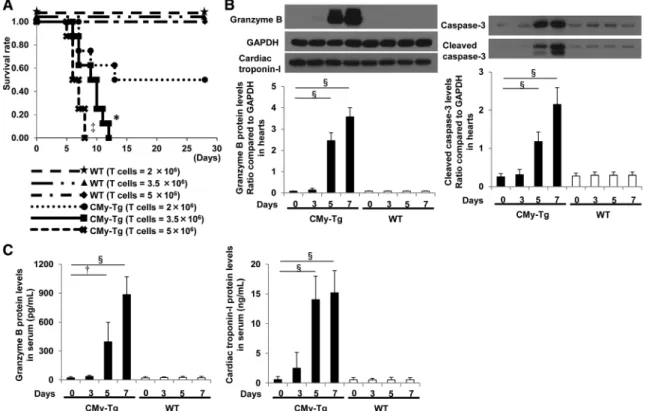
Although all WT mice survived, all CMy-Tg mice injected with 5.0×10⁶ or 3.5×10⁶ OT-1 cytotoxic T-lymphocyte cells
died within 8 to 12 days. Half the CMy-Tg mice injected with 2.0×10⁶ of OT-1 CD8+ T cells survived for 28 days
(Figure 2A). Necropsy revealed ascites, lobulated enlarged livers that associate with hepatic congestion, and foamy lungs consistent with pulmonary edema in all mice that succumbed to myocarditis. Five and 7 days after the transfer of 3.5×10⁶
OT-1 CD8+ T cells, CMy-Tg mouse hearts had higher
con-centrations of granzyme B and cleaved caspase 3 than WT mouse hearts (Figure 2B). CMy-Tg mouse serum also had higher concentrations of granzyme B and cardiac troponin-I than serum of WT mice (Figure 2C).
Dexamethasone Mitigates the Severity of Myocarditis
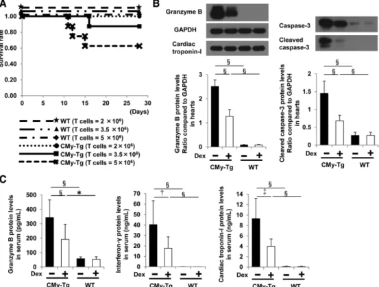
Dexamethasone (0.75 mg/kg IP) for 4 days post transfer of OT-1 cytotoxic T lymphocytes increased survival of CMy-Tg mice. Similar to WT mice, all dexamethasone-treated CMy-Tg mice injected with 2.0×10⁶ OT-1 cells survived for 28 days
(Figure 3A). The injection of 3.5×10⁶ or 5×10⁶ OT-1 cells
into dexamethasone-treated CMy-Tg mice yielded a survival
Figure 2. Characterization of CD8+T cell–mediated myocarditis. A, Kaplan–Meier survival curves of CMy-mOva transgenic (CMy-Tg)
and wild-type (WT) mice after 2×10⁶, 3.5×10⁶, or 5×10⁶ CD8+ cytotoxic T lymphocytes (each group: n=8). Time reflects days after the CD8+
T cell injection. The P value compared CMy-Tg mice receiving 2×10⁶ CD8+ T cells. B, Western blot analysis of granzyme B and cleaved
caspase-3 protein levels in the hearts of the CMy-Tg and WT mice after 0, 3, 5, or 7 days from the injection of 3.5×10⁶ CD8+T cells (each
group: n=6). The data are normalized to GAPDH. Closed bars indicate the CMy-Tg mice groups, and open bars indicate the WT mice
groups. C, Evaluation of granzyme B and cardiac troponin-I levels in the serum of mice (each group: n=6). The P value is compared with
the baseline of each group. *P<0.05, †P<0.001, ‡P<0.005, §P<0.0001.
rate between 0.6 and 0.8 (Figure 3A). Untreated mice ex-perienced a much lower survival rate (Figure 2A). CMy-Tg mouse hearts had higher protein levels of granzyme B and active caspase-3 than WT mouse hearts 5 days post trans-fer of 3.5×10⁶ OT-1 CD8+ T cells. Dexamethasone reduced
these concentrations (Figure 3B). CMy-Tg mice had higher serum concentrations of granzyme B, interferon-γ, and car-diac troponin-I than WT mice 5 days post transfer of 3.5×10⁶
OT-1 CD8+ T cells. Dexamethasone treatment limited these
increases (Figure 3C).
The hearts of CMy-Tg mice contained abundant inflam-matory cells after the transfer of 3.5×10⁶ OT-1 CD8+ T cells
(Figure 4A). CMy-Tg mice had significantly higher myocarditis grades assessed histologically than WT mice. Hearts from CMy-Tg groups harbored considerably more CD8+ T cells, CD68+
macrophages, and NIMP-R14+ neutrophils evaluated by
immu-nohistochemical examination than those from the WT groups (Figure 4B). All groups, however, revealed a similar number of scattered CD4+ T cells. Apoptotic cardiac myocytes detected as
the terminal deoxynucleotidyl transferase–mediated dUTP nick end-labeling–positive nuclei increased significantly after T cell transfer in CMy-Tg mice but not in WT mice. A reduction in inflammatory and apoptotic cells in hearts indicated that dexa-methasone mitigated myocarditis (Figure 4C).
The hearts of CMy-Tg mice had higher concentrations of mRNAs that encode cytokines, mediators of apoptosis, and adhesion molecules, such as interferon-γ, tumor necro-sis factor-α, interleukin-2, interleukin-6, caspase 3, caspase 8, BH3-interacting domain death agonist, Fas, vascular cell adhesion molecule-1, and intercellular adhesion molecule-1 than WT mice after the transfer of OT-1 CD8+ T cells.
Dexamethasone limited the expression of these mediators in the hearts (Online Figure II).
In vitro, granzyme B protein concentrations in OT-1 CD8+ T cells (Online Figure IIIA) and their culture
me-dium (Online Figure IIIB) increased above baseline after 48 hours of anti-CD3 stimulation. Dexamethasone did not mute this rise. Yet, granzyme B and cleaved caspase-3 protein levels in CMy-Tg cardiomyocytes cocultured with OT-1 CD8+ T cells (Online Figure IIIC) and granzyme B
protein concentrations in their culture medium (Online Figure IIID) increased above baseline after 24 hours. Dexamethasone suppressed these levels significantly de-pending on the duration of incubation. Dexamethasone also decreased the probe signals that colocalized with granzyme B activity in the CMy-Tg cardiomyocytes cocultured with OT-1 CD8+ T cells, as assessed by confocal microscopy
(Online Figure IIIE).
Figure 3. Dexamethasone administration attenuates CD8+T cell–mediated myocarditis. A, Kaplan–Meier survival curves of
CMy-mOva transgenic (CMy-Tg) and wild-type (WT) mice injected with 2×10⁶, 3.5×10⁶, or 5×10⁶ CD8+ cytotoxic T lymphocytes and
dexamethasone (0.75 mg/kg per day, at days 1, 2, 3, and 4 after CD8+ T cell administration; each group: n=8). Time reflects days
after the CD8+ T cell injection. The P value compares the CMy-Tg mice injected with 2×10⁶ CD8+ T cells and dexamethasone. B,
Western blot analysis of the granzyme B and cleaved caspase-3 protein levels in the hearts of the CMy-Tg and WT mice 5 days
post the injection of 3.5×10⁶ CD8+ T cells (each group of CMy-Tg mouse: n=12; each group of WT mouse: n=4). The mice received
PBS (vehicle) or dexamethasone at days 1, 2, 3, and 4 after the CD8+ T cell injection. Closed bars indicate the vehicle control
group of each mouse, and open bars indicate the dexamethasone group of each mouse. C, Evaluation of granzyme B, interferon-γ,
and cardiac troponin-I concentrations in the serum of mice (each group of CMy-Tg mouse: n=12; each group of WT mouse: n=4). The P value refers to comparison with the vehicle control of each group. *P<0.05, †P<0.001, ‡P<0.005, §P<0.0001. Dex indicates dexamesathone.
The Granzyme B-Sensitive Nanoprobe Reported on Myocarditis and the Effects of Dexamethasone
Twenty-four hours after the probe injection, CMy-Tg mice re-vealed higher signals from the probe in heart tissues and sec-tions than WT mice in the ex vivo FRI (Figure 5A) and in vivo FMT (Figure 5B). The CT fusion images permitted the anatom-ic localization of the fluorescent signals within hearts. Ex vivo FRI imaging of ProSense 680 did not demonstrate a significant difference between CMy-Tg and WT mouse hearts (Online Figure IVA and IVB). The granzyme B-sensitive nanoprobe also reported on the CD8+ T cell–mediated myocardial injury of
CMy-Tg mice injected with 2.0×10⁶ or 5.0×10⁶ OT-1 cytotoxic
T lymphocytes in the in vivo FMT (Online Figure VA and VB). The fluorescent intensities correlated linearly with myocarditis grade (R2=0.591; P<0.001;Online Figure VC). The background
signals minimized 24 hours after the probe injection in both CMy-Tg and WT mice in the in vivo FMT (Online VIA and
VIB). Plasma stability of the probe kept a baseline level ≤24 hours (Online Figure VIC). Immunofluorescence microscopy of heart sections permitted the colocalization of probe signals with CD8 and granzyme B expression (Figure 6). Dexamethasone reduced granzyme B probe activity, as detected in ex vivo FRI, in vivo FMT, and ex vivo florescence microscopy (Figure 5A, 5B, 6; Online Figure VA and VB).
The Granzyme B-Sensitive Nanoprobe Reported on Cardiac Myosin–Induced EAM
Further experiments addressed the ability of the probe to vi-sualize a more chronic autoimmune myocarditis induced by immunization with cardiac myosin. Twenty-one days after initial immunization, Balb/cByJ mice had severe myocardi-tis shown by hematoxylin and eosin staining (Figure 7A), and contained more myocardial neutrophils, macrophages, and CD4+ T cells as assessed by immunohistochemical
examination than WT mice (Figure 7B). In this form of
Figure 4. Immunohistochemical characterization of CD8+T cell–mediated myocarditis. A, Evaluation of hematoxylin and eosin
(H&E)–stained heart sections in CMy-Tg and wild-type (WT) mice 5 days after the injection of 3.5×10⁶ CD8+T cells (each group of
CMy-mOva transgenic [CMy-Tg] mice; n=12; each group of WT mice; n=4). The mice received PBS (vehicle) or dexamethasone at days
1, 2, 3, and 4 after the CD8+ T cell injection. Original magnification in the upper panels is ×20. The original magnification in the lower
panels is ×100. Bars indicate 200 μm. The figure shows grading of myocarditis grades in each group. Closed bars indicate the vehicle
control group of each mouse, and open bars indicate the dexamethasone group of each mouse. B, Immunohistochemical analysis
of heart sections in CMy-Tg and WT mice 5 days after the injection of CD8+T cells (each group of CMy-Tg mouse: n=12; each group
of WT mouse: n=4). C, Analysis of myocardiocyte apoptosis in heart sections in CMy-Tg and WT mice 5 days after the injection of
the CD8+T cells (each group of CMy-Tg mouse: n=12; each group of WT mouse: n=4). The blue signals indicate nuclei stained with
4′,6-diamidino-2-phenylindole. The red signals indicate cardiac troponin-I positive cardiomyocytes. The green signals indicate the terminal deoxynucleotidyl transferase–mediated dUTP nick end-labeling (TUNEL)–positive nuclei. The P value refers to comparison with the vehicle control of each group. †P<0.001, ‡P<0.005, §P<0.0001. IHC indicates immunohistochemistry.
chronic myocarditis, the heart sections had only scattered CD8+ cells, and lower amounts of granzyme B and cleaved
caspase-3 protein when compared with those measured in the acute CD8+ T cell–induced myocarditis (Figure 7C).
Twenty-four hours after the probe injection, Balb/cByJ mice revealed higher signals from the probe in heart tissues and sections than WT mice in the ex vivo FRI (Figure 7D). In keeping with the histological and biochemical results, the molecular imaging signal in the chronic immune myocar-ditis was less pronounced than in the acute OT-1 CD8+ T
cell–induced disease.
Specificity of the Granzyme B-Sensitive Nanoprobe In Vitro and In Vivo
In vitro, granzyme B protein concentrations in Balb/c WT CD8+ T cells increased above baseline after 48 hours of
anti-CD3 stimulation, whereas Balb/c granzyme B–deficient CD8+
T cells did not show this rise (Online Figure VIIA). The fluo-rescent signal from the probe incubated with the lysates of WT CD8+ T cells stimulated with anti–CD-3 increased above
baseline after 48 hours, yet decreased significantly, and in a concentration-dependent manner, when incubated with gran-zyme B inhibitors (Online Figure VIIB). When incubated with the lysates of granzyme B–deficient CD8+ T cells, the
nano-probe did not demonstrate a comparable increase. In vivo, nonspecific probe activation was observed in the stomach of both WT- and granzyme B–deficient mice at 24 hours after the probe injection (Online Figure VIIC).
Discussion
Endomyocardial biopsies assist in the clinical evaluation of acute myocarditis and the rejection of cardiac allografts. Yet this procedure entails the potential for complications and sampling errors. Current noninvasive imaging approaches to evaluating myocardial inflammation, including ultrasound and nuclear and magnetic resonance imaging, lack molecular specificity. This study developed, optimized, and validated a fluorogenic molecular imaging agent that visualizes granzyme B activity, a target directly involved in CD8+ T cell–mediated
myocardiocytolysis. The inclusion of a cleavage sequence
Figure 5. Investigation of the use of the nanoprobe in CD8+T cell–mediated myocarditis. A, Ex vivo fluorescence reflectance imaging
(FRI) of the heart tissues and sections, and immunofluorescent staining (IF) of heart sections in CMy-mOva transgenic (CMy-Tg) and
wild-type (WT) mice 5 days after the injection of 3.5×10⁶ CD8+T cells (each group of CMy-Tg mouse: n=12; each group of WT mice: n=4).
The mice received PBS (vehicle) or dexamethasone at days 1, 2, 3, and 4 intraperitoneally and the probe at day 4 intravenously after
the CD8+ T cell injection. The left panels show the white images and the right panels show the color images. The figure shows the mean
signal intensities. In IF, the blue signals indicate nuclei stained with 4′,6-diamidino-2-phenylindole and the red signals indicate the probe location. Closed bars indicate the vehicle control group of each mouse, and open bars indicate the dexamethasone group of each mouse.
B, In vivo fluorescent molecular tomography in conjunction with coregistered computed tomographic (FMT-CT) images for CMy-Tg and
WT mice 5 days after the injection of 3.5×10⁶ CD8+T cell (each group of CMy-Tg mouse: n=12; each group of WT mouse: n=4). The
left panels show two-dimensional (2D) images and the right panels show 3D images. The P value refers to comparison with the vehicle control of each group. *P<0.05, †P<0.001, §P<0.0001. IF indicates immunofluorescent staining; and RGB, red-green-blue color.
derived from caspase 3, one of the intracellular targets of the enzyme, enhances the signal produced by this imaging probe. Without activation by granzyme B, this probe displays mini-mal fluorescence.
The purified enzyme and medium harvested from cocul-tures of Ova-specific CD8+ cytotoxic T-lymphocyte cells and
ovalbuin-expressing cardiomyocytes isolated from CMy-Tg mice validated the imaging agent in vitro. The incubation of probes with purified granzyme B yielded an almost 4-fold increase in fluorescence signal in solution. The incubation of probes with lysates of CD8+ T cells affirmed its
selec-tivity for granzyme B. Cocultures of transgenic mouse cells allowed for further validation of the probe. The cardiomyo-cytes from the CMy-Tg mice present ovalbumin peptides in conjunction with major histocompatibility complex class-1 molecules on the cell surface, which activates the receptor on the surface of the CD8+ T lymphocytes. This process triggers
the release of granzyme B, with incubation time directly cor-relating with the extent of release. Incubation of cocultures with the fluorogenic probe caused a concentration-dependent increase in fluorescence signal. These data corroborated the results obtained with the purified enzyme.
The amount of CD8+ T cells transferred into CMy-Tg
mice related directly with myocarditis severity and lethality. The injection of fewer CD8+ T cells into CMy-Tg mice
trig-gered the development of transient heart inflammation and recovery without apparent sequelae. The transfer of a larger number of cells, however, proved lethal. Our previous inves-tigation demonstrated maximal cardiac damage at 96 to 144 hours after transfer.10 This study used conditions that
yield-ed increasyield-ed granzyme B expression and producyield-ed cardiac damage while minimizing mortality 5 days after transfer. In vivo FMT imaging of the time course of probe uptake, enzymatic cleavage, and clearance informed the choice of 24 hours postinjection to study the nanoprobe, a time that allows localization and activation while minimizing back-ground signals. The nanoprobe remains stable in serum for over 24 hours without displaying activation. The injection of myocarditic mice with the probe yielded a significant fluo-rescence signal localized to the heart via noninvasive FMT-CT imaging, whereas control WT mice exhibited no signal. In mice with mild myocarditis, the weak fluorescence signal only covered a small region of the heart. Administration of the probe to WT or granzyme B–deficient mice yielded a
Figure 6. Demonstration of probe specificity in CD8+T cell–mediated myocarditis. Immunofluorescent staining in heart sections in
CMy-mOva transgenic (CMy-Tg) and wild-type (WT) mice 5 days after the injection of 3.5×10⁶ CD8+T cells (each group of CMy-Tg mouse:
n=12; each group of WT mouse: n=4). The mice received PBS (vehicle) or dexamethasone at days 1, 2, 3, and 4 intraperitoneally and the
probe at day 4 intravenously after CD8+ T cell injection. Original magnification in the upper panels is ×100. Bars indicate 200 μm. The
blue signals indicate nuclei stained with 4′,6-diamidino-2-phenylindole. The green signals indicate the location of granzyme B, CD8, or NIMP-R14. The red signals indicate the location of the probe. The lower graph shows the probe colocalization with granzyme B, CD8, or NIMP-R14 positive area quantified using ImageJ software. Closed bars indicate the vehicle control group of each mouse, and open bars indicate the dexamethasone group of each mouse. The P value is compared with the vehicle control of each group. *P<0.05, ‡P<0.005, §P<0.0001.
fluorescence signal in the abdomen, regardless of the pres-ence of myocardial disease or the enzyme of interest, in-dicating other metabolic clearance pathways for the agent. Further investigation localized fluorescence to the stomach. The enzyme survey we conducted pointed to chymotrypsin as responsible for probe activation in the gastrointestinal tract. As demonstrated in the in vitro findings, chymotryp-sin breaks down the probe, and the enzyme is present in the intestine. Acid hydrolysis may also activate the probe in the stomach. Chymotrypsin should not give rise to a signal in the cardiovascular system, rendering this activity unlikely to confound the use of the probe described here for investi-gation of myocarditis. FRI and fluorescence microscopy ex vivo corroborated the in vivo findings. Immunofluorescent staining of heart sections further demonstrated the colocal-ization of the probe signal with both granzyme B and CD8+
T cells. Although previous studies show the expression of granzyme B by neutrophils,19 the colocalization of the probe
signal reported on fewer neutrophils than CD8+ T cells in
this study. This study also compared the capabilities of the granzyme B probe to the previously studied protease sensor, ProSense 680, which readily visualized macrophage host responses in the setting of acute rejection of mouse heart al-lografts, a close parallel the CD8+ T cell–mediated
myocar-dial injury in this study.20 The granzyme B-sensitive probe
demonstrated a substantially higher signal in this model than ProSense 680. Further study should evaluate the use of this probe in acute rejection of cardiac allografts and in viral myocarditis.
This study used dexamethasone as a model anti-inflam-matory agent to determine if the fluorogenic granzyme B nanoprobe can serve as a molecular monitor of therapeutic efficacy. Previous studies revealed that treatment with im-munosuppressive agents may improve the prognosis of giant cell myocarditis21 and that early administration of
dexametha-sone might have utility for the treatment of fulminant viral
Figure 7. Nanoprobe efficacy in experimental cardiac myosin-induced autoimmune myocarditis (EAM). A, Evaluation of hematoxylin
and eosin (H&E)–stained heart sections in the Balb/cByJ mice 21 days after immunization with a peptide derived from the murine α-myosin H chain (the EAM mice: n=16; the control mice: n=4). The control mice received PBS instead of the immunization. Original magnification in the upper panels is ×20. Original magnification in the right panels is ×100. Bars indicate 200 μm. The figure shows the
myocarditis grades of H&E staining in each group. Closed bars indicate the EAM group, and open bars indicate the control group. B,
Immunohistochemical analysis of heart sections in EAM and control mice. Total positive area was quantified using ImageJ software. C,
Western blotting analysis of the granzyme B and cleaved caspase-3 protein levels in heart extracts. D, Ex vivo fluorescence reflectance
imaging (FRI) for the heart sections in EAM and control mice. The mice received the probe intravenously at day 20. One day after the administration, the samples isolated from euthanized mice were assessed by microscopic ex vivo FRI. The left panels show the white images and the right panels show the color images. The figure shows the mean signal intensities. The P value refers to comparison to the control. *P<0.05, ‡P<0.005, §P<0.0001. RGB indicates red-green-blue color.
myocarditis.17 Dexamethasone also acts on leukocytes and
endothelial cells to attenuate the leukocyte–endothelial cell interactions and reduces the generation and release of proin-flammatory cytokines and mediators.17 In vitro, incubation of
cocultures with dexamethasone limited CD8+ T
cell–cardio-myocyte interactions and reduced granzyme B expression in the targeted cardiomyocytes, depending on the duration of ex-posure. These experiments demonstrated that dexamethasone does not modulate granzyme B expression in CD8+ T cells
after anti-CD3 stimulation. The treatment of myocarditic mice with dexamethasone and the nanoprobe yielded a significant reduction in fluorescence signal within the heart in vivo when compared with control mice receiving saline. Mice receiving this agent also displayed decreased expression of inflamma-tory cytokines, adhesion molecules, and apoptotic pathways within the heart tissue.
Experimental cardiac myosin–induced EAM results in a more chronic form of the myocarditis.10 When mice
bearing this disease were treated with the fluorogenic nanoprobe, longer exposure times were required to acquire an appropriate image via ex vivo FRI, indicating reduced probe activation. CD4+ T cells that do not release granzyme
B mainly mediate EAM. The signal observed in hearts with EAM originates from the CD8+ T cells are 6-fold less
abun-dant than CD4+ T cells, further affirming the specificity of
this probe.
In conclusion, these studies generated and validated a nov-el fluorogenic probe for the detection of granzyme B activity in vivo in mice with myocarditis. The fluorescent probe should prove useful to evaluate pathogenic mechanisms and evaluate experimental therapies in mice. With respect to clinical trans-lation, the depth dependence of fluorescence imaging current-ly limits the study of large animal or human hearts. Yet, the validation of granzyme B as a novel molecular imaging target presented here justifies future efforts to develop more readily translated magnetic resonance or radionuclide methods. This study establishes firmly the principle that a molecular target related to a particular pathophysiologic pathway involved in immune-mediated acute myocarditis can enable noninvasive imaging of this process in vivo. These results point the way toward the future development of further novel tools that can investigate of the mechanisms of immune-mediated cardiac processes, including acute cardiac transplant rejection, and evaluate the effects of therapeutic interventions.
Acknowledgments
We thank Chelsea Swallom for her editorial contributions.
Sources of Funding
This study was supported, in part, by National Institutes of Health National Heart, Lung, and Blood Institute contract HHSN268201000044C and R01HL121363-01, Grant-in-Aid for Scientific Research from the National Institutes of Health; Translational Program of Excellence in Nanotechnology, Japan Society for the Promotion of Science; Strategic Young Researcher Overseas Visits Program for Accelerating Brain Circulation, and The Uehara Memorial Foundation; Research Fellowship.
Disclosures
None.
References
1. Magnani JW, Danik HJ, Dec GW Jr, DiSalvo TG. Survival in biopsy-prov-en myocarditis: a long-term retrospective analysis of the histopathologic, clinical, and hemodynamic predictors. Am Heart J. 2006;151:463–470. doi: 10.1016/j.ahj.2005.03.037.
2. Adsett M, West MJ, Galbraith A, Duhig E, Lange A, Palka P. Eosinophilic heart: marked left ventricular wall thickening and myocardial dys-function improving with corticosteroid therapy. Echocardiography. 2003;20:369–374.
3. Dec GW, Palacios I, Yasuda T, Fallon JT, Khaw BA, Strauss HW, Haber E. Antimyosin antibody cardiac imaging: its role in the diagnosis of myocar-ditis. J Am Coll Cardiol. 1990;16:97–104.
4. Margari ZJ, Anastasiou-Nana MI, Terrovitis J, Toumanidis S, Agapitos EV, Lekakis JP, Nanas JN. Indium-111 monoclonal antimyosin cardiac scintigraphy in suspected acute myocarditis: evolution and diagnostic im-pact. Int J Cardiol. 2003;90:239–245.
5. Mahrholdt H, Goedecke C, Wagner A, Meinhardt G, Athanasiadis A, Vogelsberg H, Fritz P, Klingel K, Kandolf R, Sechtem U. Cardiovascular magnetic resonance assessment of human myocarditis: a comparison to histology and molecular pathology. Circulation. 2004;109:1250–1258. doi: 10.1161/01.CIR.0000118493.13323.81.
6. Abdel-Aty H, Boyé P, Zagrosek A, Wassmuth R, Kumar A, Messroghli D, Bock P, Dietz R, Friedrich MG, Schulz-Menger J. Diagnostic perfor-mance of cardiovascular magnetic resonance in patients with suspected acute myocarditis: comparison of different approaches. J Am Coll Cardiol. 2005;45:1815–1822. doi: 10.1016/j.jacc.2004.11.069.
7. Nahrendorf M, Sosnovik DE, Waterman P, Swirski FK, Pande AN, Aikawa E, Figueiredo JL, Pittet MJ, Weissleder R. Dual channel optical tomo-graphic imaging of leukocyte recruitment and protease activity in the heal-ing myocardial infarct. Circ Res. 2007;100:1218–1225. doi: 10.1161/01. RES.0000265064.46075.31.
8. Sosnovik DE, Nahrendorf M, Deliolanis N, Novikov M, Aikawa E, Josephson L, Rosenzweig A, Weissleder R, Ntziachristos V. Fluorescence tomography and magnetic resonance imaging of myocardial macrophage infiltration in infarcted myocardium in vivo. Circulation. 2007;115:1384– 1391. doi: 10.1161/CIRCULATIONAHA.106.663351.
9. Nahrendorf M, Waterman P, Thurber G, Groves K, Rajopadhye M, Panizzi P, Marinelli B, Aikawa E, Pittet MJ, Swirski FK, Weissleder R. Hybrid in vivo FMT-CT imaging of protease activity in atherosclerosis with custom-ized nanosensors. Arterioscler Thromb Vasc Biol. 2009;29:1444–1451. doi: 10.1161/ATVBAHA.109.193086.
10. Grabie N, Delfs MW, Westrich JR, Love VA, Stavrakis G, Ahmad F, Seidman CE, Seidman JG, Lichtman AH. IL-12 is required for differentia-tion of pathogenic CD8+ T cell effectors that cause myocarditis. J Clin
Invest. 2003;111:671–680. doi: 10.1172/JCI16867.
11. Love VA, Grabie N, Duramad P, Stavrakis G, Sharpe A, Lichtman A. CTLA-4 ablation and interleukin-12 driven differentiation synergistically augment cardiac pathogenicity of cytotoxic T lymphocytes. Circ Res. 2007;101:248–257. doi: 10.1161/CIRCRESAHA.106.147124.
12. Saito Y, Kondo H, Hojo Y. Granzyme B as a novel factor involved in cardiovascular diseases. J Cardiol. 2011;57:141–147. doi: 10.1016/j. jjcc.2010.10.001.
13. Binah O. Immune effector mechanisms in myocardial pathologies. Int J
Mol Med. 2000;6:3–16.
14. Feldman AM, McNamara D. Myocarditis. N Engl J Med. 2000;343:1388– 1398. doi: 10.1056/NEJM200011093431908.
15. Shao F, Yuan H, Josephson L, Weissleder R, Hilderbrand SA. Facile syn-thesis of monofunctional pentamethine carbocyanine fluorophores. Dyes
Pigm. 2011;90:119–122. doi: 10.1016/j.dyepig.2010.12.008.
16. Bogdanov AA Jr, Weissleder R, Frank HW, Bogdanova AV, Nossif N, Schaffer BK, Tsai E, Papisov MI, Brady TJ. A new macromol-ecule as a contrast agent for MR angiography: preparation, proper-ties, and animal studies. Radiology. 1993;187:701–706. doi: 10.1148/ radiology.187.3.8497616.
17. Nakamura H, Kunitsugu I, Fukuda K, Matsuzaki M, Sano M. Diverse stage-dependent effects of glucocorticoids in a murine mod-el of viral myocarditis. J Cardiol. 2013;61:237–242. doi: 10.1016/j. jjcc.2012.11.006.
What Is Known?
• The diagnosis and treatment of immune-mediated myocarditis and the rejection of cardiac allografts remain clinical challenges. • Endomyocardial biopsies have many drawbacks in assessing cardiac
inflammation.
• The killer T-lymphocyte enzyme granzyme B participates causally in
CD8+ T cell–mediated myocardiocytolysis.
What New Information Does This Article Contribute? • This study developed, optimized, and validated a fluorogenic
molecu-lar imaging agent that visualizes granzyme B activity in mice with experimental immune-mediated myocarditis.
• The results establish that targeting granzyme B activity can enable noninvasive imaging of immune-mediated myocarditis in vivo and monitor a therapeutic intervention.
Endomyocardial biopsies assist in the clinical evaluation of acute myocarditis and the rejection of cardiac allografts. Yet this
procedure risks of complications and sampling errors. Current noninvasive imaging approaches to imaging myocardial inflam-mation generally lack molecular specificity. This study developed, optimized, and validated a fluorogenic molecular imaging agent that visualizes granzyme B activity, a target directly involved in CD8+ T cell–mediated myocardiocytolysis. This probe emits low
fluorescence in its uncleaved, quenched form, but fluorescence brightly when cleaved by granzyme B. The probe produced a signal in hearts of mice with experimental immune-mediated myocarditis that associated with the severity of the lesions. The treatment of mice exhibiting myocarditis with a conventional anti-inflammatory agent, dexamethasone, significantly reduced the cardiac granzyme B signal. This study establishes the principle that a molecular target related to a particular pathophysiologic pathway involved in immune-mediated myocarditis could enable noninvasive imaging of this process in vivo. The findings establish the feasibility of developing molecularly targeted imaging agents to investigate the mechanisms of immune-mediated cardiac dis-eases, and evaluate the effects of therapies.
Novelty and Significance
18. Casciola-Rosen L, Garcia-Calvo M, Bull HG, Becker JW, Hines T, Thornberry NA, Rosen A. Mouse and human granzyme B have distinct tetrapeptide specificities and abilities to recruit the bid pathway. J Biol
Chem. 2007;282:4545–4552. doi: 10.1074/jbc.M606564200.
19. Wagner C, Iking-Konert C, Denefleh B, Stegmaier S, Hug F, Hänsch GM. Granzyme B and perforin: constitutive expression in human poly-morphonuclear neutrophils. Blood. 2004;103:1099–1104. doi: 10.1182/ blood-2003-04-1069.
20. Christen T, Nahrendorf M, Wildgruber M, Swirski FK, Aikawa E, Waterman P, Shimizu K, Weissleder R, Libby P. Molecular imag-ing of innate immune cell function in transplant rejection. Circulation. 2009;119:1925–1932. doi: 10.1161/CIRCULATIONAHA.108.796888. 21. Kindermann I, Barth C, Mahfoud F, Ukena C, Lenski M, Yilmaz A,
Klingel K, Kandolf R, Sechtem U, Cooper LT, Böhm M. Update on myocarditis. J Am Coll Cardiol. 2012;59:779–792. doi: 10.1016/j. jacc.2011.09.074.